. 2017 Sep 15;8(1):554.
doi: 10.1038/s41467-017-00694-2.

The catalytic subunit of DNA polymerase δ inhibits γTuRC activity and regulates Golgi-derived microtubules

Affiliations

The catalytic subunit of DNA polymerase δ inhibits γTuRC activity and regulates Golgi-derived microtubules

Yuehong Shen et al. Nat Commun. .

Abstract

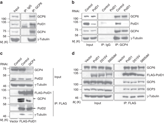
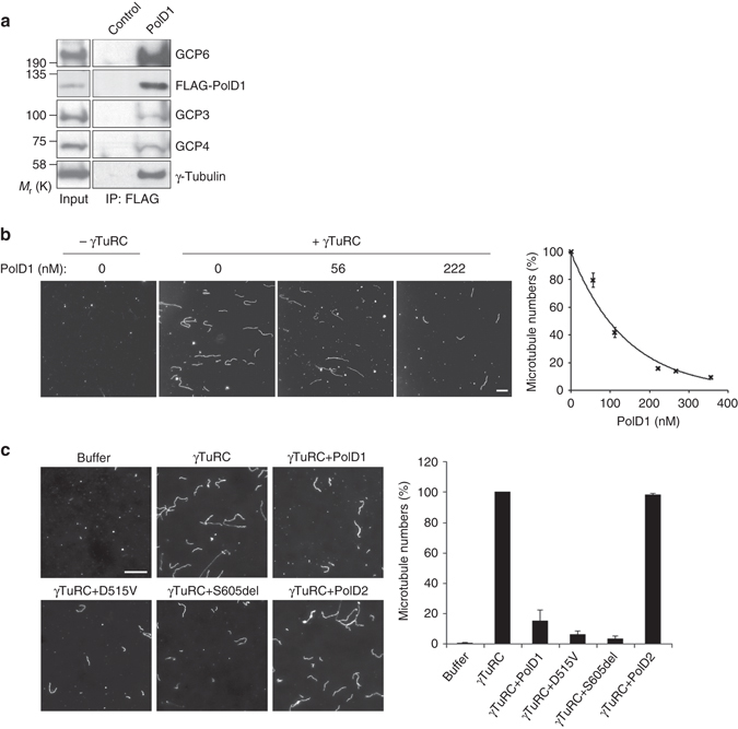
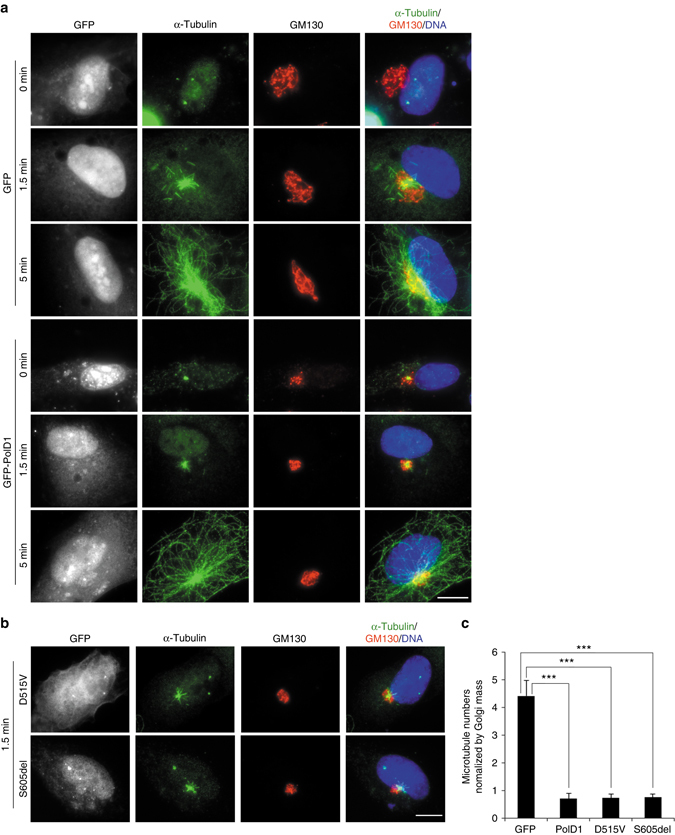
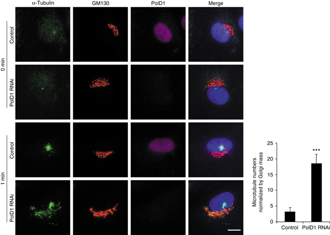
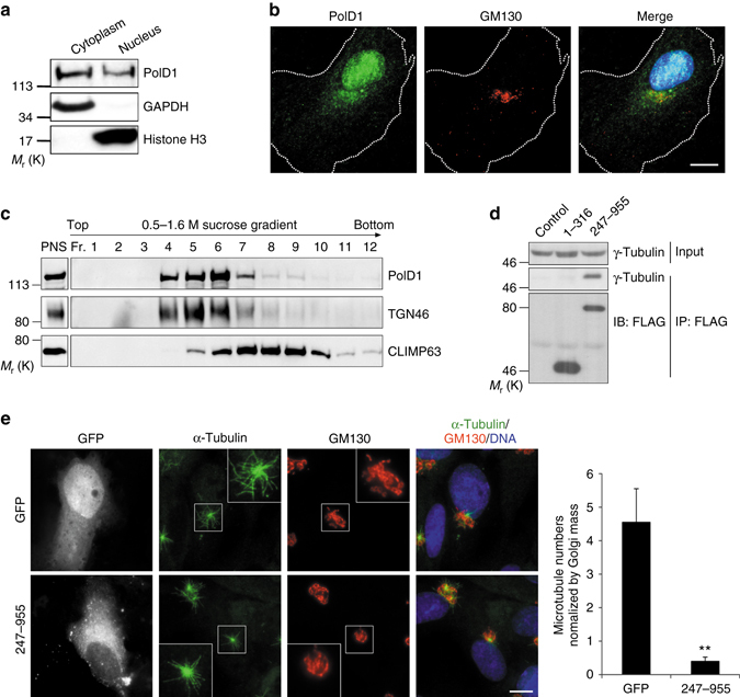
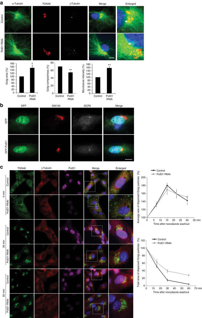
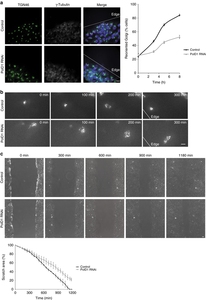
γ-Tubulin ring complexes (γTuRCs) initiate microtubule growth and mediate microtubule attachment at microtubule-organizing centers, such as centrosomes and the Golgi complex. However, the mechanisms that control γTuRC-mediated microtubule nucleation have remained mostly unknown. Here, we show that the DNA polymerase δ catalytic subunit (PolD1) binds directly to γTuRCs and potently inhibits γTuRC-mediated microtubule nucleation. Whereas PolD1 depletion through RNA interference does not influence centrosome-based microtubule growth, the depletion augments microtubule nucleation at the Golgi complex. Conversely, PolD1 overexpression inhibits Golgi-based microtubule nucleation. Golgi-derived microtubules are required for the assembly and maintenance of the proper Golgi structure, and we found that alteration of PolD1 levels affects Golgi structural organization. Moreover, suppression of PolD1 expression impairs Golgi reassembly after nocodazole-induced disassembly and causes defects in Golgi reorientation and directional cell migration. Collectively, these results reveal a mechanism that controls noncentrosomal γTuRC activity and regulates the organization of Golgi-derived microtubules.Microtubule organization requires γ-tubulin ring complexes (γTuRCs), but the mechanisms that control γTuRC-mediated microtubule nucleation are unclear. Here the authors show that the DNA polymerase δ catalytic subunit controls noncentrosomal γTuRC activity and regulates the organization of Golgi-derived microtubules.


Conflict of interest statement

The authors declare no competing financial interests.

Figures

Fig. 1
Fig. 1
PolD1 associates with γTuRCs. a Anti-GCP6 immunoprecipitation was performed using HEK293T cell extracts; normal rabbit IgG was used in the control immunoprecipitation. The immunoprecipitates were immunoblotted with the indicated antibodies. b HEK293T cells were transfected with control or pold1-targeting siRNAs, and then immunoprecipitation was performed using normal rabbit IgG or anti-GCP4 antibody. c FLAG-PolD1 was expressed in cells that were transfected with a GCP4-specific siRNA (or a control siRNA), and then anti-FLAG immunoprecipitation was performed. The immunoprecipitates and the lysate inputs were immunoblotted with the indicated antibodies. FLAG-PolD1 was detected through anti-FLAG immunoblotting. d Extracts of HEK293T cells ectopically expressing PolD1 or its mutants (D515V and S605del) were used for anti-FLAG immunoprecipitation
Fig. 2
Fig. 2
PolD1 inhibits γTuRC-induced microtubule nucleation in vitro. a The recombinant protein FLAG-PolD1 was tested for binding with purified γTuRCs. FLAG-PolD1 was absent in the control. After anti-FLAG immunoprecipitation, the input γTuRCs and the immunoprecipitates were immunoblotted. b Purified γTuRCs reconstituted with the γTuRC stimulator CDK5RAP2(51–200) were used in the microtubule-nucleating assay together with PolD1 at various concentrations. Representative microscopic fields of polymerized microtubules are shown. The data are presented as means ± s.e.m. of three independent experiments. c Purified γTuRCs were used for microtubule nucleation in the absence of CDK5RAP2(51–200). PolD1 wild-type and mutants (D515V and S605del) and PolD2 were added at an excessive amount (356 nM). Shown are representative images of polymerized microtubules, and the data are presented as means ± s.d. of three independent experiments. Scale bars, 10 μm
Fig. 3
Fig. 3
PolD1 overexpression inhibits Golgi-associated microtubule regrowth. a, b RPE1 cells were transfected with GFP or GFP-PolD1 constructs and then subject to cold-induced microtubule depolymerization, after which microtubule regrowth was performed and examined through immunostaining. Hoechst 33258 was used to stain DNA. In the cells transfected with the PolD1 mutants D515V and S605del, microtubule regrowth was for 1.5 min (b). The images shown represent the phenotypes identified from three experiments (100 cells were analyzed per sample, and cells expressing GFP, GFP-PolD1, or PolD1 mutants at similar levels were selected for analysis). Scale bars, 10 μm. c Golgi-associated microtubules at 1.5 min of regrowth were quantified. The quantified data were normalized by the Golgi mass, and are presented as means ± s.d. from three independent experiments; ***p < 0.001, two-tailed, unpaired student’s t-test
Fig. 4
Fig. 4
PolD1 depletion promotes Golgi-associated microtubule nucleation. A microtubule regrowth assay was performed on RPE1 cells transfected with control or PolD1 siRNAs. The cells were then stained for microtubules (anti-α-tubulin) and GM130. The images shown are representative of at least three experiments (100 cells analyzed per condition). Scale bar, 10 μm. Golgi-associated microtubules at 1 min of regrowth were counted and normalized by the Golgi mass. The data are presented as means ± s.d. and are representative of three independent experiments; ***p < 0.001, two-tailed, unpaired student’s t-test
Fig. 5
Fig. 5
Subcellular distribution of PolD1. a RPE1 cell lysates were fractionated into cytoplasmic (Cytoplasm) and nuclear (Nucleus) fractions, and an equal proportion of each fraction was immunoblotted. The cytoplasmic protein GAPDH and the nuclear protein histone H3 were probed as fractionation controls. b RPE1 cells were double-stained for PolD1 and GM130. The images show a representative cell of at least three experiments; the white dashed lines indicate the cell boundary. The PolD1 intensity at the Golgi is ~ 22% of that in the nucleus. c The PNS from RPE1 cells was fractionated by equilibrium centrifugation over a sucrose gradient (0.5–1.6 M). Membranes were then pelleted, and each fraction (50%) and the PNS (4%) were analyzed. d The PolD1 fragments 1–316 and 247–955 were FLAG-tagged and ectopically expressed in HEK293T cells, and the extracts were used for anti-FLAG immunoprecipitation. The lysate inputs and the immunoprecipitates were analyzed by immunoblotting (IB). Control, FLAG vector. e A microtubule regrowth assay was performed on RPE1 cells transfected with GFP or GFP fused with the PolD1 fragment 247–955. The cells were then stained for microtubules (anti-α-tubulin) and GM130. The images shown are representative of at least three experiments (20 cells expressing GFP or GFP-tagged 247–955 at similar levels were analyzed at 1.5 min of regrowth). The numbers of Golgi-associated microtubules were determined and normalized by the Golgi mass. The data are presented as means ± s.d. and are representative of three independent experiments; ***p < 0.001, two-tailed, unpaired student’s t-test. Scale bars, 10 μm
Fig. 6
Fig. 6
Alteration of PolD1 expression affects Golgi organization. a RPE1 cells were transfected with control or PolD1 siRNAs. Anti-α-tubulin fluorescence intensities were measured at the Golgi area, and the background intensity was obtained from cytoplasmic areas lacking microtubules. Here, the average microtubule intensities per pixel at the Golgi are presented after subtraction of the background. The Golgi area was outlined according to TGN46 staining by using the freehand selection option in ZEN software. Data are shown as means ± s.d. of three experiments; **p < 0.01, *p < 0.05, two-tailed, unpaired student’s t-test; control, n = 60 cells; PolD1 depletion, n = 50 cells per experiment. b Cells were transfected with GFP or GFP-PolD1 and then immunostained for GM130 and GCP6; n = 50 cells from three independent experiments. c RPE1 cells transfected with control or PolD1 siRNAs were treated with nocodazole. After nocodazole washout, Golgi reassembly was examined at the indicated time points. TGN46 immunofluorescence was used to measure the size of the Golgi particles that were not clustered in the Golgi region surrounding the centrosome. Quantification graphs show the average size of the individual particles and the total area of the particles. Data are presented as means ± s.e.m. of three experiments (30 cells analyzed per condition). Scale bars, 10 μm
Fig. 7
Fig. 7
Cell polarization is impaired in PolD1-depleted cells. a In a wound-healing assay, RPE1 cells were immunostained to analyze Golgi reorientation. DNA was stained with Hoechst 33258. White lines mark the scratch edges. The angles indicate orientation within 90° facing the scratch edge. Numbers of cells containing the reoriented Golgi are plotted as means ± s.e.m., and the graph is representative of results obtained in three independent experiments (n = 200 cells for each condition). Scale bar, 20 μm. b Movie frames of RPE1 cell imaging were selected at the indicated time points after scratching. The cells were transfected with Venus-β-1,4-galactosyltransferase. Shown are representatives of 30 observed cells per condition. Scale bar, 10 μm. c A wound-healing assay was performed using RPE1 cells; selected phase-contrast movie frames are shown. The percentages of scratch area at each time point are shown as means ± s.e.m. The graph is representative of results obtained in three independent experiments. Scale bar, 20 μm

Similar articles

Cited by

References

    1. Chabin-Brion K, et al. The Golgi complex is a microtubule-organizing organelle. Mol. Biol. Cell. 2001;12:2047–2060. doi: 10.1091/mbc.12.7.2047. - DOI - PMC - PubMed
    1. Efimov A, et al. Asymmetric CLASP-dependent nucleation of noncentrosomal microtubules at the trans-Golgi network. Dev. Cell. 2007;12:917–930. doi: 10.1016/j.devcel.2007.04.002. - DOI - PMC - PubMed
    1. Rivero S, Cardenas J, Bornens M, Rios RM. Microtubule nucleation at the cis-side of the Golgi apparatus requires AKAP450 and GM130. EMBO J. 2009;28:1016–1028. doi: 10.1038/emboj.2009.47. - DOI - PMC - PubMed
    1. Wang Z, Zhang C, Qi RZ. A newly identified myomegalin isoform functions in Golgi microtubule organization and ER-Golgi transport. J. Cell Sci. 2014;127:4904–4917. doi: 10.1242/jcs.155408. - DOI - PMC - PubMed
    1. Miller PM, et al. Golgi-derived CLASP-dependent microtubules control Golgi organization and polarized trafficking in motile cells. Nat. Cell Biol. 2009;11:1069–1080. doi: 10.1038/ncb1920. - DOI - PMC - PubMed

Publication types

MeSH terms

LinkOut - more resources