. 2017 Sep 15;8(1):548.
doi: 10.1038/s41467-017-00520-9.

Ampk phosphorylation of Ulk1 is required for targeting of mitochondria to lysosomes in exercise-induced mitophagy

Affiliations

Ampk phosphorylation of Ulk1 is required for targeting of mitochondria to lysosomes in exercise-induced mitophagy

Rhianna C Laker et al. Nat Commun. .

Abstract

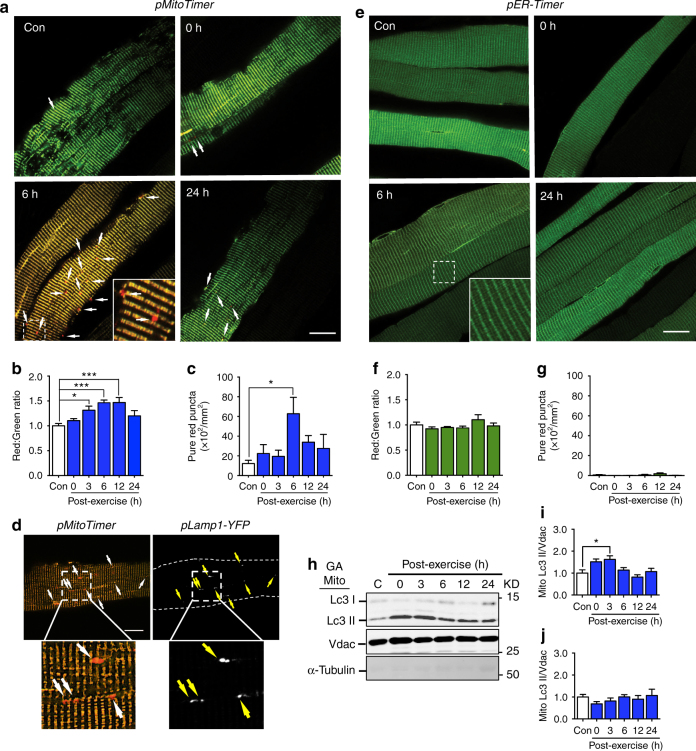
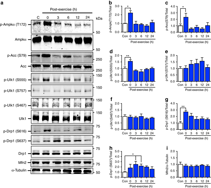
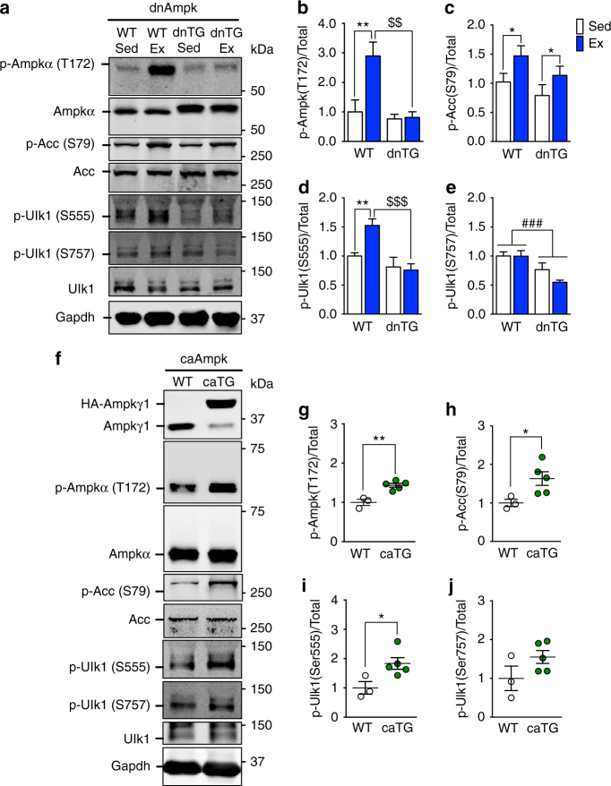
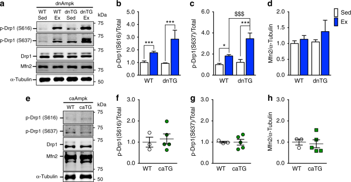
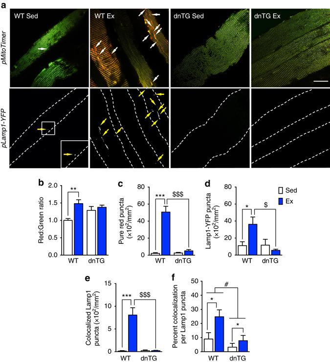
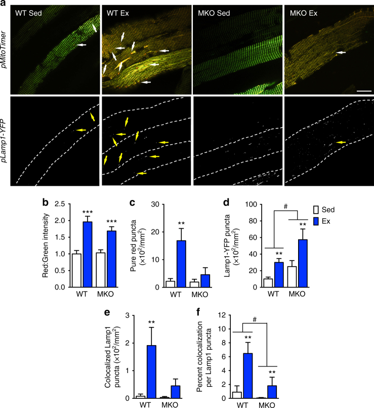
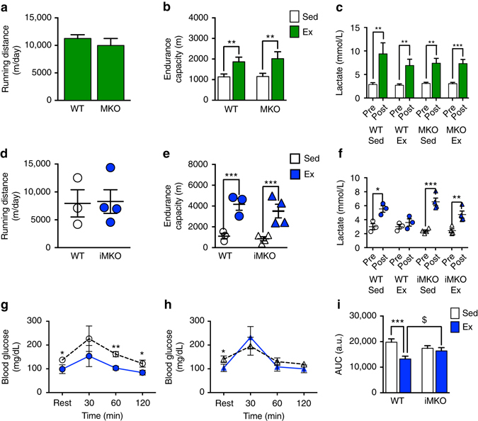
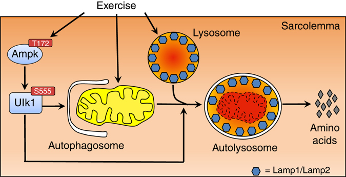
Mitochondrial health is critical for skeletal muscle function and is improved by exercise training through both mitochondrial biogenesis and removal of damaged/dysfunctional mitochondria via mitophagy. The mechanisms underlying exercise-induced mitophagy have not been fully elucidated. Here, we show that acute treadmill running in mice causes mitochondrial oxidative stress at 3-12 h and mitophagy at 6 h post-exercise in skeletal muscle. These changes were monitored using a novel fluorescent reporter gene, pMitoTimer, that allows assessment of mitochondrial oxidative stress and mitophagy in vivo, and were preceded by increased phosphorylation of AMP activated protein kinase (Ampk) at tyrosine 172 and of unc-51 like autophagy activating kinase 1 (Ulk1) at serine 555. Using mice expressing dominant negative and constitutively active Ampk in skeletal muscle, we demonstrate that Ulk1 activation is dependent on Ampk. Furthermore, exercise-induced metabolic adaptation requires Ulk1. These findings provide direct evidence of exercise-induced mitophagy and demonstrate the importance of Ampk-Ulk1 signaling in skeletal muscle.Exercise is associated with biogenesis and removal of dysfunctional mitochondria. Here the authors use a mitochondrial reporter gene to demonstrate the occurrence of mitophagy following exercise in mice, and show this is dependent on AMPK and ULK1 signaling.


Conflict of interest statement

The authors declare no competing financial interests.

Figures

Fig. 1
Fig. 1
Acute exercise causes transient mitochondrial stress and mitophagy during recovery. a Representative images of C57BL/6J mouse (10–12 weeks) FDB fibers transfected with pMitoTimer at time points following acute exercise. Images are merged red and green channels. Scale bar = 20 µm. See also Supplementary Fig. 1. b Quantification of MitoTimer red:green fluorescence intensity and c pure red puncta in n = 7–10 mice per time point. d Representative images of mouse FDB fibers co-transfected with pMitoTimer and pLamp1-YFP at 6 h post exercise illustrating that MitoTimer pure red puncta co-localized with Lamp1-YFP puncta. e Representative images of contralateral mouse FDB fibers transfected with pER-Timer at time points following acute exercise. Scale bar = 20 µm. See also Supplementary Fig. 1. f Quantification of ER-Timer red:green fluorescence intensity and g pure red puncta in n = 5–6 mice per time point. h Representative western blot for Lc3 in mitochondria enriched skeletal muscle fractions. Upper band corresponds to Lc3-I and lower band to Lc3-II, with Vdac as the loading control, at time points following acute exercise. i Quantification of Lc3-II and j Lc3-I protein abundance relative to Vdac, n = 5. See also Supplementary Fig. 5. Data presented as mean ± standard error of the mean. Results of one-way ANOVAs are *p < 0.05, **p < 0.01, and ***p < 0.001. F = 8.07 (b), F = 3.10 (c), F = 1.29 (f), F = 0.91 (g), F = 5.15 (i), F = 0.74 (j). DF = 5 MitoTimer and ER-Timer pure red puncta c, g were log transformed to account for unequal variance
Fig. 2
Fig. 2
Ampk, Ulk1 and Drp1, are transiently activated immediately following acute exercise. Ampk and its substrate Acc are phosphorylated at Thr172 a, b and S79 a, c, respectively, immediately after the cessation of exercise and return to baseline values by 3 h post-exercise in C57BL/6 J mice aged 10–12 weeks. Ulk1 is phosphorylated at Ser555, but not Ser467 or Ser757, immediately following acute exercise, which returns to basal levels by 3 h post-exercise a, df. Drp1 is phosphorylated in skeletal muscle at Ser616 a, g immediately after the cessation of exercise and Ser637 a, h reaching significance 3 and 6 h after exercise. Mfn2 protein abundance is unchanged following acute treadmill running a, i. Representative western blots are shown a and all quantification is presented as protein phosphorylation to total protein abundance ratio bf. Data presented as mean ± standard error of the mean. n = 5 (except 24 h, n = 4). See also Supplementary Figs. 6 and 7. Results of one-way ANOVAs are *p < 0.05, **p < 0.01. F = 6.44 for b, F = 3.56 for c, F = 6.50 for d, F = 0.89 for e, F = 0.33 for f, F = 3.34 for g, F = 2.76 for h, F = 0.31 i. DF = 5. p-Ampk (T172/Total) b was sqrt transformed to account for unequal variance., DF = 5. S637/total Drp1 data was log transformed to account for unequal variance
Fig. 3
Fig. 3
Ampk is necessary and sufficient for phosphorylation of Ulk1 at Ser555 in response to acute exercise. Phosphorylation of Ampk at T172 in response to acute exercise is blocked in Ampk dnTG a, b, whereas increased phosphorylation of Acc at Ser79 post-exercise is still observed a, c. In Ampk caTG mice, both T172 Ampk and S79 Acc are elevated in sedentary conditions fh. Phosphorylation of Ulk1 at Ser555 is blunted in dnTG skeletal muscle immediately after acute exercise a, d but elevated in caTG mice basally f, i. There was no effect on S757 phosphorylation in either dnTG mice post exercise a, e or sedentary caTG mice (f, j). Representative western blots are shown for dnTG mice a and caTG mice f. See also Supplementary Figs. 8 and 9. All quantification is presented as protein phosphorylation to total protein abundance ratio be, gj. Data are presented as mean ± standard error of the mean. For dnTG study n = 10 WT or 5–7 dnTG (12–13 weeks). Results of two-way ANOVAs are *p < 0.05, **p < 0.01 for sedentary vs. exercise comparisons, F = 4.57 and DF = 1 c. ### p < 0.001 for between group comparisons, F = 14.03 and DF = 1 (e). Tukey multiple comparison tests were performed when a significant interaction effect was observed, in which *p < 0.05, **p < 0.01 for WT sedentary vs. WT exercise comparisons and $$ p < 0.01, $$$ p < 0.001 for WT exercise vs. dnTG exercise comparisons, DF = 27. For caTG study n = 3 (WT) or 5 (caTG) (13–15 weeks). Results of two-tailed t-tests are *p < 0.05 and **p < 0.01 for WT to caTG comparisons, t = 4.94 (g), t = 2.57 (h), t = 2.71 (i), t = 1.72 (j), DF = 6
Fig. 4
Fig. 4
Ampk is not required for exercise-induced phosphorylation of Drp1. Phosphorylation of Drp1 at S616 and S637 is significantly increased in dnTG mice post-exercise (ac), while there was no effect of caTG Ampk on phosphorylation of Drp1 at either S616 e, f or S637 e, g under sedentary conditions. There was no effect on Mfn2 protein abundance in either dnTG mice, regardless of exercise d, or sedentary caTG mice h. Representative western blots are shown a, e and all quantification is presented as protein phosphorylation to total protein abundance ratio b, c, f, g or total protein d, h. Data are presented as mean ± standard error of the mean. For dnTG study n = 10 WT or 5–7 dnTG (12–13 weeks). See also Supplementary Fig. 10. Results of two-way ANOVAs are *p < 0.05, ***p < 0.001, for sedentary vs. exercise comparisons, F = 19.39, DF = 1 (b). Tukey multiple comparison tests were performed when a significant interaction effect was observed, in which *p < 0.05, ***p < 0.001 for WT sedentary vs. WT exercise comparisons and $$$ p < 0.001 for WT exercise vs. dnTG exercise comparisons, DF = 26. For caTG study n = 3 WT or 5 caTG (13–15 weeks). Results of two-tailed t-tests are t = 0.42 (b), t = 0.02 (c), t = 0.29 (d), DF = 6
Fig. 5
Fig. 5
Ampk is required for acute exercise-induced mitophagy and lysosomal genesis. a Representative images of WT and Ampk dnTG mouse FDB fibers co-transfected with pMitoTimer and Lamp1-YFP. Scale bar = 20 µm. Quantification of b red:green fluorescence intensity (interaction effect p = 0.03), c pure red MitoTimer puncta per fiber area (interaction effect p < 0.0001), d total Lamp11-YFP puncta per fiber area (interaction effect p = 0.01), e co-localized Lamp1-YFP puncta per fiber area (interaction effect (p = 0.0002), f percent co-localization of MitoTimer pure red puncta with Lamp1-YFP puncta. Data presented as mean ± standard error of the mean. n = 7 WT or 6 dnTG (12–13 weeks). Results of two-way ANOVAs are *p < 0.05 for sedentary vs. exercise comparisons, F = 5.97, DF = 1 f and # p < 0.05 for between group comparisons, F = 7.41, DF = 1 (f). Tukey multiple comparison tests were performed when a significant interaction effect was observed, in which *p < 0.05, **p < 0.01, ***p < 0.001 for WT sedentary vs. WT exercise comparisons and $$$ p < 0.001 for WT exercise vs. dnTG exercise comparisons, DF = 22
Fig. 6
Fig. 6
Ulk1 is required for acute exercise-induce mitophagy in skeletal muscle. a Representative images of WT and Ulk1 MKO mouse FDB fibers co-transfected with pMitoTimer and Lamp1-YFP. Scale bar = 20 µm. Quantification of b red:green fluorescence intensity, c pure red MitoTimer puncta (interaction effect p = 0.03), d total Lamp1-YFP puncta per fiber area, e co-localized Lamp1-YFP puncta per fiber area (interaction effect p = 0.1), and f percent co-localization of MitoTimer pure red puncta with Lamp1-YFP puncta (interaction effect p = 0.05). Data presented as mean ± standard error of the mean. n = 6 (12–13 weeks), **p < 0.01 and ***p < 0.001 for sedentary vs. exercise comparisons. # p < 0.05 for between group comparisons
Fig. 7
Fig. 7
Ulk1 in skeletal muscle is important for exercise training-induced metabolic adaptation. MKO and iMKO mice displayed normal adaptation to exercise training as assessed by daily running distance a, d, exhaustive exercise test b, e and blood lactate levels c, f. WT mice exhibited improved glucose tolerance following 4 weeks of exercise training as assessed by blood glucose profile during GTT g and AUC i. In contrast, iMKO mice failed to show improved glucose tolerance following exercise training during GTT h and AUC i. Data presented as mean ± standard error of the mean. For MKO study n = 7–8 WT and n = 6–7 MKO (12–13 weeks). Two-tailed t-tests were used for (A: t = 0.84 and DF = 9) and (C: WT Sed t = 3.73 and DF = 12, WT Ex t = 2.97 and DF = 14, MKO Sed t = 6.35 and DF = 10, MKO Ex t = 5.25, DF = 12). Lactate data c was 1/Y transformed to account for unequal variance. A two-way ANOVA was used for b with **p < 0.01 for sedentary vs. exercise comparison, F = 11.50, DF = 1. For iMKO study n = 3 WT and n = 4 iMKO (18–20 weeks). Two-tailed t-tests were used for (D: t = 0.10 and DF = 5), (F: WT Sed t = 3.98, DF = 4), (F: WT Ex t = 0.98, DF = 4), (F: iMKO Sed t = 7.35, DF = 6), and (F: iMKO Ex t = 3.99, DF = 6). A two-way ANOVA was used for e, i with ***p < 0.001, F = 35.16, and DF = 1 for sedentary vs. exercise comparisons. Tukey multiple comparison test was performed for the significant interaction effect in i, in which ***p < 0.001 for WT sedentary vs. WT exercise comparisons and $ p < 0.05 for WT exercise vs. iMKO exercise comparisons, DF = 10
Fig. 8
Fig. 8
Acute exercise-induced mitophagy is regulated through the Ampk-Ulk1 signaling axis. Ampk activation through phosphorylation at T172 is required for exercise-induced phosphorylation of Ulk1 S555 and lysosomal biogenesis in skeletal muscle. Simultaneously, exercise initiates mitochondrial fission, which is not dependent on Ampk activation. We demonstrate that Ulk1 is critical for targeting of the lysosome to the damage/dysfunctional mitochondrial puncta for subsequent degradation

Similar articles

Cited by

References

    1. Hollozy JO. Biochemical adaptations in muscle. Effects of exercise on mitochondrial oxygen uptake and respiratory enzyme activity in skeletal muscle. J. Biol. Chem. 1967;242:2278–2282. - PubMed
    1. Green HJ, Thomson JA, Daub WD, Houston ME, Ranney DA. Fiber composition, fiber size and enzyme activities in vastus lateralis of elite athletes involved in high intensity exercise. Eur. J. Appl. Physiol. Occup. Physiol. 1979;41:109–117. doi: 10.1007/BF00421658. - DOI - PubMed
    1. Lira VA, Benton CR, Yan Z, Bonen A. PGC-1alpha regulation by exercise training and its influences on muscle function and insulin sensitivity. Am. J. Physiol. 2010;299:E145–E161. - PMC - PubMed
    1. Romanello V, Sandri M. Mitochondrial quality control and muscle mass maintenance. Front. Physiol. 2016;6:1–21. doi: 10.3389/fphys.2015.00422. - DOI - PMC - PubMed
    1. Tseng BS, Marsh DR, Hamilton MT, Booth FW. Strength and aerobic training attenuate muscle wasting and improve resistance to the development of disability with aging. Cell Signal. 2015;50:113–119. - PubMed

Publication types

MeSH terms

Substances