. 2017 Nov;30(6):739-746.
doi: 10.1007/s10548-017-0590-y. Epub 2017 Sep 15.

Physiological Ripples (± 100 Hz) in Spike-Free Scalp EEGs of Children With and Without Epilepsy

Affiliations

Physiological Ripples (± 100 Hz) in Spike-Free Scalp EEGs of Children With and Without Epilepsy

Anne H Mooij et al. Brain Topogr. 2017 Nov.

Abstract

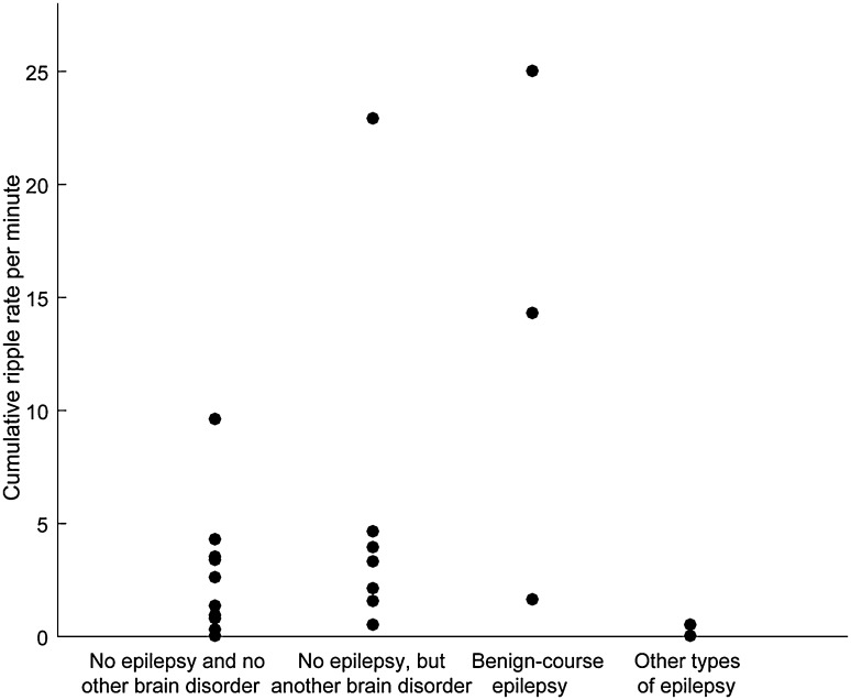
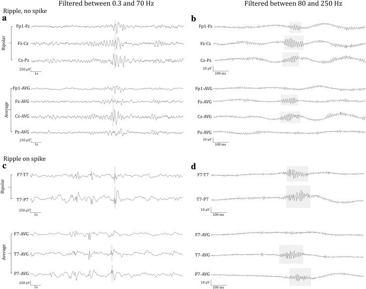
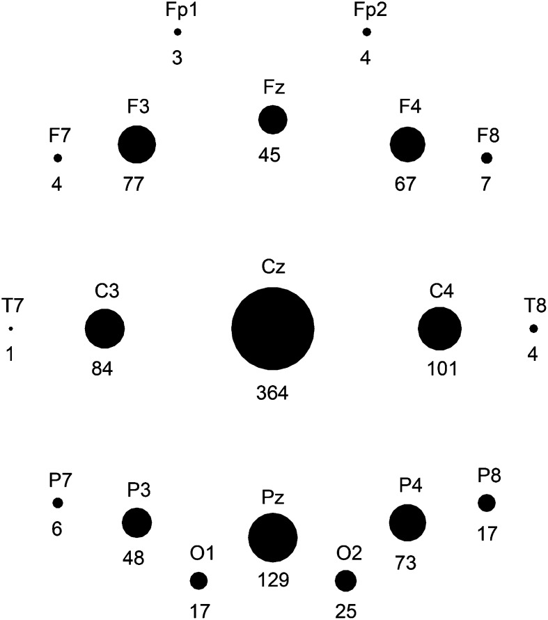
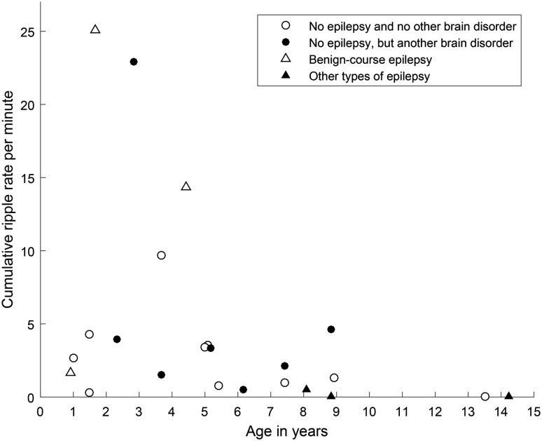
Pathological high frequency oscillations (HFOs, >80 Hz) are considered new biomarkers for epilepsy. They have mostly been recorded invasively, but pathological ripples (80-250 Hz) can also be found in scalp EEGs with frequent epileptiform spikes. Physiological HFOs also exist. They have been recorded invasively in hippocampus and neocortex. There are no reports of spontaneously occurring physiological HFOs recorded with scalp EEG. We aimed to study ripples in spike-free scalp EEGs. We included 23 children (6 with, 17 without epilepsy) who had an EEG without interictal epileptiform spikes recorded during sleep. We differentiated true ripples from spurious ripples such as filtering effects of sharp artifacts and high frequency components of muscle artifacts by viewing ripples simultaneously in bipolar and average montage and double-checking the unfiltered signal. We calculated mean frequency, duration and root mean square amplitude of the ripples, and studied their shape and distribution. We found ripples in EEGs of 20 out of 23 children (4 with, 16 without epilepsy). Ripples had a regular shape and occurred mostly on central and midline channels. Mean frequency was 102 Hz, mean duration 70 ms, mean root mean square amplitude 0.95 µV. Ripples occurring in normal EEGs of children without epilepsy were considered physiological; the similarity in appearance suggested that the ripples occurring in normal EEGs of children with epilepsy were also physiological. The finding that it is possible to study physiological neocortical ripples in scalp EEG paves the way for investigating their occurrence during brain development and their relation with cognitive functioning.

Keywords: Childhood; HFOs; High-frequency oscillations; Seizures; Surface EEG.


Figures

Fig. 1
Fig. 1
Plot of the ripple rate per child in each diagnostic category. Cumulative ripple rate per minute was calculated by taking the total number of ripples (in the 10 min with the maximum number of ripples for that child) and dividing it by 10 min (minus the duration of artifacts during those 10 min). Ripples occurred across all diagnostic categories
Fig. 2
Fig. 2
Examples of ripples marked in this study (b), which do not occur on spikes (see corresponding grey area in (a) and of ripples (d) that occur on epileptic spikes (see corresponding grey area in (c). The EEG with epileptic spikes was not included in this study, the examples are shown for comparison with the ripples shown in (b). The EEG traces in a and c are filtered between 0.3 and 70 Hz, time scale is 15 s per page, amplitude scale is 30 µV per mm. The EEG traces in b and d are filtered between 80 and 250 Hz, time scale is 1 s per page, amplitude scale is 1 µV per mm. Channels are shown in bipolar and average montage. Note the difference in appearance of the regular ripples in b and the more irregular ripples in d
Fig. 3
Fig. 3
Schematic overview of the number of ripples per channel, marked in average montage. Number of ripples is written underneath the black circle that represents the channel, and reflected in the size of the black circle. Ripples occurred most frequently in central and midline channels, particularly Cz
Fig. 4
Fig. 4
Scatterplot of age versus cumulative ripple rate. Ripple rates of children without a neurological diagnosis are plotted as open circles, ripple rates of children without epilepsy, but with another brain disorder as closed circles. Ripple rate of children with benign-course epilepsy are plotted as open triangles, ripple rate of children with other types of epilepsy as closed triangles

References

    1. Alkawadri R, Gaspard N, Goncharova II, Spencer DD, Gerrard JL, Zaveri H, Duckrow RB, Blumenfeld H, Hirsch LJ. The spatial and signal characteristics of physiologic high frequency oscillations. Epilepsia. 2014;55:1986–1995. doi: 10.1111/epi.12851. - DOI - PMC - PubMed
    1. Andrade-Valenca LP, Dubeau F, Mari F, Zelmann R, Gotman J. Interictal scalp fast oscillations as a marker of the seizure onset zone. Neurology. 2011;77:524–531. doi: 10.1212/WNL.0b013e318228bee2. - DOI - PMC - PubMed
    1. Averkin RG, Szemenyei V, Bordé S, Tamás G. Identified cellular correlates of neocortical ripple and high-gamma oscillations during spindles of natural sleep. Neuron. 2016;92:916–928. doi: 10.1016/j.neuron.2016.09.032. - DOI - PMC - PubMed
    1. Buzsáki G. Memory consolidation during sleep: a neurophysiological perspective. J Sleep Res. 1998;7(Suppl 1):17–23. doi: 10.1046/j.1365-2869.7.s1.3.x. - DOI - PubMed
    1. Buzsáki G, Lopes da Silva F. High frequency oscillations in the intact brain. Prog Neurobiol. 2012;98:41–49. doi: 10.1016/j.pneurobio.2012.02.004. - DOI - PMC - PubMed