. 2017 Sep 15:8:291-299.
doi: 10.1016/j.omtn.2017.06.021. Epub 2017 Jul 3.

LncRNA TRERNA1 Function as an Enhancer of SNAI1 Promotes Gastric Cancer Metastasis by Regulating Epithelial-Mesenchymal Transition

Affiliations

LncRNA TRERNA1 Function as an Enhancer of SNAI1 Promotes Gastric Cancer Metastasis by Regulating Epithelial-Mesenchymal Transition

Huazhang Wu et al. Mol Ther Nucleic Acids. .

Abstract

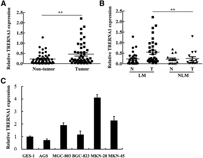
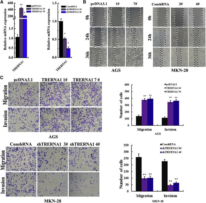
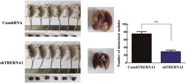
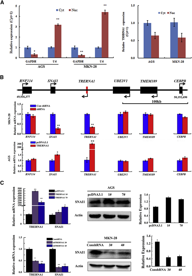
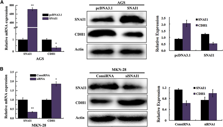
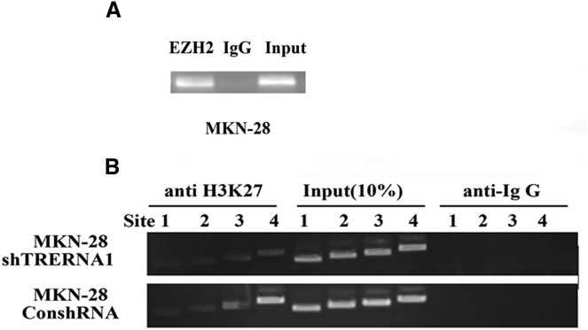
Long noncoding RNA (lncRNA) has been implicated in cancer, but little is known about the role of lncRNAs as regulators of tumor metastasis. In the present study, we demonstrate that lncRNA TRERNA1 acts like an enhancer of SNAI1 to promote cell invasion and migration and to contribute to metastasis of gastric cancer (GC). TRERNA1 is significantly unregulated in GCs and GC cell lines. Increased TRERNA1 is positively correlated with lymph node metastasis of GCs. RNA immunoprecipitation (RIP) and chromatin immunoprecipitation (ChIP) assays revealed that TRERNA1 functions as a scaffold to recruit EZH2 to epigenetically silence epithelial-mesenchymal transition marker CDH1 by H3K27me3 of its promoter region. TRERNA1 knockdown markedly reduced GC cell migration, invasion, tumorigenicity, and metastasis. Depletion of TRERNA1 reduced cell metastasis of GCs in vivo. Taken together, our findings indicated that TRERNA1 serves as a critical effector in GC progression by regulating CDH1 at the transcription level. It is implied that TRERNA1/CDH1 is a new potential target for GC therapy.

Keywords: CDH1; SNAI1; TRERNA1; gastric cancer; metastasis.


Figures

Figure 1
Figure 1
LncRNA TRERNA1 Is Upregulated in GC Tissues and Cells (A) Relative expression of lncRNA TRERNA1 in GC tissues compared with the corresponding non-tumor tissues (n = 48). (B) Patients with LM showed high levels of lncRNA TRERNA1 compared with NLM patients. (C) qRT-PCR analysis of relative expression of lncRNA TRERNA1 in GC cells. lncRNA TRERNA1 expression levels were normalized to β-actin. Data are means ± SD. **p < 0.01. LM, lymph node metastasis; N, non-tumor tissues; NLM, no lymph node metastasis; T, tumor tissues.
Figure 2
Figure 2
TRERNA1 Regulates Gastric Cancer Cell Migration and Invasion In Vitro (A) Overexpression and knockdown efficiency of TRERNA1 after transfection of the overexpression and shRNA construct in AGS and MKN-28 cells. (B) Wound healing assay showed that the enforced expression of TRERNA1 displayed a notably faster recovery compared with control cells in AGS cells (left); conversely, there was a slower recovery in TRERNA1 knockdown clones compared with control in MKN-28 cells (right). (C) TRERNA1 modulated GC cell migration and invasion in vitro by transwell migration and invasion assays in AGS and MKN-28 cells. *p < 0.05; **p < 0.01.
Figure 3
Figure 3
TRERNA1 Stable Knockdown in MKN-28 Cells Significantly Reduced the Number of Visible Nodules on the Lung Surface after Tail Vein Injection in Nude Mice Lungs from each group are shown (left). The middle panel is representative of pulmonary nodules, and the right panel is a statistical analysis of the number of nodules from two groups. Bar graph represents the number of visible nodules on the lung surface. Error bars represent mean ± SD. p < 0.05, **p < 0.01.
Figure 4
Figure 4
TRERNA1 Regulates SNIA1 Function Like an Enhancer (A) The relative distribution of TRERNA1 in the cytoplasm and nucleus as identified using qRT-PCR; GAPDH was used as a cytosolic marker, and U6 was used as a nuclear marker in AGS and MKN-28 cells. (B) TRERNA1 is a regulator of SNAI1 expression. The relative position of the adjacent gene to the TRERNA1 (upper) and the effects on mRNA levels for the surrounding genes after knockdown or enforced expression of TRERNA1 in MKN-28 and AGS cells as determined by qRT-PCR (middle and lower panels) are shown. Scale bar, 100 kb. (C) Western blot assays detected the expression of SNAI1 after the transfection of the overexpression and shRNA TRERNA1 plasmid in GC cell lines. *p < 0.05, **p < 0.01. Cyt, cytoplasm; Nuc, nucleus.
Figure 5
Figure 5
Transcription Factor SNAI1 Represses the Expression of CDH1 (A and B) The expression of CDH1 after transfection of the overexpression construct (A) and siRNA (B) of SNAI1 in AGS and MKN-28 cells detected by qRT-PCR and western blot assays. *p < 0.05; **p < 0.01.
Figure 6
Figure 6
TRERNA1 Regulates EMT Marker Expression in GC Cells (A) qRT-PCR was used to measure expression levels of EMT-related markers (CDH1, N-cadherin, Vimentin, and β-catenin) in AGS and MKN-28 cells, which transfected with construct (pcDNA3.1+TRERNA1) or shRNA-TRERNA and normalized to β-actin expression. (B) TRERNA1 affects the expression of EMT-related genes in GC cells as determined by western blot analysis. *p < 0.05; **p < 0.01.
Figure 7
Figure 7
TRERNA1 Represses Transcription of CDH1 by Recruiting PRC2 Subunits EZH2 to the CDH1 Promoter (A) The TRERNA1 directly interacts with PRC2 subunits EZH2 by RIP assay (RIP experiments using anti-EZH2 antibody and IgG as a negative control were performed in MKN-28 cells, and the coprecipitated RNA was subjected to PCR for TRERNA1). (B) ChIP-PCR of H3K27me3 enrichment of the promoter region of the CDH1 after knockdown TRERNA1 in MKN-28 cells.

References

    1. Ferlay J., Soerjomataram I., Dikshit R., Eser S., Mathers C., Rebelo M., Parkin D.M., Forman D., Bray F. Cancer incidence and mortality worldwide: sources, methods and major patterns in GLOBOCAN 2012. Int. J. Cancer. 2015;136:E359–E386. - PubMed
    1. Bass A.J., Thorsson V., Shmulevich I., Reynolds S.M., Miller M., Bernard B., Hinoue T., Laird P.W., Curtis C., Shen H., Cancer Genome Atlas Research Network Comprehensive molecular characterization of gastric adenocarcinoma. Nature. 2014;513:202–209. - PMC - PubMed
    1. Gupta G.P., Massagué J. Cancer metastasis: building a framework. Cell. 2006;127:679–695. - PubMed
    1. Redlak M.J., Power J.J., Miller T.A. Prevention of deoxycholate-induced gastric apoptosis by aspirin: roles of NF-kappaB and PKC signaling. J. Surg. Res. 2008;145:66–73. - PubMed
    1. Safe S., Abdelrahim M. Sp transcription factor family and its role in cancer. Eur. J. Cancer. 2005;41:2438–2448. - PubMed