

Regulation of mitochondrial bioenergetics by the non-canonical roles of mitochondrial dynamics proteins in the heart
- PMID: 28918113
- PMCID: PMC5851799
- DOI: 10.1016/j.bbadis.2017.09.004
Regulation of mitochondrial bioenergetics by the non-canonical roles of mitochondrial dynamics proteins in the heart
Abstract
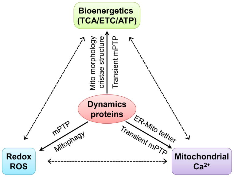
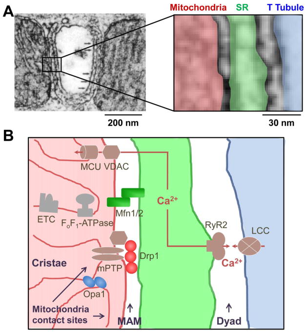
Recent advancement in mitochondrial research has significantly extended our knowledge on the role and regulation of mitochondria in health and disease. One important breakthrough is the delineation of how mitochondrial morphological changes, termed mitochondrial dynamics, are coupled to the bioenergetics and signaling functions of mitochondria. In general, it is believed that fusion leads to an increased mitochondrial respiration efficiency and resistance to stress-induced dysfunction while fission does the contrary. This concept seems not applicable to adult cardiomyocytes. The mitochondria in adult cardiomyocytes exhibit fragmented morphology (tilted towards fission) and show less networking and movement as compared to other cell types. However, being the most energy-demanding cells, cardiomyocytes in the adult heart possess vast number of mitochondria, high level of energy flow, and abundant mitochondrial dynamics proteins. This apparent discrepancy could be explained by recently identified new functions of the mitochondrial dynamics proteins. These "non-canonical" roles of mitochondrial dynamics proteins range from controlling inter-organelle communication to regulating cell viability and survival under metabolic stresses. Here, we summarize the newly identified non-canonical roles of mitochondrial dynamics proteins. We focus on how these fission and fusion independent roles of dynamics proteins regulate mitochondrial bioenergetics. We also discuss potential molecular mechanisms, unique intracellular location, and the cardiovascular disease relevance of these non-canonical roles of the dynamics proteins. We propose that future studies are warranted to differentiate the canonical and non-canonical roles of dynamics proteins and to identify new approaches for the treatment of heart diseases. This article is part of a Special issue entitled Cardiac adaptations to obesity, diabetes and insulin resistance, edited by Professors Jan F.C. Glatz, Jason R.B. Dyck and Christine Des Rosiers.
Keywords: Cardiac bioenergetics; Metabolic heart disease; Mitochondria associated membranes; Mitochondrial dynamics proteins.
Copyright © 2017 Elsevier B.V. All rights reserved.
Figures
Similar articles
-
Mitochondrial dynamism and cardiac fate--a personal perspective.Circ J. 2013;77(6):1370-9. doi: 10.1253/circj.cj-13-0453. Epub 2013 Apr 25. Circ J. 2013. PMID: 23615052 Review.
-
A novel fission-independent role of dynamin-related protein 1 in cardiac mitochondrial respiration.Cardiovasc Res. 2017 Feb;113(2):160-170. doi: 10.1093/cvr/cvw212. Epub 2016 Oct 29. Cardiovasc Res. 2017. PMID: 27794519 Free PMC article.
-
Mitochondrial-Shaping Proteins in Cardiac Health and Disease - the Long and the Short of It!Cardiovasc Drugs Ther. 2017 Feb;31(1):87-107. doi: 10.1007/s10557-016-6710-1. Cardiovasc Drugs Ther. 2017. PMID: 28190190 Free PMC article. Review.
-
Mitochondrial dynamics and cell death in heart failure.Heart Fail Rev. 2016 Mar;21(2):123-36. doi: 10.1007/s10741-016-9530-2. Heart Fail Rev. 2016. PMID: 26872674 Review.
-
Binding of FUN14 Domain Containing 1 With Inositol 1,4,5-Trisphosphate Receptor in Mitochondria-Associated Endoplasmic Reticulum Membranes Maintains Mitochondrial Dynamics and Function in Hearts in Vivo.Circulation. 2017 Dec 5;136(23):2248-2266. doi: 10.1161/CIRCULATIONAHA.117.030235. Epub 2017 Sep 23. Circulation. 2017. PMID: 28942427 Free PMC article.
Cited by
-
Posttranslational modifications of mitochondrial fission and fusion proteins in cardiac physiology and pathophysiology.Am J Physiol Cell Physiol. 2019 May 1;316(5):C583-C604. doi: 10.1152/ajpcell.00523.2018. Epub 2019 Feb 13. Am J Physiol Cell Physiol. 2019. PMID: 30758993 Free PMC article. Review.
-
Neurohormonal connections with mitochondria in cardiomyopathy and other diseases.Am J Physiol Cell Physiol. 2022 Aug 1;323(2):C461-C477. doi: 10.1152/ajpcell.00167.2022. Epub 2022 Jun 27. Am J Physiol Cell Physiol. 2022. PMID: 35759434 Free PMC article. Review.
-
Mitochondria as a therapeutic target for cardiac ischemia‑reperfusion injury (Review).Int J Mol Med. 2021 Feb;47(2):485-499. doi: 10.3892/ijmm.2020.4823. Epub 2020 Dec 16. Int J Mol Med. 2021. PMID: 33416090 Free PMC article. Review.
-
DRP1 contributes to head and neck cancer progression and induces glycolysis through modulated FOXM1/MMP12 axis.Mol Oncol. 2022 Jul;16(13):2585-2606. doi: 10.1002/1878-0261.13212. Epub 2022 Apr 15. Mol Oncol. 2022. PMID: 35313071 Free PMC article.
-
Sevoflurane postconditioning attenuates cardiomyocytes hypoxia/reoxygenation injury via PI3K/AKT pathway mediated HIF-1α to regulate the mitochondrial dynamic balance.BMC Cardiovasc Disord. 2024 May 29;24(1):280. doi: 10.1186/s12872-024-03868-1. BMC Cardiovasc Disord. 2024. PMID: 38811893 Free PMC article.
References
Publication types
MeSH terms
Substances
Grants and funding
LinkOut - more resources
Full Text Sources
Other Literature Sources
Medical
