Review
. 2017 Dec:208:15-28.
doi: 10.1016/j.autneu.2017.09.001. Epub 2017 Sep 5.

Homeostatic systems, biocybernetics, and autonomic neuroscience

Affiliations
Review

Homeostatic systems, biocybernetics, and autonomic neuroscience

David S Goldstein et al. Auton Neurosci. 2017 Dec.

Abstract

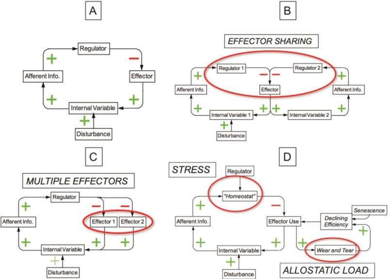
In this review we describe a series of major concepts introduced during the past 150years that have contributed to our current understanding about how physiological processes required for well-being and survival are regulated. One can theorize that hierarchical networks involving input-output relationships continuously orchestrate and learn adaptive patterns of observable behaviors, cognition, memory, mood, and autonomic systems. Taken together, these networks function as "good regulators" determining levels of internal variables and act as if there were homeostatic comparators ("homeostats"). The consequences of models with vs. without homeostats remain the same in terms of allostatic load and the eventual switch from stabilizing negative feedback loops to destabilizing, pathogenic positive feedback loops. Understanding this switch seems important for comprehending senescence-related, neurodegenerative disorders that involve the autonomic nervous system. Our general proposal is that disintegration of homeostatic systems causes disorders of regulation in degenerative diseases and that medical cybernetics can inspire and rationalize new approaches to treatment and prevention.

Keywords: Allostasis; Autonomic; Biocybernetics; Homeostasis; Stress.


Conflict of interest statement

Conflicts of interest: None.

Figures

Figure 1
Figure 1. (A) Cannon’s concept diagram for homeostasis of blood glucose, and (B) concept diagram depicting homeostasis of a regulated variable by two complementary effectors
Cannon’s diagrammed how what he called the “sympathico-adrenal” and complementary “vago-insular” systems maintain homeostasis of blood sugar. Increases in blood glucose stimulate pancreatic islet cells to release insulin, which facilitates glucose uptake by organs such as the liver. Decreases in blood glucose reflexively stimulate adrenomedullary cells to release adrenaline, which augments glucose formation from liver glycogen and evokes glucose release into the bloodstream. The double-y graph at the bottom left is a more generalized depiction of Cannon’s schema. Increases in the level of the regulated variable beyond a certain limit (green) evoke effects at multiple levels of the neuraxis that tend to return the level of the regulated variable to within bounds, and decreases in the level of regulated variable (red) evoke effects that also tend to return the level of the regulated variable to within bounds. One effector (e.g., insulin) responds to an increase and one (e.g., the sympathetic adrenergic system) to a decrease in the level of the regulated variable (e.g., serum glucose). The level of the regulated variable is kept within bounds by the input-output relationship, without a homeostatic comparator (e.g., a “glucostat”). Cannon’s diagram is reproduced with permission from the American Physiological Society.
Figure 2
Figure 2. Conceptual model of homeostasis by negative feedback (error control), anticipatory (feed-forward) control, and buffering
In the model, the effectors are both autonomic and non-autonomic. Effector responses to a disturbance are determined by three forms of regulation. First, effector activities are determined from input-output (afferent-efferent) curves relating sensory input to effector output (error control by negative feedback). Second, via exteroceptive or interoceptive input, effector activities are altered by instinct or imprinting, in advance of a change in the level of the regulated variable. Third, via exteroceptive input, effector activities are altered by associative learning (classical or instrumental conditioning), also in advance of a change in the level of the regulated variable. The extent of sensor activation in response to a disturbance is modulated by buffering. Buffering is a means of diminishing the intensity of an external disturbance, thereby reducing the required use of reflexive homeostatic mechanisms Effector responses can also modify buffering in advance of a disturbance, via instinct or learned behaviors (e.g., piloerection during cold exposure; donning a jacket before entering a cold environment).
Figure 3
Figure 3. Concept diagram showing mechanisms of anticipatory and error-controlled regulation
The autonomic responses seem to reflect a hierarchical arrangement, with the lowest level sensory-effector mechanisms in the target cells (e.g., glucose sensing by pancreatic islet cells). Superimposed on these are spinal- and brainstem-mediated reflexes, based on input from interoceptors (here designated “controllers”). The brainstem reflexes in turn are modulated by hypothalamic and limbic centers, also dependent on sensory input (“commanders”). Hypothalamic activity patterns are modulated by the limbic centers, which reflect emotions, habituation, sensitization, imprinting, and classical conditioning. Finally, higher cortical centers are responsible for cognition, instrumentally conditioned learned behaviors, predictions of future events, and interpretations of environmental and social stimuli. Under ordinary circumstances, levels of the regulated variable are kept within bounds by anticipatory (learned) control. When this gives way, error-controlled (reflexive) regulation comes into play. Being reactive, error-controlled regulation is associated with increased variability of the regulated variable. The curved arrows indicate the multiple input-output relationships at ascending strata in the neuraxis, from the target organ (e.g., the pancreatic islet cells) to the autonomic effectors, to lower brainstem “controller” sites mediating reflexes (e.g., the nucleus of the solitary tract, rostral ventrolateral medulla and dorsal motor nucleus of the vagus nerve), to hypothalamic/upper brainstem “commander” sites mediating vigilance, automatic motor behaviors, and patterned instinctive responses (e.g., locus ceruleus, substantia nigra), to limbic sites involving emotional memory and classically conditioned learning (e.g., hippocampus, amygdala), to cortical sites involving social consciousness, restraint of lower centers, instrumentally conditioned learning, and interactions with the environment (e.g., orbitofrontal cortex, anterior cingulate cortex, insular cortex).
Figure 4
Figure 4. Systems biologic and integrative physiologic views of homeostatic regulation of blood pressure in response to infusion of a vasoconstrictor
This concept diagram conveys a way neurocybernetics and medical biocybernetics can be linked. Neurocybernetics is focused on the complex systems biologic network, and medical biocybernetics deals with the negative feedback regulation that is necessary for homeostasis. This Figure depicts homeostatic regulation of blood pressure during infusion of a vasoconstrictor. (A) Reflexive responses to a vasoconstrictor involve increased baroreceptor afferent traffic to the nucleus of the solitary tract (NTS), followed by activation of the caudal ventrolateral medulla (CVLM). Release of the inhibitory neurotransmitter GABA from the CVLM terminating in the rostral ventrolateral medulla (RVLM) inhibits RVLM outflow to the sympathetic pre-ganglionic neurons (SPN) in the intermediolateral columns of the spinal cord, resulting in decreased post-ganglionic sympathetic noradrenergic system (SNS) traffic and decreased delivery of norepinephrine (NE) to its receptors on vascular smooth muscle cells. Green positive signs indicate positive relationships, and the red negative sign indicates a negative relationship. Because there is a single negative relationship in the feedback loop, the blood pressure attains a steady-state value lower than that produced by the vasoconstrictor in the absence of the feedback loop. (B) Nodes and relationships in the central autonomic network that mediate modulation of the blood pressure response. (C) Input-output curves at ascending levels of the neuraxis. Locus ceruleus (LC) activation as part of vigilance shifts the relationship between activity of rostral ventrolateral medulla (RVLM) neurons and blood pressure; a defence reaction evoked at the level of the hypothalamus shifts the curve relating LC activity to blood pressure; classically conditioned fear shifts the relationship between paraventricular nucleus (PVN) activity and blood pressure; and instrumentally conditioned avoidance shifts the relationship between cortical activity and blood pressure. (D) Hypothetical pathway by which release from cortical restraint exerts a feed-forward inhibition of baroreflex regulation of blood pressure. (E) The adjustments in the input-output curves have the net effect of resetting a hypothetical “barostat.” Other abbreviations: Class. Cond.=classical conditioned; Instr. Cond.=instrumentally conditioned.
Figure 5
Figure 5. Concept diagrams for negative feedback regulation of internal variables
(A) Simple negative feedback loop. In response to a constant disturbance, the level of the internal variable attains a steady state value. (B) Diagram of multiple effectors. Having multiple effectors extends the range of control, enables compensatory activation when one effector fails, and provides a basis for patterning of stress responses. (C) Diagram of effector sharing. Effector sharing can account for phenomena such as hyperglycemia in shock and hyponatremia in decompensated congestive heart failure. (D) Homeostatic definitions of stress and allostatic load. Stress is a condition in which the internal variable remains out of bounds despite allostatic adjustments.
Figure 6
Figure 6. Hypothetical allostatic adjustments in compensated heart failure
Normally blood pressure is kept within bounds (between the vertical dashed lines) by increases in SNS outflow when the blood pressure falls and increases in cardiovagal outflow when the blood pressure rises. In compensated heart failure the curves are shifted, such that blood pressure and SNS outflow are higher and cardiovagal outflow lower. In this setting, when the blood pressure is increased (e.g., by injection of a vasoconstrictor), there is little or no increase in cardiovagal outflow.

Similar articles

Cited by

References

    1. Abel JJ, Crawford AC. On the blood-pressure-raising constituent of the suprarenal capsule. Bull Johns Hopkins Hosp. 1897;8:151–157.
    1. Ahlquist RP. A study of adrenotropic receptors. Am J Physiol. 1948;153:586–600. - PubMed
    1. Appenzeller O, Minko T, Pozharov V, Bonfichi M, Malcovati L, Gamboa J, Bernardi L. Gene expression in the Andes; relevance to neurology at sea level. J Neurol Sci. 2003;207:37–41. - PubMed
    1. Ashby WR. An Introduction to Cybernetics. Chapman & Hall; London, UK: 1956.
    1. Aston-Jones G, Chiang C, Alexinsky T. Discharge of noradrenergic locus coeruleus neurons in behaving rats and monkeys suggests a role in vigilance. Prog Brain Res. 1991;88:501–520. - PubMed

Publication types