. 2017 Dec;22(12):1532-1542.
doi: 10.1007/s10495-017-1423-x.

The muscle regulatory transcription factor MyoD participates with p53 to directly increase the expression of the pro-apoptotic Bcl2 family member PUMA

Affiliations

The muscle regulatory transcription factor MyoD participates with p53 to directly increase the expression of the pro-apoptotic Bcl2 family member PUMA

Terri J Harford et al. Apoptosis. 2017 Dec.

Abstract

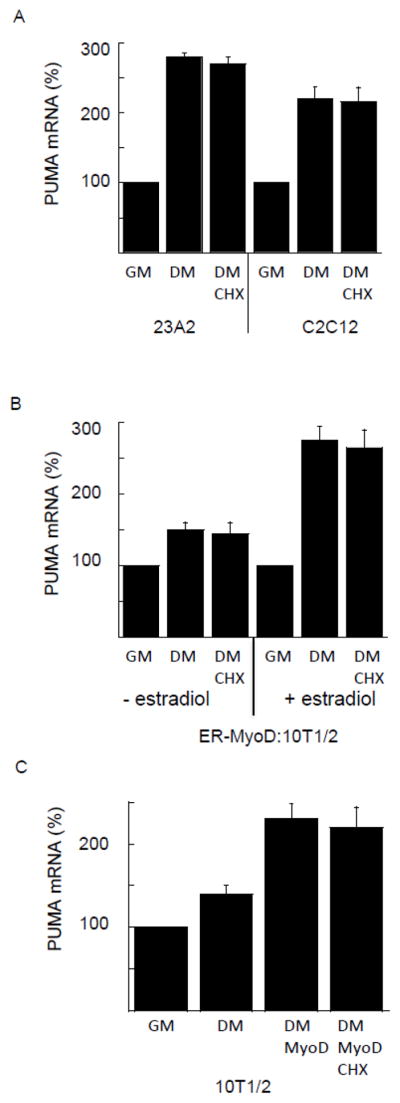
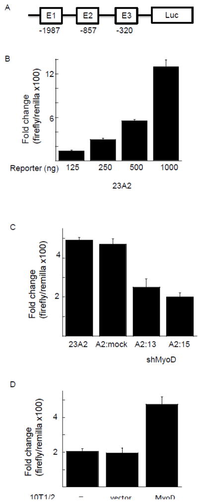
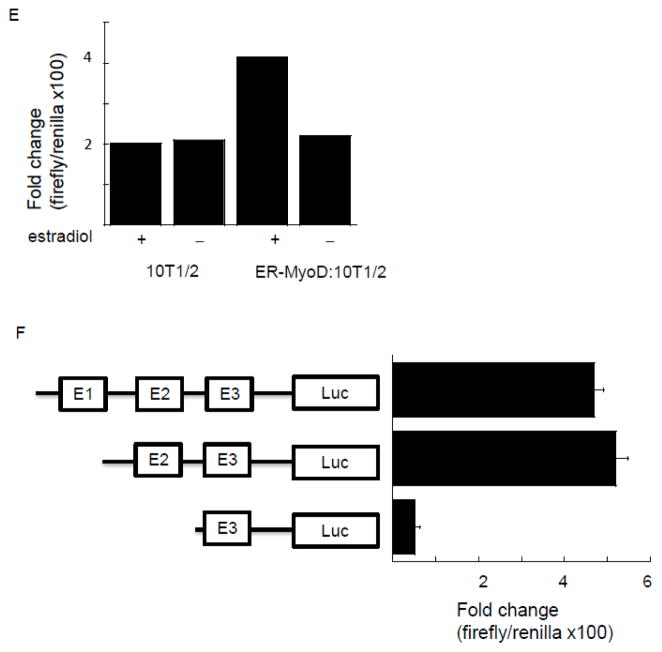
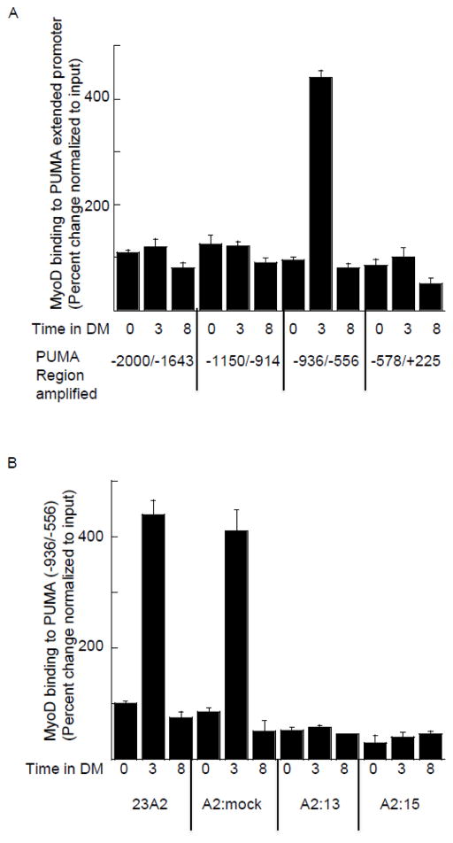
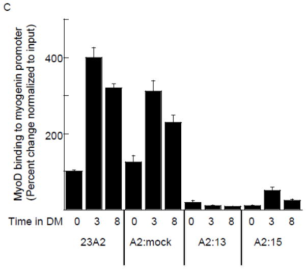
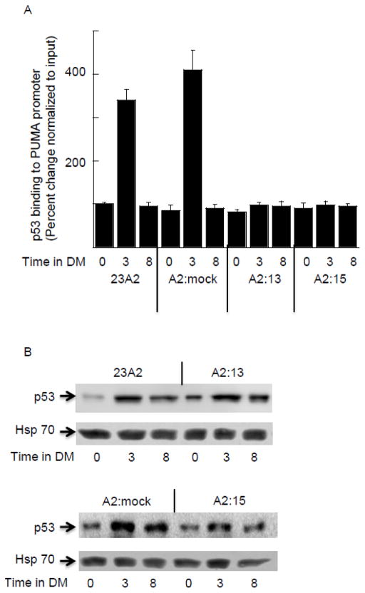
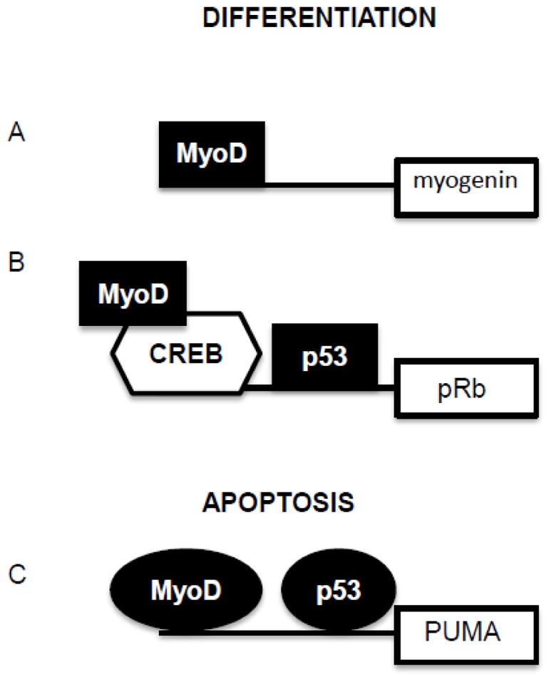
The muscle regulatory transcription factor MyoD is a master regulator of skeletal myoblast differentiation. We have previously reported that MyoD is also necessary for the elevated expression of the pro-apoptotic Bcl2 family member PUMA, and the ensuing apoptosis, that occurs in a subset of myoblasts induced to differentiate. Herein, we report the identification of a functional MyoD binding site within the extended PUMA promoter. In silico analysis of the murine PUMA extended promoter revealed three potential MyoD binding sites within 2 kb of the transcription start site. Expression from a luciferase reporter construct containing this 2 kb fragment was enhanced by activation of MyoD in both myoblasts and fibroblasts and diminished by silencing of MyoD in myoblasts. Experiments utilizing truncated versions of this promoter region revealed that the potential binding site at position - 857 was necessary for expression. Chromatin immunoprecipitation (ChIP) analysis confirmed binding of MyoD to the DNA region encompassing position - 857. The increase in MyoD binding to the PUMA promoter as a consequence of culture in differentiation media (DM) was comparable to the increase in MyoD binding at the myogenin promoter and was diminished in myoblasts silenced for MyoD expression. Finally, ChIP analysis using an antibody specific for the transcription factor p53 demonstrated that, in myoblasts silenced for MyoD expression, p53 binding to the PUMA promoter was diminished in response to culture in DM. These data indicate that MyoD plays a direct role in regulating PUMA expression and reveal functional consequences of MyoD expression on p53 mediated transcription of PUMA.

Keywords: Apoptosis; MyoD; P53; PUMA.


Figures

Figure 1
Figure 1
New protein synthesis is not required for increased PUMA mRNA in response to serum withdrawal. In (A) equal cell numbers of 23A2 or C2C12 myoblasts were plated and the next day switched to fresh GM or DM with or without 10 g/ml cycloheximide (CHX) for three hours. In (B) equal cell numbers of ER-MyoD:10T1/2 fibroblasts were plated and the next day pretreated with estradiol (1 M) before switching to fresh GM or DM with or without CHX with or without estradiol as indicated for three hours. In (C), equal cell numbers of 10T1/2 fibroblasts were plated and the next day transfected with 1 g pcMyoD. Two days later, cultures were switched to fresh GM or DM with or without CHX for three hours. Quantitative RT-PCR was performed as described in Methods. Shown is an average of 3 experiments (mean +/− SEM).
Figure 2
Figure 2
Localization of MyoD responsive element in the 5′ extended promoter of PUMA. (A) Schematic representation of construct used in (B–E). In (B), equal cell numbers of 23A2 myoblasts were plated and the next day transfected with the indicated amount of a luciferase reporter construct containing −2000/+225 of the PUMA gene. The next day, cells were switched to DM for three hours prior to the determination of luciferase activity as described in Methods. In (C), equal cell numbers of 23A2 myoblasts, 23A2 myoblasts mock silenced for MyoD expression (A2:mock), or 23A2 myoblasts silenced for MyoD expression (clones A2:13 and A2:15) were plated and the next day transfected with 500ng of the luciferase reporter construct containing −2000/+225 of the PUMA gene. The next day, cells were switched to DM for three hours prior to the determination of luciferase activity as described in Methods. In (D), equal cell numbers of 10T1/2 fibroblasts were plated and the next day transfected as described in Methods with 500 ng of the luciferase reporter construct containing −2000/+225 of the PUMA gene with or without pcMyoD. The next day, cultures were switched to DM for three hours prior to the determination of luciferase activity as described in Methods. In (E), equal cell numbers of ER-MyoD:10T1/2 fibroblasts or 10T/12 fibroblasts were plated and the next day transfected as described in Methods with 500 ng of the luciferase reporter construct containing −2000/+225 of the PUMA gene. The next day, cultures were pretreated with estradiol (1 M) as indicated before switching to DM with or without estradiol as indicated for three hours prior to the determination of luciferase activity as described in Methods. In (F), equal cell numbers of 23A2 myoblasts were plated and the next day transfected with the luciferase reporter construct containing −2000/+225 of the PUMA gene or the indicated deletion mutant (500 ng). The next day, cells were switched to DM for three hours prior to the determination of luciferase activity as described in Methods. Shown for each (B–F) is an average of at least 2 experiments (mean +/− SEM).
Figure 2
Figure 2
Localization of MyoD responsive element in the 5′ extended promoter of PUMA. (A) Schematic representation of construct used in (B–E). In (B), equal cell numbers of 23A2 myoblasts were plated and the next day transfected with the indicated amount of a luciferase reporter construct containing −2000/+225 of the PUMA gene. The next day, cells were switched to DM for three hours prior to the determination of luciferase activity as described in Methods. In (C), equal cell numbers of 23A2 myoblasts, 23A2 myoblasts mock silenced for MyoD expression (A2:mock), or 23A2 myoblasts silenced for MyoD expression (clones A2:13 and A2:15) were plated and the next day transfected with 500ng of the luciferase reporter construct containing −2000/+225 of the PUMA gene. The next day, cells were switched to DM for three hours prior to the determination of luciferase activity as described in Methods. In (D), equal cell numbers of 10T1/2 fibroblasts were plated and the next day transfected as described in Methods with 500 ng of the luciferase reporter construct containing −2000/+225 of the PUMA gene with or without pcMyoD. The next day, cultures were switched to DM for three hours prior to the determination of luciferase activity as described in Methods. In (E), equal cell numbers of ER-MyoD:10T1/2 fibroblasts or 10T/12 fibroblasts were plated and the next day transfected as described in Methods with 500 ng of the luciferase reporter construct containing −2000/+225 of the PUMA gene. The next day, cultures were pretreated with estradiol (1 M) as indicated before switching to DM with or without estradiol as indicated for three hours prior to the determination of luciferase activity as described in Methods. In (F), equal cell numbers of 23A2 myoblasts were plated and the next day transfected with the luciferase reporter construct containing −2000/+225 of the PUMA gene or the indicated deletion mutant (500 ng). The next day, cells were switched to DM for three hours prior to the determination of luciferase activity as described in Methods. Shown for each (B–F) is an average of at least 2 experiments (mean +/− SEM).
Figure 3
Figure 3
Confirmation of elevated or silenced MyoD levels. For (A), equal cell numbers of ER-MyoD:10T1/2 cells were plated and the next day cultured with or without estradiol (1 M) as indicated. In (B), equal cell numbers of 10T1/2 cells were plated and the next day transfected as described in Figure legend 1. In (C), equal cell numbers of 23A2 myoblasts, 23A2 myoblasts mock silenced for MyoD expression (A2:mock), or 23A2 myoblasts silenced for MyoD expression (clones A2:13 and A2:15) were plated. In (A–C), after treatment or transfection as indicated, lysates were prepared and Western analysis to measure MyoD levels was performed as described in Methods.
Figure 4
Figure 4
Binding of MyoD to a region in the extended PUMA promoter containing E2. For (A–C), equal cell numbers were plated and the next day cultured as indicated. Chromatin Immunoprecipitation was performed on each cell sample using EZ ChIP Chromatin Immunoprecipitation Kit (Upstate) per manufacturer’s instructions using 5 g anti-MyoD (M-318) (sc-760 Santa Cruz Biotechnology) or IgG control (Sigma-Aldrich). Quantitative PCR was used to assay for the relative levels of the indicated DNA as described in Methods. Data was normalized to the signal detected from the input of each sample. Error bars represent mean +/− SEM of triplicates.
Figure 4
Figure 4
Binding of MyoD to a region in the extended PUMA promoter containing E2. For (A–C), equal cell numbers were plated and the next day cultured as indicated. Chromatin Immunoprecipitation was performed on each cell sample using EZ ChIP Chromatin Immunoprecipitation Kit (Upstate) per manufacturer’s instructions using 5 g anti-MyoD (M-318) (sc-760 Santa Cruz Biotechnology) or IgG control (Sigma-Aldrich). Quantitative PCR was used to assay for the relative levels of the indicated DNA as described in Methods. Data was normalized to the signal detected from the input of each sample. Error bars represent mean +/− SEM of triplicates.
Figure 5
Figure 5
Increased binding of p53 to the extended PUMA promoter in response to culture in DM does not occur in the absence of MyoD binding. For all, equal cell numbers were plated and the next day cultured as indicated. In (A) Chromatin Immunoprecipitation was performed on each cell sample using EZ ChIP Chromatin Immunoprecipitation Kit (Upstate) per manufacturer’s instructions using 5 g anti-p53 (OP03-Calbiochem) or IgG control (Sigma-Aldrich). Quantitative PCR was used to assay for the relative levels of the PUMA (−936/−556). Data was normalized to the signal detected from the input of each sample. Error bars represent mean +/− SEM of triplicates. In (B) Western analysis to measure p53 levels was performed as described in Methods. Shown are results from one experiment that are representative of three independent experiments.
Figure 6
Figure 6
Proposed model for the coordinated regulation of differentiation and apoptosis by MyoD and p53. MyoD is well known as a pioneer transcription factor responsible for the differentiation of skeletal myoblasts. MyoD drives transcription necessary for differentiation through both direct (A) and indirect (B) binding to DNA. p53 is well known for its role in tumor suppression as a pivotal transcription factor responsible for interpreting the extent of DNA damage into either cell cycle arrest or apoptosis (C). Shown are two examples where both p53 and MyoD sites have been confirmed. We propose that post-translational modification(s) or distinct binding partners, portrayed herein as shape changes, could explain the mutually exclusive, dual, biological roles in differentiation or apoptosis for both of these key transcription factors.

Similar articles

Cited by

References

    1. Ceafalan LC, Popescu BO, Hinescu ME. Cellular players in muscle regeneration. Biomed Res Int. 2014 doi: 10.1155/2014/957014. - DOI - PMC - PubMed
    1. Ten Broek RW, Grefte S, Von den Hoff JW. Regulatory factors and cell populations involved in skeletal muscle regeneration. J Cell Physiol. 2010;224(1):7–16. - PubMed
    1. Partridge TA, Morgan JE. Multiple insights from myogenic cell transplants. Hum Gene Ther. 2014;25(5):404–5. - PubMed
    1. Grenier G, Rudnicki MA. The potential use of myogenic stem cells in regenerative medicine. Handb Exp Pharmacol. 2006;174:299–317. - PubMed
    1. Skuk D, Caron NJ, Goulet M, Roy B, Tremblay JP. Resetting the problem of cell death following muscle-derived cell transplantation: detection, dynamics and mechanisms. J Neuropathol Exp Neurol. 2003;62:951–67. - PubMed

MeSH terms

LinkOut - more resources