. 2017 Sep 25;42(6):626-639.e5.
doi: 10.1016/j.devcel.2017.08.009. Epub 2017 Sep 14.

Phospho-Regulation of Soma-to-Axon Transcytosis of Neurotrophin Receptors

Affiliations

Phospho-Regulation of Soma-to-Axon Transcytosis of Neurotrophin Receptors

Naoya Yamashita et al. Dev Cell. .

Abstract

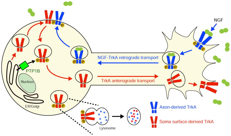
Axonal targeting of signaling receptors is essential for neuronal responses to extracellular cues. Here, we report that retrograde signaling by target-derived nerve growth factor (NGF) is necessary for soma-to-axon transcytosis of TrkA receptors in sympathetic neurons, and we define the molecular underpinnings of this positive feedback regulation that enhances neuronal sensitivity to trophic factors. Activated TrkA receptors are retrogradely transported in signaling endosomes from distal axons to cell bodies, where they are inserted on soma surfaces and promote phosphorylation of resident naive receptors, resulting in their internalization. Endocytosed TrkA receptors are then dephosphorylated by PTP1B, an ER-resident protein tyrosine phosphatase, prior to axonal transport. PTP1B inactivation prevents TrkA exit from soma and causes receptor degradation, suggesting a "gatekeeper" mechanism that ensures targeting of inactive receptors to axons to engage with ligand. In mice, PTP1B deletion reduces axonal TrkA levels and attenuates neuron survival and target innervation under limiting NGF (NGF+/-) conditions.

Keywords: ER; PTP1B; TrkA signaling endosomes; axon transport; neurotrophins; protein tyrosine phosphatase; sympathetic neuron development; transcytosis.


Figures

Figure 1
Figure 1. Trk activity and endocytosis in distal axons is necessary for transcytosis
(A) Antibody feeding to monitor soma-to-axon transcytosis of FLAG-TrkB:A receptors in compartmentalized cultures. Cell body compartments were live-labeled with FLAG antibody and distal axons were stimulated with BDNF (100 ng/ml, 4 hr) in the presence of inhibitors of Trk receptor kinase activity or endocytosis added to axon compartments. (B–G) Ligand-induced transcytosis of FLAG antibody-bound Trk receptors is suppressed by inhibition of Trk activity (1NMPP1) or PLCγ-calcineurin-dynamin-mediated endocytosis in distal axons. Arrowheads indicate soma surface-derived FLAG-labeled receptors in axons. Nuclei were stained by DAPI (blue). Scale bars, 5 μm. (H, I) Quantification of FLAG-Trk punctae in cell bodies (H) or axons (I). **p<0.01 relative to “no ligand” condition, two-way ANOVA and Tukey-Kramer post-hoc test. Results are means ± SEM from 3 independent experiments. 15 cell bodies (H) or 20 axons (I) were counted per experiment.
Figure 2
Figure 2. Axon- and soma surface-derived Trk receptors interact in cell bodies
(A) Antibody feeding to detect exocytosis of axon-derived FLAG-TrkB:A receptors in cell bodies. Axon compartments were live-labeled with FLAG antibody, distal axons stimulated with BDNF (100 ng/ml, 1hr), and neurons incubated with fluorescent secondary antibody without permeabilization. (B,C) Retrogradely transported Trk receptors undergo exocytosis on soma membranes. GFP is co-expressed with FLAG-TrkB:A. Scale bar, 5 μm. (D) Quantification of FLAG punctae per neuron. **p<0.01, t-test. Results are means ± SEM from 3 independent experiments. 20 neurons were analyzed per condition per experiment. (E) Dual color antibody feeding for simultaneous detection of axon and soma surface Trk receptors. Axon and cell body compartments were live-labeled with mouse anti-FLAG antibody (red) and rabbit anti-FLAG antibody (green), respectively and distal axons were stimulated with BDNF (100 ng/ml, 1hr). (F–K) Axon- and soma surface-derived Trk receptors co-localize near the cell body perimeter. GFP is co-expressed with FLAG-TrkB:A. Scale bar, 5 μm. (L) Quantification of co-localization of axon and soma surface-derived Trk receptors. **p<0.01, t-test. n=45 neurons each for un-stimulated and ligand treatments, from 3 independent experiments. (M, N) Interactions between axon- and soma surface-derived Trk receptors are visualized by in situ PLA. GFP is co-expressed with FLAG-TrkB:A. Scale bar, 5 μm. Representative images are from at least 20 neurons analyzed per treatment, from 3 independent experiments. (O–Q) Soma surface-resident TrkA receptors are phosphorylated by retrograde NGF signaling. Cell bodies in compartmentalized cultures were biotin-labeled before stimulating distal axons with NGF (100 ng/ml, 1 hr). Cell body lysates were subjected to neutravidin precipitation and immunoblotting using anti-P-TrkAY794. P-TrkA immunoblots were stripped and reprobed for TrkA for normalization of protein amounts. (Q) Densitometric quantification of P-TrkA levels normalized to total TrkA. Results are means ± SEM from 3 independent experiments, and expressed relative to the un-stimulated condition. **p<0.01 t-test.
Figure 3
Figure 3. PTP1B dephosphorylates endocytosed soma surface-derived Trk receptors
(A) NGF-induced association of TrkA with PTP1BY46F/D181A substrate trapping mutant, but not PTP1BWT, in sympathetic neurons. Adenoviruses under Tet-On regulation were used to express PTP1BWT-Myc or PTP1BY46F/D181A-Myc in sympathetic neurons. No exogenous PTP1B-Myc expression is observed in absence of doxycycline (Dox). Immunoprecipitation was done with anti-Myc, and immunoblotting with anti-Myc or anti-TrkA antibodies. (B) NGF-induced tyrosine phosphorylation of TrkA is suppressed by PTP1BWT over-expression, and enhanced by PTP1BY46F/D181A substrate-trapping mutant. (C) Densitometric quantification of P-TrkA levels normalized to total TrkA. **p<0.01, *p<0.05, two-way ANOVA and Tukey-Kramer post-hoc test. Results are mean ± SEM from 3 independent experiments. (D) Visualization of soma surface FLAG-TrkB:A receptors and mCherry-PTP1BY46F/D181A in compartmentalized neurons. (E–P) Peripheral co-localization of intracellular Trk receptors, originating from soma surfaces, and mCherry-PTP1BY46F/D181A in BDNF-stimulated neurons. Magnified images of dashed boxed areas in (G) and (M) are shown in lower panels (H–J and N–P). Co-localization of FLAG-TrkB:A and PTP1B is shown in white, using Image J co-localization highlighter (G, J, M, P). GFP is co-expressed with FLAG-TrkB:A. Scale bars, 5 μm. (Q) Quantification of co-localization of TrkB:A and mCherry-PTP1BY46F/D181A. **p<0.01 t-test. Results are mean ± SEM from 3 independent experiments. Total of 45 neurons per condition were analyzed.
Figure 4
Figure 4. PTP1B activity in cell bodies is required for soma-to-axon Trk transcytosis
(A) Antibody feeding to monitor transcytosis of FLAG-Trk receptors in compartmentalized cultures with PTP1B inhibitor (200 nM) added either to cell body or axon compartments. Distal axons were stimulated with BDNF (100 ng/ml, 4 hr). (B–E) PTP1B activity in cell bodies, but not distal axons, is required for Trk soma-to-axon transcytosis. Arrowheads indicate soma surface-derived FLAG-labeled receptors in axons. Nuclei were labeled by DAPI (blue). Scale bars, 5 μm. (F) Quantification of FLAG-Trk punctae in axons. **p<0.01 relative to “no ligand” condition, two-way ANOVA and Tukey-Kramer post-hoc test. Results are means ± SEM from 3 independent experiments. 20 axons per condition were counted per experiment. (G–J) Mutant FLAG-TrkB:AR685A receptors, unable to bind PTP1B and undergo dephosphorylation, show impaired soma-to-axon transcytosis. Compartmented sympathetic neurons were infected with adenoviruses for FLAG-TrkB:AR685A or control FLAG-TrkB:A receptors. Arrowheads indicate soma surface-derived FLAG-labeled receptors in axons. Nuclei were labeled by DAPI (blue). Scale bars, 5 μm. (K) Quantification of FLAG-Trk punctae in axons. **p<0.01 relative to “no ligand for FLAG-TrkB:A” condition, two-way ANOVA and Tukey-Kramer post-hoc test. Results are means ± SEM from 3 independent experiments. 20 axons were counted per experiment.
Figure 5
Figure 5. Soma surface-derived Trk receptors undergo lysosomal degradation upon PTP1B inhibition
(A) FLAG antibody feeding and LAMP-1 immunostaining to detect soma surface FLAG-TrkB:A receptors and lysosomes in compartmentalized neurons. Distal axons were stimulated with BDNF (100 ng/ml, 2 hr), and PTP1B inhibitor was locally applied to cell body compartments. (B–D) Soma-specific inhibition of PTP1B promotes lysosomal targeting of soma surface-derived Trk receptors. Co-localization of FLAG-TrkB:A and LAMP-1 is shown in white using Image J co-localization highlighter. Magnified images of dashed boxed areas in (B) and (C) are shown in inserts on top right. Scale bar, 5 μm. (D) Quantification of co-localization between TrkB:A and anti-LAMP1. **p<0.01, t-test. Results are mean ± SEM from 3 independent experiments. Total 45 neurons were analyzed. (E) Inhibition of lysosome acidification prevents the degradation of Trk receptors elicited by PTP1B inactivation. Cell body compartments were live-labeled with FLAG antibody in the presence of chloroquine (50 μM) and PTP1B inhibitor (200 nM), while distal axons were stimulated with BDNF (100 ng/ml, 4 hr). (F–I) Chloroquine treatment of cell bodies antagonizes the PTP1B inhibitor-mediated disappearance of soma surface-derived Trk receptors. FLAG antibody-bound Trk receptors accumulate in neuronal cell bodies and are also transported to axons in neurons treated with chloroquine + PTP1B inhibitor locally on cell bodies and BDNF on distal axons. Arrowheads indicate soma surface-derived FLAG-labeled receptors in axons. Nuclei were stained by DAPI (blue). Scale bars, 5 μm. (J, K) Quantification of FLAG-Trk punctae in cell bodies (J) or axons (K). **p<0.01 relative to “no ligand” condition, two-way ANOVA and Tukey-Kramer post-hoc test. Results are means ± SEM from 3 independent experiments. 15 cell bodies (J) or 20 axons (K) were counted per experiment.
Figure 6
Figure 6. PTPN1 deletion decreases axonal TrkA levels and disrupts NGF-dependent trophic functions
(A) Conditional PTPN1 deletion in sympathetic neurons attenuates TrkA protein levels in axon terminals innervating salivary glands, but not in cell bodies residing in superior cervical ganglia (SCG). Sympathetic ganglia and salivary glands from P0.5 TH-CRE;PTPN1f/f and control PTPN1f/f mice were harvested and immunoblotted for TrkA, followed by stripping and reprobing for Tyrosine Hydroxylase (TH). (B) Densitometric quantification of TrkA levels normalized to TH. **p<0.01 t-test. Results are means ± SEM from 5 mice per genotype. (C–G) PTPN1 deletion exacerbates sympathetic neuron loss in NGF+/− mice. New-born NGF+/− mice have a 31% decrease in SCG numbers which is further reduced by PTPN1 loss. SCGs were visualized by Nissl staining and cell counts were performed on Nissl stained tissue sections. Scale bar, 200 μm. **p<0.01, *p<0.05, ANOVA and Tukey-Kramer post-hoc test. Results are mean ± SEM from 4 mice per genotype. (H–L) PTPN1 loss aggravates sympathetic innervation deficits in NGF+/− mice, assessed by TH immunohistochemistry of P0.5 salivary gland tissue sections. Normalized innervation density (% of PTPN1f/f) is shown in (L). Scale bar, 100 μm. **p<0.01, *p < 0.05, one-way ANOVA. Results are mean ± SEM from 4 mice per genotype.
Figure 7
Figure 7. Retrograde control of soma-to-axon transcytosis of Trk receptors
NGF signaling, initiated in axon terminals, is necessary for anterograde transport of new TrkA receptors from soma surfaces. Active TrkA receptors, retrogradely transported in NGF-TrkA-harboring signaling endosomes, are inserted on soma surfaces where they elicit phosphorylation and subsequent endocytosis of naive soma surface-resident TrkA receptors. The endocytosed receptors are then dephosphorylated by PTP1B, an ER-resident protein tyrosine phosphatase, to ensure axon targeting of inactive receptors to engage with ligand. PTP1B inactivation in soma results in lysosomal degradation of TrkA receptors. In vivo, PTP1B is necessary for replenishing axonal TrkA levels and for NGF-mediated trophic support of sympathetic neurons under suboptimal NGF concentrations. These results identify phospho-regulatory mechanisms that underlie a positive feedback loop where target-derived NGF recruits its own receptors to nerve terminals to amplify neuronal responsiveness.

Comment in

  • TrkA Bumps into Its Future Self.
    Barford K, Keeler A, Deppmann C, Winckler B. Barford K, et al. Dev Cell. 2017 Sep 25;42(6):557-558. doi: 10.1016/j.devcel.2017.09.002. Dev Cell. 2017. PMID: 28950095

Similar articles

Cited by

References

    1. Arimura N, Kimura T, Nakamuta S, Taya S, Funahashi Y, Hattori A, Shimada A, Menager C, Kawabata S, Fujii K, et al. Anterograde transport of TrkB in axons is mediated by direct interaction with Slp1 and Rab27. Developmental cell. 2009;16:675–686. - PubMed
    1. Ascano M, Richmond A, Borden P, Kuruvilla R. Axonal targeting of Trk receptors via transcytosis regulates sensitivity to neurotrophin responses. The Journal of neuroscience: the official journal of the Society for Neuroscience. 2009;29:11674–11685. - PMC - PubMed
    1. Baumdick M, Bruggemann Y, Schmick M, Xouri G, Sabet O, Davis L, Chin JW, Bastiaens PI. EGF-dependent re-routing of vesicular recycling switches spontaneous phosphorylation suppression to EGFR signaling. eLife. 2015;4 - PMC - PubMed
    1. Bence KK, Delibegovic M, Xue B, Gorgun CZ, Hotamisligil GS, Neel BG, Kahn BB. Neuronal PTP1B regulates body weight, adiposity and leptin action. Nature medicine. 2006;12:917–924. - PubMed
    1. Bhattacharyya A, Watson FL, Pomeroy SL, Zhang YZ, Stiles CD, Segal RA. High-resolution imaging demonstrates dynein-based vesicular transport of activated Trk receptors. Journal of neurobiology. 2002;51:302–312. - PubMed

Publication types

MeSH terms

Substances