. 2017 May 11;6(8):e1320630.
doi: 10.1080/2162402X.2017.1320630. eCollection 2017.

Umbilical cord blood CD34+ progenitor-derived NK cells efficiently kill ovarian cancer spheroids and intraperitoneal tumors in NOD/SCID/IL2Rgnull mice

Affiliations

Umbilical cord blood CD34+ progenitor-derived NK cells efficiently kill ovarian cancer spheroids and intraperitoneal tumors in NOD/SCID/IL2Rgnull mice

Janneke S Hoogstad-van Evert et al. Oncoimmunology. .

Abstract

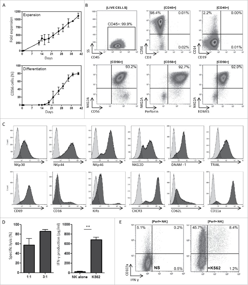
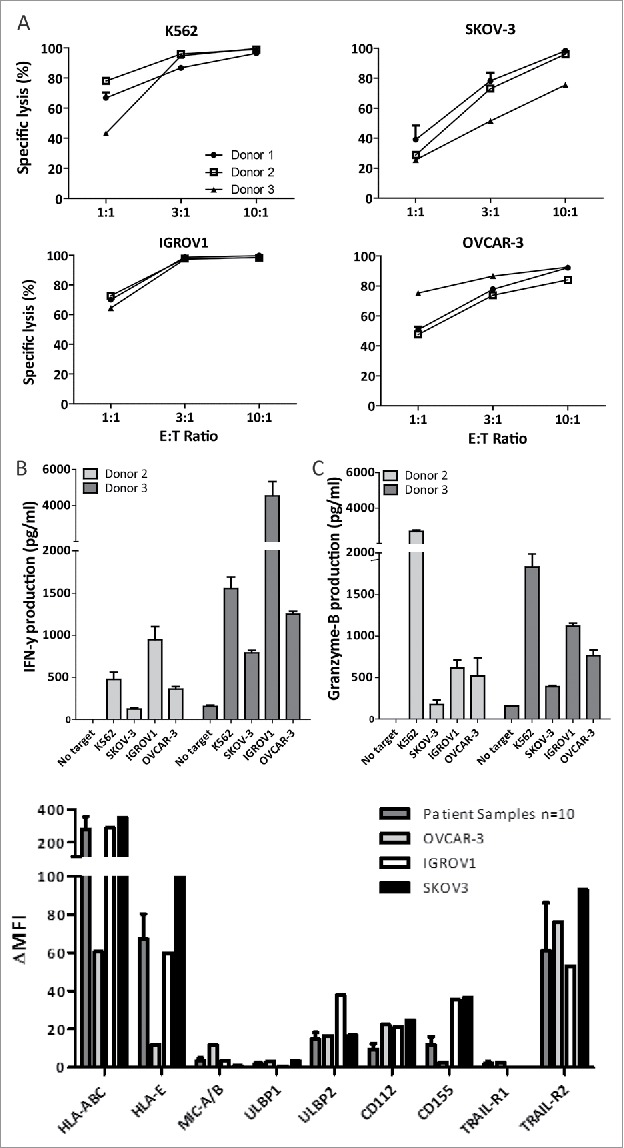
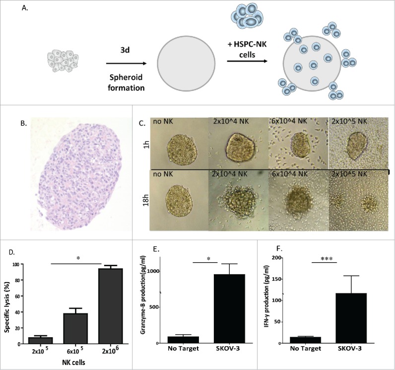
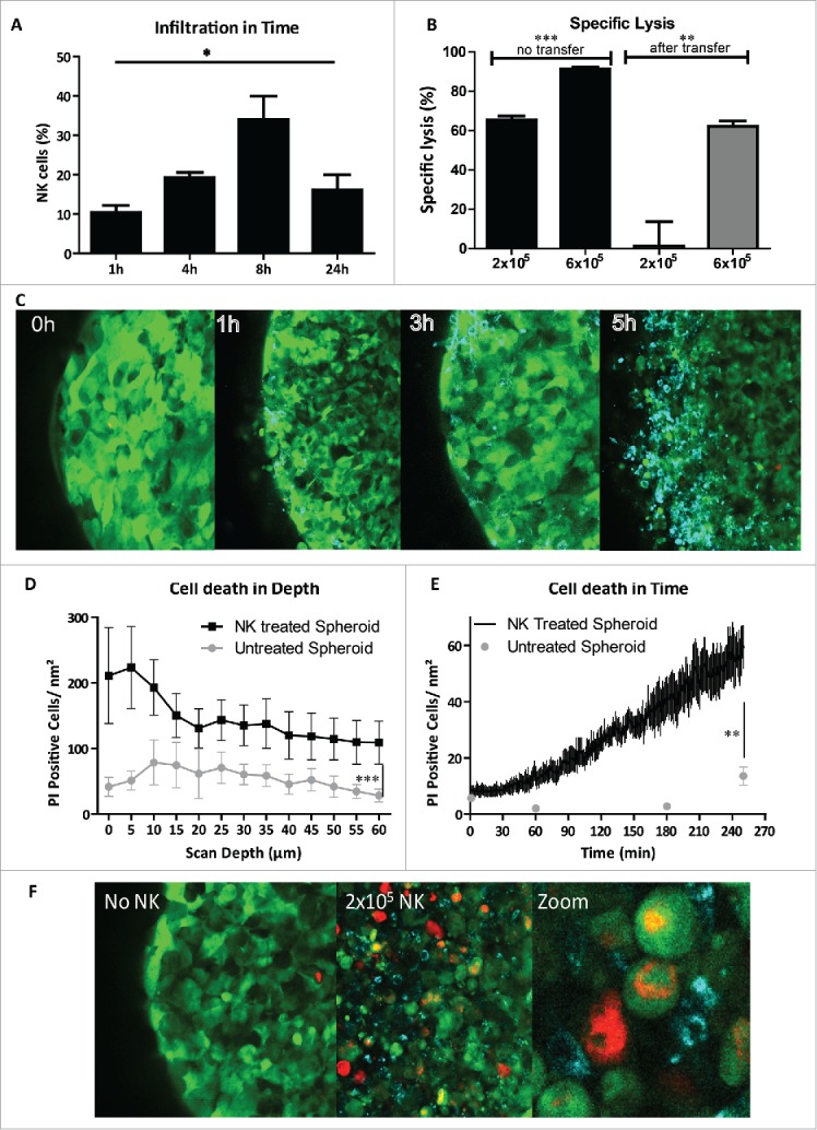
Adoptive transfer of allogeneic natural killer (NK) cells is an attractive therapy approach against ovarian carcinoma. Here, we evaluated the potency of highly active NK cells derived from human CD34+ haematopoietic stem and progenitor cells (HSPC) to infiltrate and mediate killing of human ovarian cancer spheroids using an in vivo-like model system and mouse xenograft model. These CD56+Perforin+ HSPC-NK cells were generated under stroma-free conditions in the presence of StemRegenin-1, IL-15, and IL-12, and exerted efficient cytolytic activity and IFNγ production toward ovarian cancer monolayer cultures. Live-imaging confocal microscopy demonstrated that these HSPC-NK cells actively migrate, infiltrate, and mediate tumor cell killing in a three-dimensional multicellular ovarian cancer spheroid. Infiltration of up to 30% of total HSPC-NK cells within 8 h resulted in robust tumor spheroid destruction. Furthermore, intraperitoneal HSPC-NK cell infusions in NOD/SCID-IL2Rγnull (NSG) mice bearing ovarian carcinoma significantly reduced tumor progression. These findings demonstrate that highly functional HSPC-NK cells efficiently destruct ovarian carcinoma spheroids in vitro and kill intraperitoneal ovarian tumors in vivo, providing great promise for effective immunotherapy through intraperitoneal HSPC-NK cell adoptive transfer in ovarian carcinoma patients.

Keywords: Adoptive immunotherapy; NK cells; mouse ovarian cancer xenograft; ovarian cancer; tumor spheroid infiltration.


Figures

Figure 1.
Figure 1.
SR1/IL-15/IL-12-induced HSPC-NK cells can be generated at high numbers and are highly functional. (A) Fold expansion of seven HSPC-NK cell products cultured for 40 d depicted as the mean with SD in the upper half. In the lower half, the percentage of CD56+ cells counted by flow cytometry is depicted as the mean with SD. (B) Representative FCM dot plots of an SR1/IL-15/IL-12-generated HSPC-NK cell product with high frequency of CD56+, NKG2A+, perforin+, and EOMES+ conventional NK cells. (C) Histograms illustrating the high expression level of activating receptors on SR1/IL-15/IL-12 generated HSPC-NK cell products. (D) SR1/IL-15/IL-12-induced HSPC-NK cells demonstrated high cytolytic activity (left figure) and IFNγ production (right figure) against K562 cells at a low E:T ratio of 1:1, t-test 0.001. (E) Reactivity is shown at the single cell level with the induction of significant proportions of degranulating CD107a+ and IFNγ+ NK cells upon short-term stimulation with K562 target cells in a representative FCM dot-plot.
Figure 2.
Figure 2.
HSPC-NK cells are effective killers of ovarian cancer cell monolayers. (A) Percentage of specific lysis after 24 h co-culture of HSPC-NK cells with different OC cell lines. Graphs represent data of three different UCB-donors. (B) IFNγ production by HSPC-NK cells from different donors against OC cell lines after 24 h co-culture. (C) Release of GzmB by HSPC-NK cells against OC cell lines after 24 h co-culture. Graphs represent the mean ± SEM of three experiments. (D) Delta of mean fluorescence index of NK-activation markers of SKOV-3 cells vs. tumor cells (EpCAM+) of 10 patient ascites samples. Data of EPCAM+ tumor cells of 10 patients are depicted as the mean ± SEM.
Figure 3.
Figure 3.
HSPC-NK cells have high cytolytic activity against SKOV-3 spheroids. (A) Experimental design illustrating the formation of SKOV-3 spheroids, NK cell transfer, and different functional assays performed. (B) H&E staining at 72 h on 4 µm thick section of the SKOV-3 spheroid. (C) Overview of SKOV-3 spheroid co-cultures without (i.e., medium control) and with 2 × 10⁴, 6 × 10⁴, and 20 × 104 HSPC-NK cells. Pictures by light microscopy were taken after 1 h co-culture (top panel) and after 18 h co-culture (bottom panel) . (D) Specific lysis by HSPC NK cells of SKOV-3 tumor cells within the spheroids at different E:T ratios. Data are shown as mean ± SEM of a three experiment. Two-way ANOVA p = 0.02 (E) and (F) GzmB and IFNγ production of HSPC-NK cells after 24 h co-culture with SKOV-3 spheroids (unpaired t-test IFNγ p = 0.04, GzmB p = 0.0009).
Figure 4.
Figure 4.
HSPC-NK cells infiltrate and mediate efficient intratumoral killing in SKOV-3 spheroids. (A) Percentage of infiltrated NK cells in SKOV-3 spheroids was measured by FCM after co-culture with 2 × 10⁵ NK cells and trypsinization on four different time points. Bars represent mean ± SEM of four experiments. Two-way ANOVA p = 0.018 (B) spheroids with infiltrated HSPC-NK cells after 5 h of incubation were transferred to a new well to measure the cytotoxic capacity of the infiltrated HSPC-NK cells. On the left, the percentage specific lysis without transfer is shown, and on the right, the specific HSPC-NK cell mediated lysis is depicted after 24 h incubation following transfer to a new well. Unpaired t-test comparing different HSPC-NK cell dosages without transfer shows a p-value of 0.0003, and a p-value of 0.008 comparing the transferred spheres. (C) Confocal images of an SKOV-3 spheroid incubated with HSPC-NK cells after four time points. The green cells are GFP+ SKOV-3 cells and the blue cells are HSPC-NK cells infiltrating in the spheroid. (D) Amount of PtdIns positive SKOV-3 cells in the spheroids following incubation with HSPC-NK cells (in black), and in untreated spheroids (in gray), per scan depth in µm. Data are shown as mean ± SD of a representative experiment. 60 µm is the maximum scan depth for the used confocal microscopy. In the NK-treated spheres, the amount of PtdIns+ cells is significantly higher (two-way anova; p <0.0001). (E) Amount of PtdIns positive SKOV-3 cells in time within HSPC-NK cell treated spheroid was calculated with the assistance of Fiji image analysis (in black). As control three time points of an untreated SKOV-3 spheroid is depicted in gray. SKOV-3 cell death is significantly increased within the HSPC-NK cell treated spheroid compared with untreated spheroid (unpaired t-test with the Welch correction; p = 0.004). (F) Confocal image of SKOV-3 spheroid incubated 5 h with HSPC-NK cells, with addition of PtdIns. On the left, a spheroid without NK addition is shown, where few PtdIns positive cells are seen, in the middle a spheroid with HSPC-NK cells, where we see more dead cells, and on the left, a zoomed in picture of a part of the spheroid illustrating the infiltration of HSPC-NK cells causing cell death in an SKOV-3 spheroid.
Figure 5.
Figure 5.
Intraperitoneal infusion of HSPC-NK cells slows down tumor growth and improves survival of OC-bearing NSG mice. (A) Experimental design of i.p. SKOV-3 injection and subsequent adoptive transfer of two HSPC-NK cell infusions combined with rhIL-15 administration till day 21. Tumor load was followed by BLI imaging till day 56 and mice were followed for survival till day 92. (B) Ki67 immunohistochemical staining of intraperitoneal SKOV-3 tumor nodules of NSG mice bearing SKOV-3 tumors, showing high tumor cell proliferation. (C) Results of the high tumor (1 × 10⁶ SKOV-3 cells i.p.) dose experiment of the tumor size evaluation by the quantification of the bioluminescent signal (in photons per second per cm2) in a standardized ROI. In red, the BLI signal of mice treated with intraperitoneal HSPC-NK infusions and in black the untreated mice. HSPC-NK cell treatment significantly reduced tumor growth (two-way ANOVA; p = 0.016). (D) Radiance in photons per second per cm2 of the lower tumor dose experiment with 0.2 × 10⁶ SKOV-3 cells i.p. HSPC-NK cell treatment significantly reduced tumor growth at this lower tumor burden (two-way ANOVA p = 0.0007). (E) Percentage survival of the NSG mice of the low tumor dose experiment. Kaplan meier with the log rank Mantel Cox test showed a significance of p = 0.023 and a hazard ratio of 6. (F) Macroscopic tumor score of the NSG mice after sacrificing at day 92 with a significantly lower tumor score on peritoneal surfaces after HSPC-NK cell treatment (Paired t-test; p = 0.034).

References

    1. Siegel RL, Miller KD, Jemal A. Cancer statistics, 2015. CA Cancer J Clin 2015; 65(1):5-29; PMID:25559415; https://doi.org/10.3322/caac.21254 - DOI - PubMed
    1. Geller MA, Knorr DA, Hermanson DA, Pribyl L, Bendzick L, McCullar V, Miller JS, Kaufman DS. Intraperitoneal delivery of human natural killer cells for treatment of ovarian cancer in a mouse xenograft model. Cytotherapy 2013; 15(10):1297-306; PMID:23993303; https://doi.org/10.1016/j.jcyt.2013.05.022 - DOI - PMC - PubMed
    1. Hermanson DL, Bendzick L, Pribyl L, McCullar V, Vogel RI, Miller JS, Geller MA, Kaufman DS. Induced pluripotent stem cell-derived natural killer cells for treatment of ovarian cancer. Stem Cells 2016; 34(1):93-101; PMID:26503833; https://doi.org/10.1002/stem.2230 - DOI - PMC - PubMed
    1. Davis ZB, Felices M, Verneris MR, Miller JS. Natural killer cell adoptive transfer therapy: Exploiting the first line of defense against cancer. Cancer J 2015; 21(6):486-491; PMID:26588681; https://doi.org/10.1097/PPO.0000000000000156 - DOI - PMC - PubMed
    1. Miller JS, Soignier Y, Panoskaltsis-Mortari A, McNearney SA, Yun GH, Fautsch SK, McKenna D, Le C, Defor TE, Burns LJ et al.. Successful adoptive transfer and in vivo expansion of human haploidentical NK cells in patients with cancer. Blood 2005; 105(8):3051-7; PMID:15632206; https://doi.org/10.1182/blood-2004-07-2974 - DOI - PubMed

Publication types

LinkOut - more resources