

doi: 10.21699/jns.v6i3.581.
eCollection 2017 Jul-Sep.
A Rare Case of Thoracoschisis
Affiliations
- PMID: 28920025
- PMCID: PMC5593484
- DOI: 10.21699/jns.v6i3.581
Item in Clipboard
A Rare Case of Thoracoschisis
J Neonatal Surg.
.
Abstract
A term male baby, after delivery, was found to have a 3-centimeter beefy-red mass protruding from the left chest wall, adjacent to the left nipple. Radiological imaging suggested it's origin from the left lateral liver segment. A diagnostic laparoscopy confirmed the isolated connection to the liver, elevated left hemidiaphragm, and protrusion between the ribs. The mass was excised using electrocautery, and pathologic examination showed normal liver tissue.
Keywords: Exophytic liver; Neonate; Thoracoschisis.
Figures
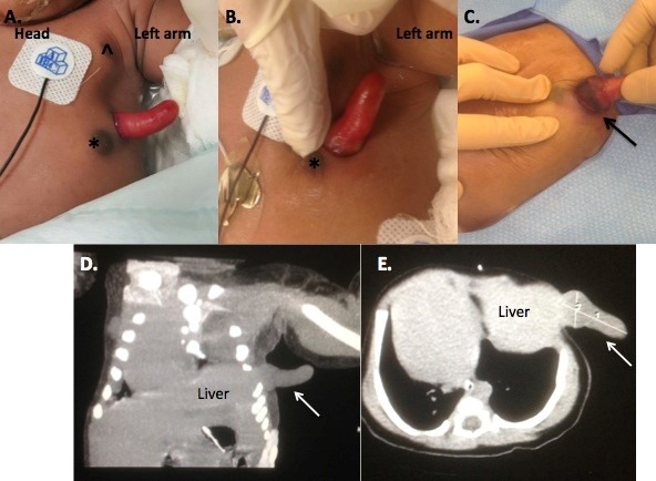
Figure 1: A. Appearance of the exophytic mass on initial examination. The mass was located on the left lateral chest between the normal (^) and accessory (*) left nipples. B. Upon close inspection, the mass was pink with a moist surface. C. Hyperemia and ischemia/necrosis at the base of the mass (black arrow) on day of life two. Coronal (D) and axial image (E) from computed tomography scan demonstrating extension of this mass from the liver (white arrow).
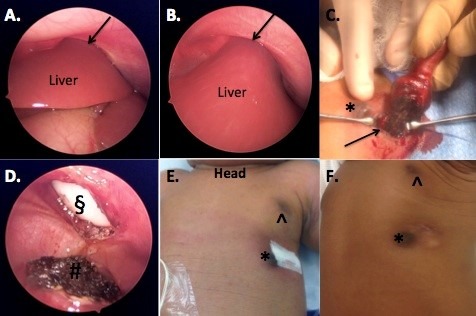
Figure 2: A and B. Laparoscopic images showing the left lateral segment of the liver in continuity with the external portion of the mass. The black arrow demonstrated the liver exiting through the chest wall. The left hemidiaphram was significantly elevated. C. Electrocautery was used to amputate the mass where it bordered the normal liver (black arrow). D. Post amputation of the exophytic mass demonstrated a circular chest wall defect (§), and hemostatic liver edge (#). E. Fascial closure and skin closure without further defect noted. Normal (^) and accessory left nipple (*) F. Three-month follow up demonstrating well-healed incision and mild chest wall deformity.
References
-
- Davies MR, Rode H, Cywes S. "Thoracoschisis" associated with an ipsilateral distal phocomelia and an anterolateral diaphragmatic hernia--a case report. J Pediatr Surg. 1977;12:755-7. - PubMed
-
- McKay JD, Parker CM, Loewen J, Cundiff CA, Herman HK, Abramowsky CR, et al. Thoracoschisis: A case report and review of literature. Fetal Pediatr Pathol. 2015;34:307-14. - PubMed
-
- Karaman I, Karaman A, Erdogan D, Cavusoglu YH, Ozguner IF. The first male with thoracoschisis: case report and review of the literature. J Pediatr Surg. 2011;46:2181-3. - PubMed
Grants and funding
LinkOut - more resources
Full Text Sources
Other Literature Sources