Review
. 2017 Dec;17(12):733-745.
doi: 10.1038/nri.2017.101. Epub 2017 Sep 18.

γδ T cells in homeostasis and host defence of epithelial barrier tissues

Affiliations
Review

γδ T cells in homeostasis and host defence of epithelial barrier tissues

Morten M Nielsen et al. Nat Rev Immunol. 2017 Dec.

Abstract

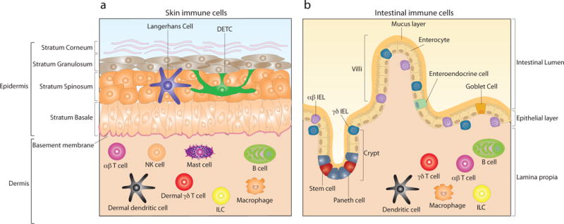
Epithelial surfaces line the body and provide a crucial interface between the body and the external environment. Tissue-resident epithelial γδ T cells represent a major T cell population in the epithelial tissues and are ideally positioned to carry out barrier surveillance and aid in tissue homeostasis and repair. In this Review, we focus on the intraepithelial γδ T cell compartment of the two largest epithelial tissues in the body - namely, the epidermis and the intestine - and provide a comprehensive overview of the crucial contributions of intraepithelial γδ T cells to tissue integrity and repair, host homeostasis and protection in the context of the symbiotic relationship with the microbiome and during pathogen clearance. Finally, we describe epithelium-specific butyrophilin-like molecules and briefly review their emerging role in selectively shaping and regulating epidermal and intestinal γδ T cell repertoires.


Figures

Figure 1
Figure 1. Skin and intestinal epithelial composition with immune cell distribution
a) Skin is composed of two major compartments, the epidermis and the dermis, which are separated by the basement membrane. The outer most layer is the epidermis, which is further subdivided into four layers; stratum corneum, strata granulosum, strata spinosum and stratum basale. The cellular composition of epidermis is dominated by keratinocytes, accounting for ~95% of all cells in the epidermal compartment. Among additional cells residing in murine epidermis are Langerhans cells and dendritic epidermal γδ T cells (DETC). Due to the presence of blood and lymphatic vessels, through which circulating lymphocytes traffic to and from skin, the cellular composition of the dermal compartment is more diverse than the epidermis. Dermal residing immune cells include dermal dendritic cells, γδ T cells, αβ T cells, innate lymphoid cells (ILC), mast cells, macrophages, B cells and NK cells. b) The epithelium of the gastrointestinal tract is composed of a single cell-layer which separates the intestinal lumen from the underlying lamina propia. The intestinal epithelial layer forms both crypts and villi. Pluripotent stem cells are located at the base of crypts and give rise to 4 distinct epithelial cell subsets; enterocytes, which represent the majority of epithelial cells, Goblet cells, enteroendocrine cells and Paneth cells. Both γδ and αβ intraepithelial lymphocytes (IEL) are interspersed throughout the epithelium and are positioned both within and directly below the epithelium. Beneath the intestinal epithelium, the lamina propia is home to a plethora of immune cells which include dendritic cells, γδ T cells, αβ T cells, ILC, macrophages and B cells.
Figure 2
Figure 2. Thymic development and epithelial migration
In the fetal thymus, Vγ3Vδ1+ dendritic epidermal T cell (DETC) progenitors mature by receiving adequate stimuli through the T cell receptor and by conditioning by Skint1. Thymic DETC precursor maturation includes up-regulation of sphingosine-1 phosphate receptor 1 (S1PR1) which facilitates thymic egress. The chemokine receptor, CCR10, is also up-regulated and aids in directing the migration of DETC precursors to the epidermis through the recognition of the CCR10 ligand, CCL27, expressed by keratinocytes. Once positioned in the epidermis, DETC down-regulate CCR10 corresponding with up-regulation of the chemokine receptor CCR4. Development of intestinal γδ intraepithelial lymphocytes (IEL) in can occur both within the adult thymus and extra-thymic, and gives rise to γδ IEL expressing the chemokine receptor CCR9. Unlike DETC precursors, thymic egress of γδ IEL is independent of S1PR1. γδ IEL migration to intestinal epithelium is directed by CCR9 and the recognition of the ligand CCL25 expressed by intestinal epithelial cells which allows entry and seeding within the epithelium. An early proliferative boost of γδ IEL is then facilitated by the timely expression of butyrophilin-like molecule 1 (Btnl1) on enterocytes whereby homeostatic γδ IEL numbers are obtained.
Figure 3
Figure 3. Epithelial maintenance and repair
a) In healthy tissue, dendritic epidermal T cells (DETC) are highly dendritic with dendrites extending both basally and apically towards the stratum corneum. Maintenance of DETC homeostatic numbers is dependent upon IL-15, produced by keratinocytes, insulin-like growth factor 1 (IGF1) produced by the DETC themselves and ligand activation and cell intrinsic signaling through the transcription factor aryl hydrocarbon receptor (AhR). Additionally, IGF1 acts on keratinocytes to increase proliferation and reduce apoptosis. The sensing of stressed or damaged keratinocytes leads to DETC activation which, in vivo, is visualized by the dramatic morphological change in DETC from dendritic to round. Upon activation, DETC at the wound edge produce IGF1 and keratinocyte growth factor 1 and 2 (KGF1/2), which all facilitate keratinocyte proliferation and timely wound closure. A subset of activated DETC further produces IL-17A. Acting on keratinocytes, IL-17A induces production of the antimicrobial peptides (AMP) Regenerating islet-derived protein 3 gamma (RegIII3γ) and β-defensin thereby providing antimicrobial protection to the damaged tissue. In addition, RegIIIγ acts directly on keratinocytes to induce proliferation and mediate re-epitheliazation of wounded skin. b) Intestinal γδ intraepithelial lymphocyte (IEL) survival and retention in healthy tissue depends on cell-intrinsic AhR activation and IL-15 production by neighboring epithelial cells. In return, γδ IEL secrete KGF1 which induces intestinal epithelial cell (IEC) proliferation and increases barrier integrity, while further facilitating epithelial repair following tissue damage. Through production of tumor growth factor β-1 (TGFβ-1) γδ IEL also act to dampen the pro-inflammatory (IFNγ) and cytolytic (Granzyme B) potential αβ IEL thereby reducing additional damage and tissue destruction caused by an excessive inflammatory immune response.
Figure 4
Figure 4. Proposed model of DETC and intestinal γδ IEL participation in microbial tolerance and clearance
a) Dendritic epidermal T cells (DETC) are capable of responding to bacterial insult and directly respond to Gram-negative bacteria via recognition of the lipopolysaccharide (LPS) component of the bacterial cell membrane, which leads to production of effector cytokines granulocyte-macrophage colony-stimulating factor (GM-CSF) and IL-2. DETC also respond to Staphylococcus aureus infection and aid in the recruitment of neutrophils by producing IL-17A. The abundant presence of the skin commensal, Staphylococcus epidermis (S. epidermis) tunes IL-1 signaling in the skin by inducing keratinocyte production of IL-1α and IL-1 Receptor antagonist (IL-1RA), which in turn induce both DETC and dermal γδ T cells to produce IL-17A. In doing so, S. epidermis mediates effector T cell responses in the skin and promotes protection against Leishmania major infection. S. epidermis also aids proper wound healing by providing an optimal inflammatory environment through the production of small molecules that act on keratinocytes via Toll-like receptor 2 (TLR2) to dampen the TLR3-induced production of the pro-inflammatory cytokines TNFα and IL-6. Germ-free mice have faster wound healing and no scarring when compared to conventionally housed mice, indicating that the skin microbiota has a negative effect on wound healing and scaring. To date, the role of the host microbiome in DETC function is unknown. b) The microbiome conditions γδ IEL effector functions through an intestinal epithelial cell-intrinsic myeloid differentiation primary response gene 88 (MyD88) pathway. Microbiome conditioning of γδ IEL leads to production of several AMP, including Regenerating islet-derived protein 3 gamma (RegIIIγ) and collectively facilitate early protection against invasion by intestinal-resident bacteria. Activated γδ IEL further induce an anti-viral response by producing Type I/III IFNs which in turn induce up-regulation of anti-viral IFN-responsive genes in intestinal epithelial cells. Upon intestinal epithelial damage, γδ IEL again aid in host protection by mounting an anti-bacterial response which includes RegIIIγ production. This response is also shaped by the microbiome which acts directly on γδ IEL, through a MyD88-dependent mechanism, to limit early penetration by opportunistic bacteria following tissue injury. γδ IEL further produce pro-inflammatory cytokines (IL-1β) and chemokines that recruit additional effector cells to the site of damage.

References

    1. Chien YH, Meyer C, Bonneville M. gammadelta T cells: first line of defense and beyond. Annu Rev Immunol. 2014;32:121–155. - PubMed
    1. Jameson JM, Sharp LL, Witherden DA, Havran WL. Regulation of skin cell homeostasis by gamma delta T cells. Front Biosci. 2004;9:2640–2651. - PubMed
    1. Roberts N, Horsley V. Developing stratified epithelia: lessons from the epidermis and thymus. Wiley Interdiscip Rev Dev Biol. 2014;3:389–402. - PMC - PubMed
    1. van der Flier LG, Clevers H. Stem cells, self-renewal, and differentiation in the intestinal epithelium. Annu Rev Physiol. 2009;71:241–260. - PubMed
    1. Cheroutre H, Lambolez F, Mucida D. The light and dark sides of intestinal intraepithelial lymphocytes. Nat Rev Immunol. 2011;11:445–456. - PMC - PubMed

MeSH terms