. 2018 Mar;53(3):449-460.
doi: 10.1007/s00535-017-1390-6. Epub 2017 Sep 18.

Basophils activated via TLR signaling may contribute to pathophysiology of type 1 autoimmune pancreatitis

Affiliations

Basophils activated via TLR signaling may contribute to pathophysiology of type 1 autoimmune pancreatitis

Masato Yanagawa et al. J Gastroenterol. 2018 Mar.

Erratum in

Abstract

Background: Pathophysiology of type 1 autoimmune pancreatitis (AIP) is still unclear. We previously reported that M2 macrophages might play an important role in type 1 AIP. Recently, it has been reported that basophils regulate differentiation to M2 macrophages. In this study, we investigated basophils from the pancreatic tissue and peripheral blood of individuals with type 1 AIP.

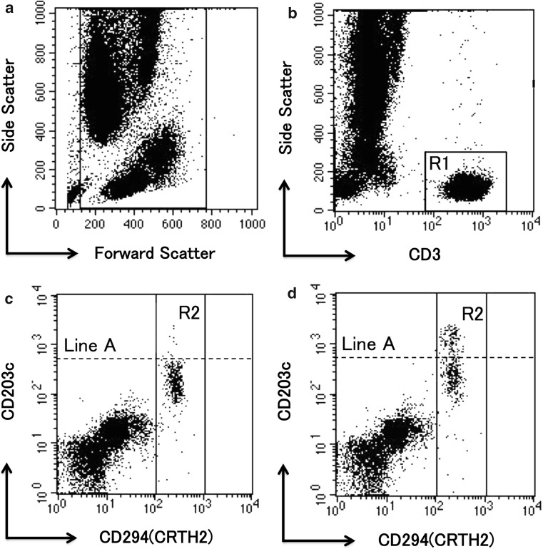
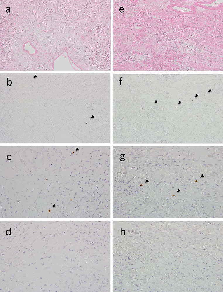
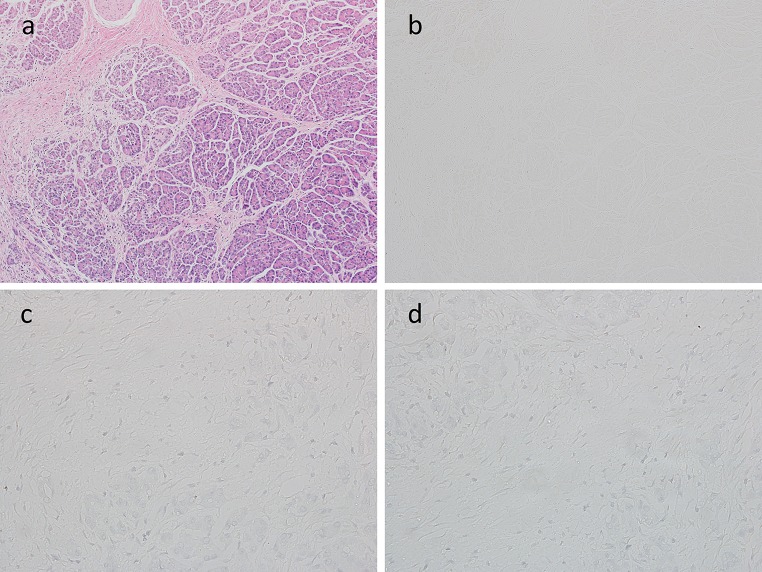
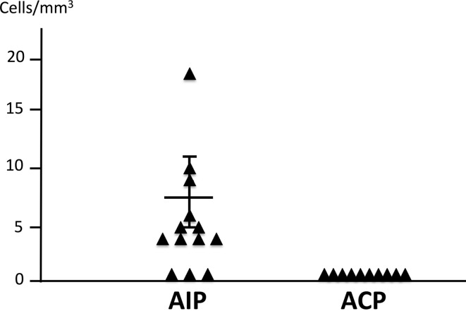
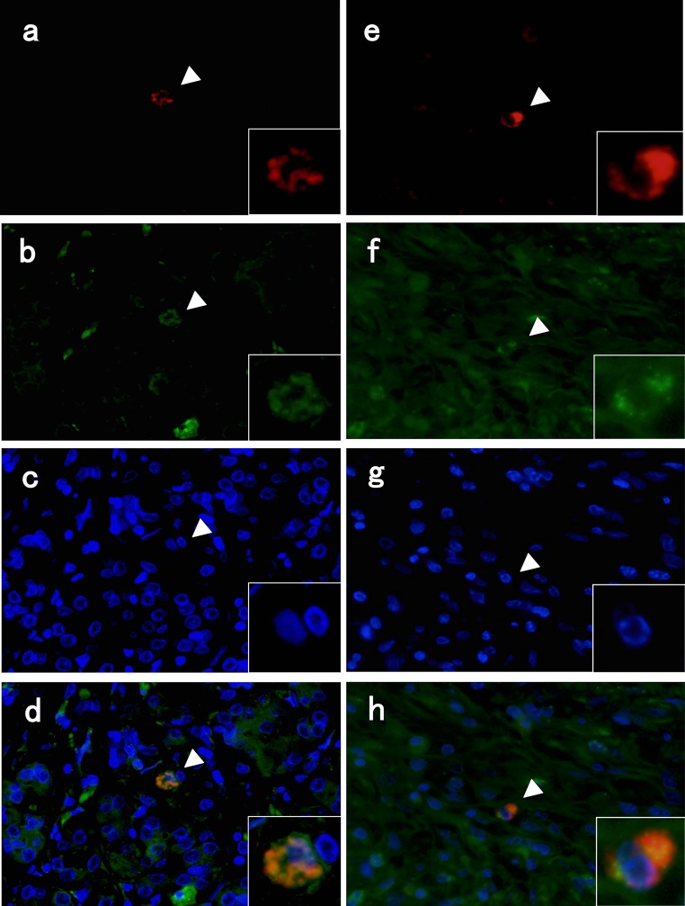
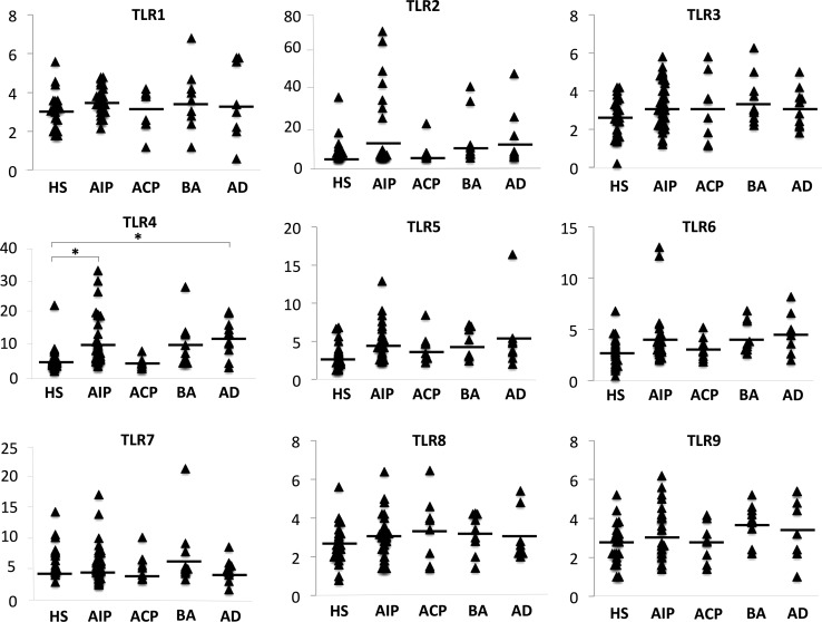
Methods: By using immunohistochemistry, we investigated basophils in pancreatic tissue from 13 patients with type 1 AIP and examined expression of toll-like receptors (TLRs) by these cells. Additionally, we obtained peripheral blood samples from 27 healthy subjects, 40 patients with type 1 AIP, 8 patients with alcoholic chronic pancreatitis, 10 patients with bronchial asthma, and 10 patients with atopic dermatitis, and analyzed activation of basophils by stimulating them with ligands of TLR1-9. We also compared TLR expression in basophils from the tissue and blood samples.

Results: Basophils were detected in pancreatic tissues from 10 of 13 patients with type 1 AIP. Flow cytometric analysis revealed that the ratios of basophils activated by TLR4 stimulation in type 1 AIP (9.875 ± 1.148%) and atopic dermatitis (11.768 ± 1.899%) were significantly higher than those in healthy subjects (5.051 ± 0.730%; P < 0.05). Levels of basophils activated by TLR2 stimulation were higher in seven type 1 AIP cases. Furthermore, stimulation of TLR2 and/or TLR4, which were expressed by basophils in pancreas, activated basophils in peripheral blood.

Conclusions: Basophils activated via TLR signaling may play an important role in the pathophysiology of type 1 AIP.

Keywords: Autoimmune pancreatitis; Basophil; M2 macrophage; TLR.


Conflict of interest statement

The authors declare that they have no conflicts of interest.

Figures

Fig. 1
Fig. 1
Identification of resting and activated basophils from peripheral blood by flow cytometry. All leukocytes were stained with CD3, CD294, and CD203c. All leukocytes formed discrete FSC/SSC populations after fixation and lysis (a). CD3-positive events (R1) were removed due to the isolation of lymphocytes and monocytes (b). Basophils were defined as CD294 (CRTH2)-positive events (R2) (c). CD203c is a specific marker of activated basophils. Line A was drawn so that 2–5% of basophils were included above it, and we defined them as activated basophils. We compared the percentage of basophils above line A before and after the stimulation (d). These experiments were performed using the Allergenicity Kit (Beckman Coulter Company, Brea, CA, USA)
Fig. 2
Fig. 2
H&E staining (a, e), immunohistochemical findings for basophils [low magnification view, ×100] (b, f), [high magnification view, ×400] (c, g), and negative controls [high magnification view, ×400] (d, h) in pancreatic tissue samples of two cases with type 1 autoimmune pancreatitis (AIP). Infiltration of basophils was confirmed by using mouse anti-basophil antibodies (2D7)
Fig. 3
Fig. 3
H&E staining (a), immunohistochemical findings for basophils [low magnification view, ×100] (b), [high magnification view, ×400] (c), and negative controls [high magnification view, ×400] (d) in a pancreatic tissue sample with alcoholic chronic pancreatitis (ACP)
Fig. 4
Fig. 4
Comparison of the total number of basophils in three different 1-mm2 fields of view in pancreatic tissue samples from patients with type 1 AIP or alcoholic chronic pancreatitis (ACP). The average number of basophils in samples from patients with type 1 AIP was 8.615 ± 2.528 (n = 13). However, basophils were not detected in the pancreatic tissue of patients with ACP (n = 10)
Fig. 5
Fig. 5
Double-immunofluorescence staining for basophils, TLR2, and TLR4 in pancreatic tissue of patients with type 1 autoimmune pancreatitis. Images show staining for basophils (2D7; red, a, e), TLR2 (green, b), TLR4 (green, f), and 4′,6-diamidino-2-phenylindole (DAPI, blue, c, g). Merged image of basophils and TLR2 (d)/TLR4 (h)
Fig. 6
Fig. 6
The ratio of basophils activated by the stimulation of TLR1–TLR9. We analyzed peripheral basophils in healthy subjects (n = 27), patients with type 1 autoimmune pancreatitis (AIP, n = 40), patients with alcoholic chronic pancreatitis (ACP, n = 8), patients with bronchial asthma (n = 10), and patients with atopic dermatitis (n = 10). Basophils were stimulated with the TLR1–9 ligands, Pam3CSK4 (400 ng/mL, InvivoGen, San Diego, CA, USA), HKLM (108 cells/mL, InvivoGen), poly:IC (50 μg/mL, Sigma, St. Louis, MO, USA), LPS (1 μg/mL, Sigma), FLA-ST (10 μg/mL, InvivoGen), FSL-1 (5 μg/mL, InvivoGen), imiquimod (5 μg/mL, InvivoGen), ssRNA40/LyoVec (5 μg/mL, InvivoGen), or ODN2006 (10 μg/mL, AdipoGen, San Diego, CA, USA), respectively. Data are presented as the mean ± standard error of the mean. Statistical comparisons of quantitative data were carried out by the Tukey–Kramer test. Differences were considered to be significant when the value of P was less than 0.05 (P < 0.05)

Comment in

Similar articles

Cited by

References

    1. Okazaki K, Uchida K, Miyoshi H, et al. Recent concepts of autoimmune pancreatitis and IgG4-related disease. Clin Rev Allergy Immunol. 2011;41(2):126–138. doi: 10.1007/s12016-010-8214-2. - DOI - PubMed
    1. Uchida K, Okazaki K, Konishi Y, et al. Clinical analysis of autoimmune-related pancreatitis. Am J Gastroenterol. 2000;95(10):2788–2794. doi: 10.1111/j.1572-0241.2000.03187.x. - DOI - PubMed
    1. Uchida K, Miyoshi H, Ikeura T, et al. Clinical and pathophysiological issues associated with type 1 autoimmune pancreatitis. Clin J Gastroenterol. 2016;9(1):7–12. doi: 10.1007/s12328-016-0628-9. - DOI - PubMed
    1. Sarles H, Sarles JC, Muratore R, et al. Chronic inflammatory sclerosis of the pancreas—an autonomous pancreatic disease? Am J Dig Dis. 1961;6:688–698. doi: 10.1007/BF02232341. - DOI - PubMed
    1. Kawaguchi K, Koike M, Tsuruta K, et al. Lymphoplasmacytic sclerosing pancreatitis with cholangitis: a variant of primary sclerosing cholangitis extensively involving pancreas. Hum Pathol. 1991;22(4):387–395. doi: 10.1016/0046-8177(91)90087-6. - DOI - PubMed

LinkOut - more resources