. 2019 Mar;49(6):824-833.
doi: 10.1111/ejn.13715. Epub 2017 Oct 6.

Selective remodeling of glutamatergic transmission to striatal cholinergic interneurons after dopamine depletion

Affiliations

Selective remodeling of glutamatergic transmission to striatal cholinergic interneurons after dopamine depletion

Jose de Jesus Aceves Buendia et al. Eur J Neurosci. 2019 Mar.

Abstract

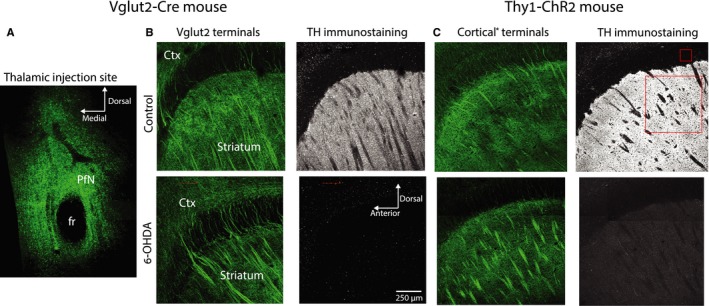
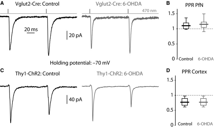
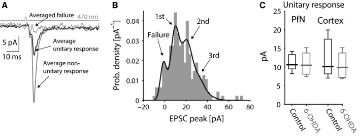
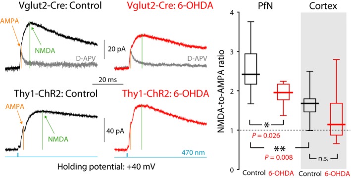
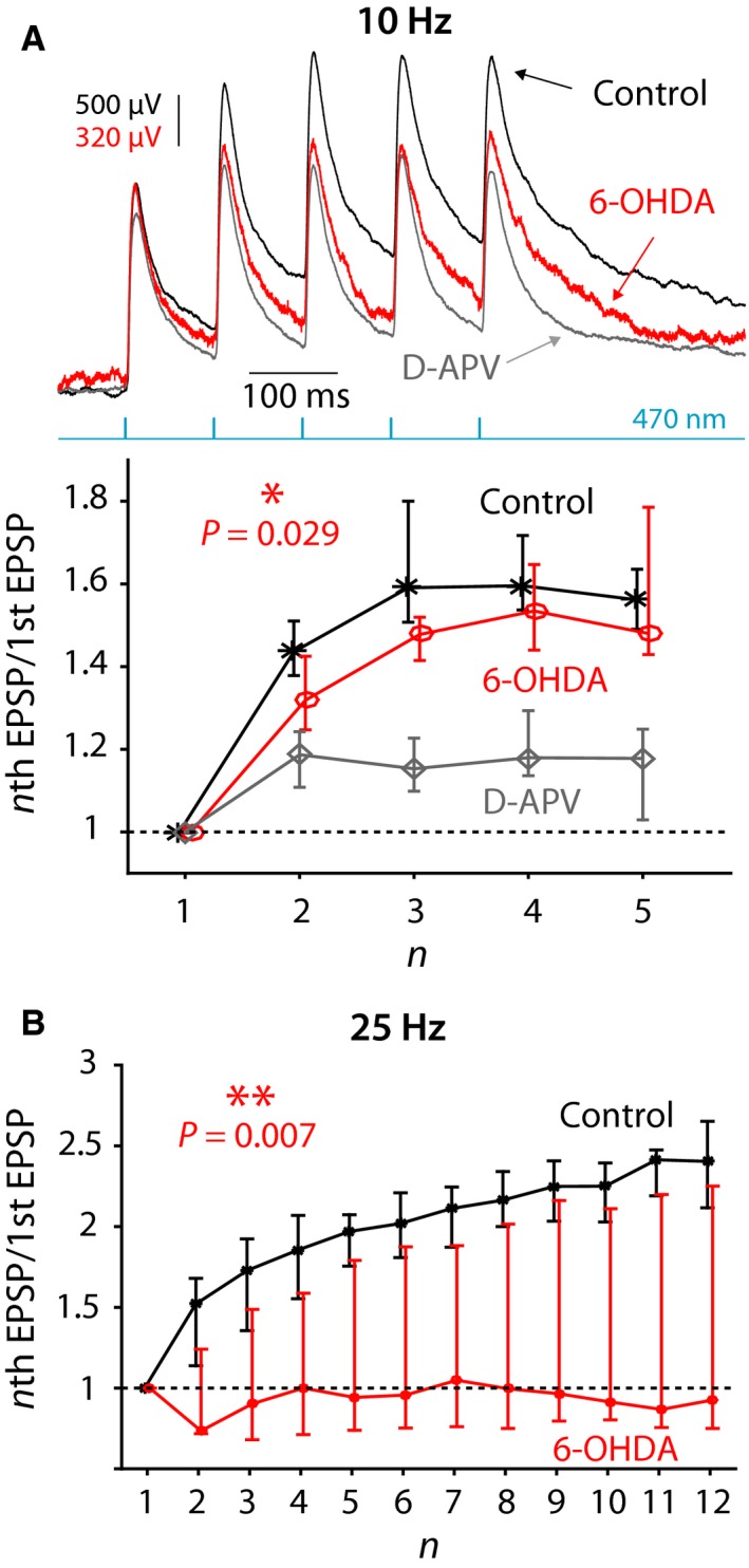
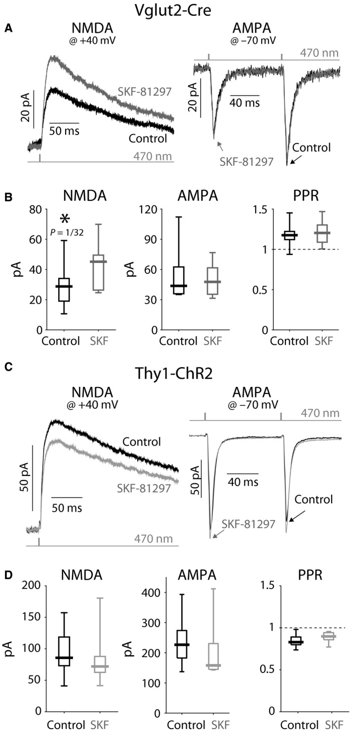
The widely held view that the pathophysiology of Parkinson's disease arises from an under-activation of the direct pathway striatal spiny neurons (dSPNs) has gained support from a recently described weakening of the glutamatergic projection from the parafascicular nucleus (PfN) to dSPNs in experimental parkinsonism. However, the impact of the remodeling of the thalamostriatal projection cannot be fully appreciated without considering its impact on cholinergic interneurons (ChIs) that themselves preferentially activate indirect pathway spiny neurons (iSPNs). To study this thalamostriatal projection, we virally transfected with Cre-dependent channelrhodopsin-2 (ChR2) the PfN of Vglut2-Cre mice that were dopamine-depleted with 6-hydroxydopamine (6-OHDA). In parallel, we studied the corticostriatal projection to ChIs in 6-OHDA-treated transgenic mice expressing ChR2 under the Thy1 promoter. We found the 6-OHDA lesions failed to affect short-term synaptic plasticity or the size of unitary responses evoked optogenetically in either of these projections. However, we found that NMDA-to-AMPA ratios at PfN synapses-that were significantly larger than NMDA-to-AMPA ratios at cortical synapses-were reduced by 6-OHDA treatment, thereby impairing synaptic integration at PfN synapses onto ChIs. Finally, we found that application of an agonist of the D5 dopamine receptors on ChIs potentiated NMDA currents without affecting AMPA currents or short-term plasticity selectively at PfN synapses. We propose that dopamine depletion leads to an effective de-potentiation of NMDA currents at PfN synapses onto ChIs which degrades synaptic integration. This selective remodeling of NMDA currents at PfN synapses may counter the selective weakening of PfN synapses onto dSPNs in parkinsonism.

Keywords: 6-OHDA; basal ganglia; optogenetics; slice electrophysiology.


Conflict of interest statement

The authors declare no conflict of interest.

Figures

Figure 1
Figure 1
Distribution of fibers arising from the parafascicular nucleus (PfN) or the cortex in control and dopamine‐depleted striata. (A) Confocal image of a coronal slice through the site of AAV injections in the PfN of Vglut2‐Cre mice. The green signal arises from the eYFP conjugated to the ChR2 harbored in the AAV. The dark spot is the fasciculus retroflexus (fr). (B) Left: Expression of eYFP in Vglut2 fibers arising from the PfN in sagittal slices of the striatum of control (top) and 6‐OHDA‐lesioned (bottom) Vglut2‐Cre mice. Ctx—cortex. Right: Tyrosine hydroxylase (TH) immunoreactivity in control (top) and 6‐OHDA‐lesioned (bottom) Vglut2‐Cre mice. Left: Expression of eYFP in nominally cortical (hence the asterisk) fibers in sagittal slices of the striatum of control (top) and 6‐OHDA‐lesioned (bottom) Thy1‐ChR2 mice. C. Right: TH immunoreactivity in control (top) and 6‐OHDA‐lesioned (bottom) Thy1‐ChR2 mice. Small and large red boxes represent the areas within the corpus callosum and the striatum, respectively, used to assess the extent of the reduction in TH immunoreactivity after dopamine depletion.
Figure 2
Figure 2
Optogenetic PPRs in ChIs from control and dopamine‐depleted striata. (A) Averaged EPSCs evoked in ChIs by a pair of 470‐nm light pulses (100‐ms inter‐pulse interval) that activate ChR2‐laden PfN fibers from a control (left) and 6‐OHDA‐lesioned (right) Vglut2‐Cre mouse. (B) Boxplot of PPRs at PfN synapses onto ChIs in control and 6‐OHDA‐lesioned mice. (C) Averaged EPSCs evoked in ChIs by a pair of 470‐nm light pulses that activate ChR2‐laden fibers from a control (left) and 6‐OHDA‐lesioned (right) Thy1‐ChR2 mouse. (D) Boxplot of PPRs at nominally cortical synapses onto ChIs in control and 6‐OHDA‐lesioned mice.
Figure 3
Figure 3
Minimal optogenetic stimulation of glutamatergic inputs to ChIs from control and dopamine‐lesioned striata. (A) Averaged unitary (black) and non‐unitary (dark gray) responses using a minimal stimulation protocol where the presence or absence (light gray) of a response is stochastic. (B) Empirical probability density function (pdf) of the amplitude of the stochastically generated EPSCs (corresponding to the experiment depicted in panel A) is fit by a binomial mixture of Gaussian model. (C) Boxplot of unitary response extracted from the value of the first mode of the pdf fit to each experiment demonstrates a lack of difference between PfN and cortical synapses and a lack of change following dopamine depletion.
Figure 4
Figure 4
NMDA currents are relatively larger for PfN than for cortical input to ChIs and are reduced at PfN synapses onto ChIs following dopamine depletion. Left: Averaged optogenetic EPSC evoked in ChIs clamped at +40 mV due to PfN input in a control Vglut2‐Cre mouse (top, left), and in a 6‐OHDA‐lesioned Vglut2‐Cre mouse (top, right); nominally cortical input in a control Thy1‐ChR2 mouse (bottom, left), and in a 6‐OHDA‐lesioned Thy1‐ChR2 mouse (bottom, right). Light gray traces are the responses after application of D‐APV, an NMDAR antagonist. Right: Boxplots of NMDA‐to‐AMPA ratios at cortical and PfN synapses in control mice, and 6‐OHDA‐lesioned mice, reveal that the NMDA component at PfN synapses is significantly larger than at cortical synapses onto ChIs and is reduced following 6‐OHDA lesions. n.s.—not significant.
Figure 5
Figure 5
Synaptic integration at PfN synapses onto ChIs is degraded following 6‐OHDA treatment. (A) Top: Synaptic integration at PfN synapses onto ChIs in response to five 470‐nm pulses at 10 Hz in a control mouse before (black) and after (gray) application of D‐APV and in 6‐OHDA‐lesioned mouse (red). Note the difference in scale between the red vs. the other traces chosen so as to align the amplitude of the first EPSP. Bottom: Ratio of the amplitude of nth EPSP to the 1st EPSP in control mice before (black) and after (gray) application of D‐APV and in 6‐OHDA‐lesioned mice (red). (B) Same as in panel B, bottom, except that 12 pulses are given at 25 Hz. The curves in panels B and C depict medians and confidence intervals given by the 50%× (1 ± 1/√k) quantiles, where k is the sample size (Lasser‐Katz et al., 2017).
Figure 6
Figure 6
NMDAR currents at PfN—but not cortical—synapses onto ChIs are potentiated by D5R activation. (A) Averaged optogenetic EPSC from PfN fibers in Vglut2‐Cre mice evoked in ChIs clamped at +40 mV (left) and ‐70 mV (right) before (black) and after (gray) application of SKF‐81297, a D1‐like receptor agonist. (B) Boxplots of the amplitude of the NMDA component (left, measured at +40 mV); the AMPA component (middle, 1st EPSC, measured at ‐70 mV); and the PPRs (right) demonstrate a significant amplification of the NMDA current without a concurrent change in the AMPA current or a change in the PPRs. (C) Averaged optogenetic EPSC from nominally cortical fibers in Thy1‐ChR2 mice evoked in ChIs clamped at +40 mV (left) and ‐70 mV (right) before (black) and after (gray) application of SKF‐81297. B. Boxplots of the amplitude of the NMDA component (left, measured at +40 mV); the AMPA component (middle, 1st EPSC, measured at −70 mV); and the PPRs (right) failed to demonstrate any change in the NMDA or AMPA currents or a change in the PPRs.

Similar articles

Cited by

References

    1. Albin, R.L. , Young, A.B. & Penney, J.B. (1995) The functional anatomy of disorders of the basal ganglia. Trends Neurosci., 18, 63–64. - PubMed
    1. Aoki, S. , Liu, A.W. , Zucca, A. , Zucca, S. & Wickens, J.R. (2015) Role of striatal cholinergic interneurons in set‐shifting in the rat. J. Neurosci., 35, 9424–9431. - PMC - PubMed
    1. Aosaki, T. , Graybiel, A.M. & Kimura, M. (1994) Effect of the nigrostriatal dopamine system on acquired neural responses in the striatum of behaving monkeys. Science, 265, 412–415. - PubMed
    1. Aosaki, T. , Kimura, M. & Graybiel, A.M. (1995) Temporal and spatial characteristics of tonically active neurons of the primate's striatum. J. Neurophysiol., 73, 1234–1252. - PubMed
    1. Arenkiel, B.R. , Peca, J. , Davison, I.G. , Feliciano, C. , Deisseroth, K. , Augustine, G. , Ehlers, M.D. & Feng, G. (2007) In vivo light‐induced activation of neural circuitry in transgenic mice expressing channelrhodopsin‐2. Neuron, 54, 205–218. - PMC - PubMed

Publication types