. 2017 Oct 1;27(10):4971-4987.
doi: 10.1093/cercor/bhx185.

The Transcription Factors COUP-TFI and COUP-TFII have Distinct Roles in Arealisation and GABAergic Interneuron Specification in the Early Human Fetal Telencephalon

Affiliations

The Transcription Factors COUP-TFI and COUP-TFII have Distinct Roles in Arealisation and GABAergic Interneuron Specification in the Early Human Fetal Telencephalon

Ayman Alzu'bi et al. Cereb Cortex. .

Abstract

In human telencephalon at 8-12 postconceptional weeks, ribonucleic acid quantitative sequencing and immunohistochemistry revealed cortical chicken ovalbumin upstream promotor-transcription factor 1 (COUP-TFI) expression in a high ventro-posterior to low anterior gradient except for raised immunoreactivity in the anterior ventral pallium. Unlike in mouse, COUP-TFI and SP8 were extensively co-expressed in dorsal sensory neocortex and dorsal hippocampus whereas COUPTFI/COUPTFII co-expression defined ventral temporal cortex and ventral hippocampus. In the ganglionic eminences (GEs) COUP-TFI immunoreactivity demarcated the proliferative zones of caudal GE (CGE), dorsal medial GE (MGE), MGE/lateral GE (LGE) boundary, and ventral LGE whereas COUP-TFII was limited to ventral CGE and the MGE/LGE boundary. Co-labeling with gamma amino butyric acidergic interneuron markers revealed that COUP-TFI was expressed in subpopulations of either MGE-derived (SOX6+) or CGE-derived (calretinin+/SP8+) interneurons. COUP-TFII was mainly confined to CGE-derived interneurons. Twice as many GAD67+ cortical cells co-labeled for COUP-TFI than for COUP-TFII. A fifth of COUP-TFI cells also co-expressed COUP-TFII, and cells expressing either transcription factor followed posterior or anterio-lateral pathways into the cortex, therefore, a segregation of migration pathways according to COUP-TF expression as proposed in mouse was not observed. In cultures differentiated from isolated human cortical progenitors, many cells expressed either COUP-TF and 30% also co-expressed GABA, however no cells expressed NKX2.1. This suggests interneurons could be generated intracortically from progenitors expressing either COUP-TF.

Keywords: SP8; cerebral cortex development; ganglionic eminences; hippocampus development; interneuron migration; ventral pallium.


Figures

Figure 1.
Figure 1.
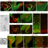
Gradients of COUP-TFI, COUP-TFII, ROBO1, SP8, and FGFR3 expression across the cortex by RNAseq. (A) The location of four cortical sampling cortical regions; anterior (A), central (C), posterior (P), and temporal (T). (B) COUP-TFI expression significantly higher in temporal and posterior regions at all ages. (C) Highest expression of COUP-TFII in the temporal regions, with no significant difference in the expression between anterior, central, and posterior regions at 9–10 PCW, although there were differences at 11–12 PCW. (D and E) ROBO1 and SP8 both expressed in decreasing anterior posterior gradients. (F) FGFR3 expressed in increasing anterior posterior gradient. Asterisk(s) represent statistically significant differences between regions (1-way ANOVA, Tukey's post hoc comparison, *P < 0.05, **P < 0.01). Error bars represent the standard error of the mean.
Figure 2.
Figure 2.
Gradients of COUP-TFI and COUP-TFII immunoreactivity across the telencephalon. (A). (AA”) Parasagittal sections from medial to lateral parts of the 8 PCW fetal brain; COUP-TFI highly expressed across the GE except the dLGE, strong expression also observed in proliferative zones and CP of posterior, lateral and temporal cortex, and anterior ventral pallium. (BB”) COUPT-FII mainly expressed in the vCGE and at the MGE/LGE boundary (arrow, B) and temporal ventral pallium (VP, B’, B”) and amygdala (Amy, B’) with dispersed scattered cells throughout rest of GE. Ant, anterior (=rostral); pos, posterior (=caudal); cent, central; Crx, cortex; PCrx, piriform cortex; CP, choroid plexus; d and vHip, dorsal and ventral hippocampus; CH, cortical hem; vCGE, ventral caudal ganglionic eminence; VP, ventral pallium; Th, thalamus; Pr Th, pre thalamus; Amy, amygdala. Scale bars = 2 mm.
Figure 3.
Figure 3.
COUP-TFI immunoreactivity in the cortical wall. (A) Distinct posterior to anterior gradient in COUP-TFI expression across cortex in sagittal section 8 PCW. (B) Only a few scattered COUP-TFI+ cells observed in anterior cortex VZ, moderate to weak immunoreactivity in the SVZ/IZ/CP. (C) Moderate immunoreactivity across cortical wall of central cortex. (D) Strongest immunoreactivity in posterior cortex, relatively stronger in proliferative than in postmitotic zones. (E) Similar expression gradient at 12 PCW. (F) Strong COUP-TFI expression in proliferative and postmitotic zones of ventral pallium (VP). (G) COUP-TFI+ cells broadly distributed to all cortical layers of anterior cortex at 12 PCW. (HJ) COUP-TFI expression increased gradually in central, posterior, and temporal (ventral) regions, respectively. Boxed areas in A and E show where images (BD and FJ) were taken. Ant, anterior; pos, posterior; VZ, ventricular zone; SVZ, subventricular zone; MZ, marginal zone; cp, choroid plexus; Pia, pia matter; VP, ventral pallium; Th, thalamus; Pr Th, pre thalamus. Scale bars = 500 μm in A, E; 100 μm in D (and for B, C); 100 μm in F; 100 μm in J (and for G, H, I).
Figure 4.
Figure 4.
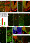
Multiple progenitor domains in the GE of early human fetal brain. (AC) Medial parasagittal sections of 8 PCW human fetal brain. NKX2.1 expression mainly confined to MGE (A). PAX6 expressed in gradient with higher expression in cortical proliferative zone to lower expression in the LGE (arrows in A and B indicate boundary between MGE and LGE). (B) COUP-TFI showed partially overlapped expression with NKX2.1 and OLIG2 in MGE, arrow head marks the boundary between dorsal and ventral MGE (C). (DH) Horizontal sections of 10 PCW human fetal brain; NKX2.1 expression confined to MGE and its caudal extension (D). PAX6 complementarily expressed with NKX2.1 in GE (E, E’). COUP-TFI expressed in the VZ/SVZ of the MGE, CGE, vLGE, but only in migrating cells in dLGE (F, F’). COUPT-FII highly expressed in CGE, the MGE/LGE boundary (arrowhead), and in a stream of cells entering the cortex from dLGE (G, G’). SP8 expressed in the SVZ of CGE, and in an increasing gradient from vLGE to dLGE (H, H’). Boxed areas in E shows where images (E’–H’) were taken. Ant, anterior; pos, posterior; Lat, lateral, Med, medial; Crx, cortex; IC, internal capsule; Sep, septum; v and d LGE, ventral and dorsal LGE; Th, thalamus. Scale bars = 500 μm in AH, 200 μm in E’–H’.
Figure 5.
Figure 5.
(A) Counter gradients of SP8 and COUP-TFI expression in cortical VZ broadly overlapped (yellow) including in posterior cortex (Pos) and the hippocampal primordium dorsal to the cortical hem (dHip). Only anterior cortex (Ant) predominantly expressed SP8, and only ventral temporal cortex including ventral hippocampus (vHip) exclusively expressed COUP-TFI. Likewise counter gradient of SP8 expression in VZ and COUP-TFI in SVZ was apparent in GE. The medial amygdala (Amy) predominantly expressed SP8, whereas ventral pallium (VP) and piriform cortex (PCrx) expressed COUP-TFI. (B) COUP-TFI expression in the anterior ventral pallium (VP) with SP8 confined to LGE. (C) In cortex SP8 and COUP-TFII show abrupt expression boundaries; COUP-TFII confined to ventral temporal lobe. (D) Posterior regions of medial amygdala populated by SP8+/COUP-TFII+ cells. (E) the dLGE/cortex boundary was sharply delineated by SP8 and COUP-TFI expression. (F) Relatively small numbers of SP8+/COUPTFI- cells migrate from LGE-Like CGE (LCGE) into the cortex at this stage. (G) Low density of COUP-TFII+ cells in SP8+ dLGE. (H) High density of SP8 + /COUP-TFII+ cells in SVZ of the LGE-Like CGE. (I and J) SP8+/COUP-TFI+ cells predominantly seen in cortical VZ although double-labeled cells present in SVZ and IZ (I). SP8+/COUP-TFII+ double-labeled cells predominantly observed in SVZ and IZ (J). (K and L) SP8 Co-expressed with both COUP-TFI and COUP-TFII in ventral CGE. (M, N, O) Extensive co-expression of SP8 and CalR in the vCGE (M) but also in the LGE and cortex (N, O). Crx, cortex; d and vHip, dorsal and ventral hippocampus; Amy, amygdala; VP, ventral pallium; dLGE, dorsal LGE; L and vCGE, lateral and ventral CGE; VZ, ventricular zone. Scale bars = 2 mm in A, C; 500 μm in K, L, M; 200 μm in EH, N, O; 100 μm in B, D, I, J.
Figure 6.
Figure 6.
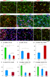
COUPT-FI and COUP-TFII expression in cortical GABAergic interneurons. (AC) Double labeling for COUPT-FI and GAD67 in cortical wall at 8 PCW. COUP-TFI+/GAD67+ cells broadly distributed in the cortical wall, mostly either just above the thin CP in the marginal zone or just below in the pre-subplate/IZ; (A, B) A considerable number also found in proliferative (VZ/SVZ) zone of anterior and posterior cortex with radial nuclear morphology (arrows). (C) A stream of COUP-TFI+/GAD67+ cells enter temporal cortex from vCGE, migrating tangentially mainly in the SVZ/IZ. (D, E) A smaller proportion of GAD67+ cells co-express COUP-TFII. (F) The percentage of COUP-TFI+ and COUP-TFII+ cells from all GAD67+ cells in the proliferative (VZ/SVZ) and postmitotic zones (IZ/CP) of 8 PCW cortical wall. (G, H) Double labeling for COUPT-FI and COUP-TFII with calretinin (CalR) in cortical wall of 12 PCW fetal brain. (I, K) Double labeling for COUPT-FI and COUP-TFII with calbindin (CalB) in 12 PCW cortical wall. (L, M) Double labeling for SOX6 and GAD67 in the 8 PCW cortical wall. The majority of SOX6+ cells co-expressed GAD67 this stage (L), while a proportion of SOX6+ cells also co-expressed COUP-TFI (M). (N) Double labeling for SOX6 and NKX2.1 in the GE of of 10 PCW fetal brain. SOX6 mainly expressed in NKX2.1+ cells in SVZ of MGE and in migrating cells through the LGE mantle zone; SOX6 also expressed in the cortical and dLGE VZ. (OQ) Double labeling for COUPT-FI or COUP-TFII with SOX6 in GE at 10 PCW; COUP-TFI highly expressed in SOX6+ cells in MGE and in cells entering cortex from LGE (O), no similar double labeling with COUP-TFII (P, Q). Insets with boxed areas show where images (AE and GM) were taken. Images NQ are taken from a similar section to Figure 4I. VZ, ventricular zone; Pia, pia matter; Temp crx, temporal cortex; Sep, septum; dLGE, dorsal LGE. Scale bars = 100 μm in B, D (and for A); 200 μm in C, E; 100 μm in K (and for G, H, I); 100 μm in M (and for L) 500 μm in P (and for N, O); 100 μm in Q.
Figure 7.
Figure 7.
Examples of double immunofluorescent staining of cultured cells derived from human fetal cortex (Crx, AC, EH) and GEs (GE, D, I, J) showing that GABA+ neurons can be generated from cortically-derived progenitor cells. Counts were made from these cultures and the results plotted as bar charts; for instance K shows the% of B-tubulin expressing neurons expressing GABA in cultures derived from GE anterior cortex (ACx) posterior cortex (PCx). Error bars represent the standard error of the mean, 1 asterisk denotes a statistically significant difference P < 0.05, 2 asterisks P < 0.0005, 2-tailed t-test.

References

    1. Aggleton JP, Wright NF, Vann SD, Saunders RC. 2012. Medial temporal lobe projections to the retrosplenial cortex of the macaque monkey. Hippocampus. 22:1883–1900. - PMC - PubMed
    1. Alfano C, Magrinelli E, Harb K, Hevner RF, Studer M. 2014. a. Postmitotic control of sensory area specification during neocortical development. Nature Commun. 5:5632. - PubMed
    1. Alfano C, Magrinelli E, Harb K, Studer M. 2014. b. The nuclear receptors COUP-TF: a long-lasting experience in forebrain assembly. Cell Molec Life Sci. 71:43–62. - PMC - PubMed
    1. Alfano C, Viola L, Heng JIT, Pirozzi M, Clarkson M, Flore G, De Maio A, Schedl A, Guillemot F, Studer M. 2011. COUP-TFI promotes radial migration and proper morphology of callosal projection neurons by repressing Rnd2 expression. Development. 138:4685–4697. - PubMed
    1. Al-Jaberi N, Lindsay S, Sarma S, Bayatti N, Clowry GJ. 2015. The early fetal development of human neocortical gabaergic interneurons. Cereb Cortex. 25:635–651. - PMC - PubMed

Publication types

MeSH terms

Substances