Growing Membranes In Vitro by Continuous Phospholipid Biosynthesis from Free Fatty Acids
- PMID: 28922922
- PMCID: PMC5778391
- DOI: 10.1021/acssynbio.7b00265
Growing Membranes In Vitro by Continuous Phospholipid Biosynthesis from Free Fatty Acids
Abstract
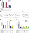
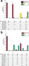
One of the key aspects that defines a cell as a living entity is its ability to self-reproduce. In this process, membrane biogenesis is an essential element. Here, we developed an in vitro phospholipid biosynthesis pathway based on a cascade of eight enzymes, starting from simple fatty acid building blocks and glycerol 3-phosphate. The reconstituted system yields multiple phospholipid species that vary in acyl-chain and polar headgroup compositions. Due to the high fidelity and versatility, complete conversion of the fatty acid substrates into multiple phospholipid species is achieved simultaneously, leading to membrane expansion as a first step toward a synthetic minimal cell.
Keywords: enzymatic conversion; membrane proteins; membranes; phospholipid biosynthesis; synthetic cell.
Conflict of interest statement
The authors declare no competing financial interest.
Figures


Similar articles
-
Cell-Free Phospholipid Biosynthesis by Gene-Encoded Enzymes Reconstituted in Liposomes.PLoS One. 2016 Oct 6;11(10):e0163058. doi: 10.1371/journal.pone.0163058. eCollection 2016. PLoS One. 2016. PMID: 27711229 Free PMC article.
-
Mechanism of phospholipid biosynthesis in Escherichia coli: acyl-CoA synthetase is not required for the incorporation of intracellular free fatty acids into phospholipid.Biochem Biophys Res Commun. 1976 Mar 22;69(2):506-13. doi: 10.1016/0006-291x(76)90550-7. Biochem Biophys Res Commun. 1976. PMID: 773377 No abstract available.
-
Lipid composition of membranes of Escherichia coli by liquid chromatography/tandem mass spectrometry using negative electrospray ionization.Rapid Commun Mass Spectrom. 2007;21(11):1721-8. doi: 10.1002/rcm.3013. Rapid Commun Mass Spectrom. 2007. PMID: 17477452
-
[Regulation of the synthesis of phospholipid molecular species in Escherichia coli membranes (author's transl)].Seikagaku. 1977;49(12):1301-15. Seikagaku. 1977. PMID: 342643 Review. Japanese. No abstract available.
-
Molecular genetics of membrane phospholipid synthesis.Annu Rev Genet. 1986;20:253-95. doi: 10.1146/annurev.ge.20.120186.001345. Annu Rev Genet. 1986. PMID: 3545060 Review.
Cited by
-
Synthetic Minimal Cell: Self-Reproduction of the Boundary Layer.ACS Omega. 2019 Mar 31;4(3):5293-5303. doi: 10.1021/acsomega.8b02955. Epub 2019 Mar 13. ACS Omega. 2019. PMID: 30949617 Free PMC article.
-
A synthetic metabolic network for physicochemical homeostasis.Nat Commun. 2019 Sep 18;10(1):4239. doi: 10.1038/s41467-019-12287-2. Nat Commun. 2019. PMID: 31534136 Free PMC article.
-
Photochemical synthesis of natural lipids in artificial and living cells.Nat Commun. 2025 May 31;16(1):5068. doi: 10.1038/s41467-025-60358-4. Nat Commun. 2025. PMID: 40450019 Free PMC article.
-
ATP Recycling Fuels Sustainable Glycerol 3-Phosphate Formation in Synthetic Cells Fed by Dynamic Dialysis.ACS Synth Biol. 2022 Jul 15;11(7):2348-2360. doi: 10.1021/acssynbio.2c00075. Epub 2022 Apr 4. ACS Synth Biol. 2022. PMID: 35377147 Free PMC article.
-
The hallmarks of living systems: towards creating artificial cells.Interface Focus. 2018 Oct 6;8(5):20180023. doi: 10.1098/rsfs.2018.0023. Epub 2018 Aug 17. Interface Focus. 2018. PMID: 30443324 Free PMC article. Review.
References
Publication types
MeSH terms
Substances
LinkOut - more resources
Full Text Sources
Other Literature Sources

