. 2018 Jan:134:93-107.
doi: 10.1016/j.prostaglandins.2017.09.005. Epub 2017 Sep 18.

Human retinal endothelial cells and astrocytes cultured on 3-D scaffolds for ocular drug discovery and development

Affiliations

Human retinal endothelial cells and astrocytes cultured on 3-D scaffolds for ocular drug discovery and development

Kay D Beharry et al. Prostaglandins Other Lipid Mediat. 2018 Jan.

Abstract

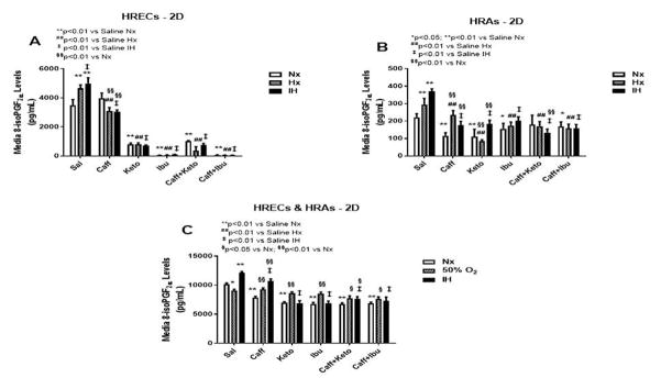
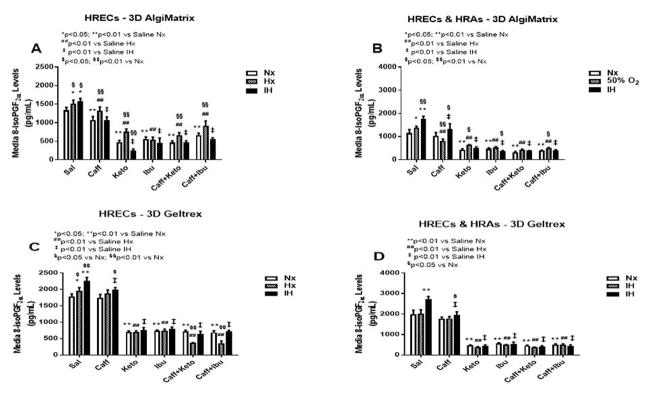
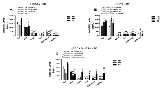
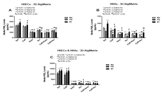
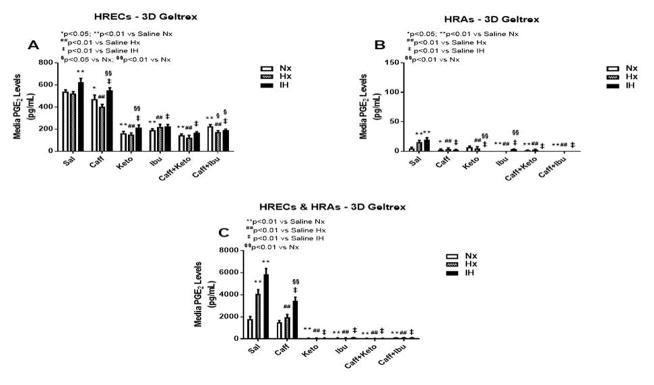
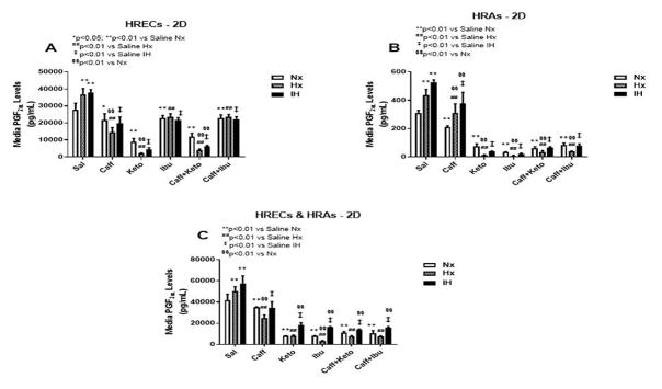
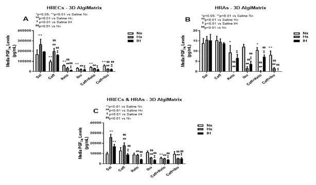
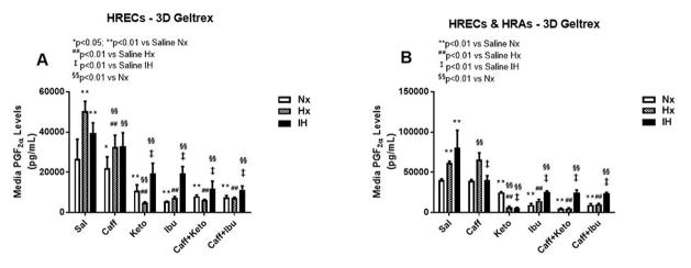
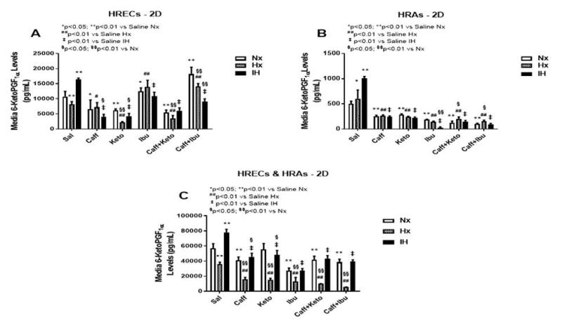
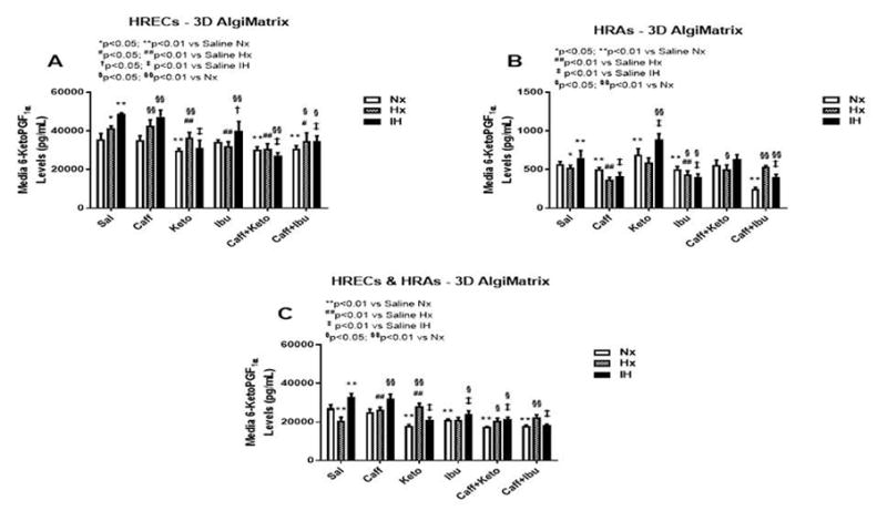
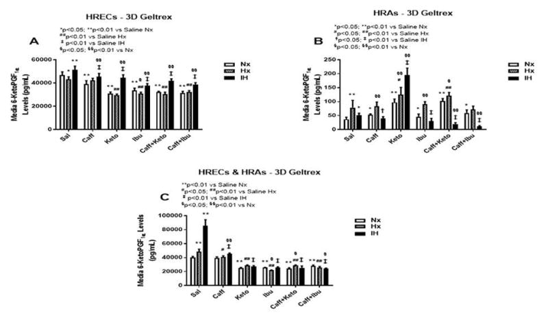
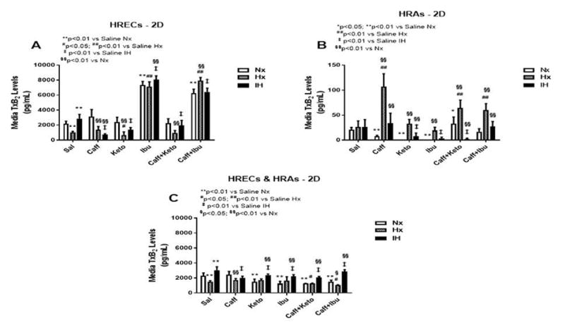
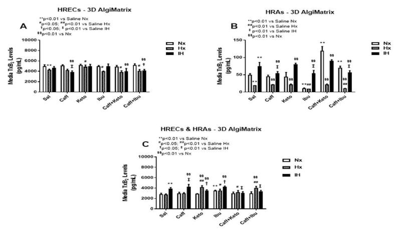
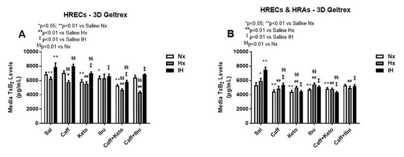
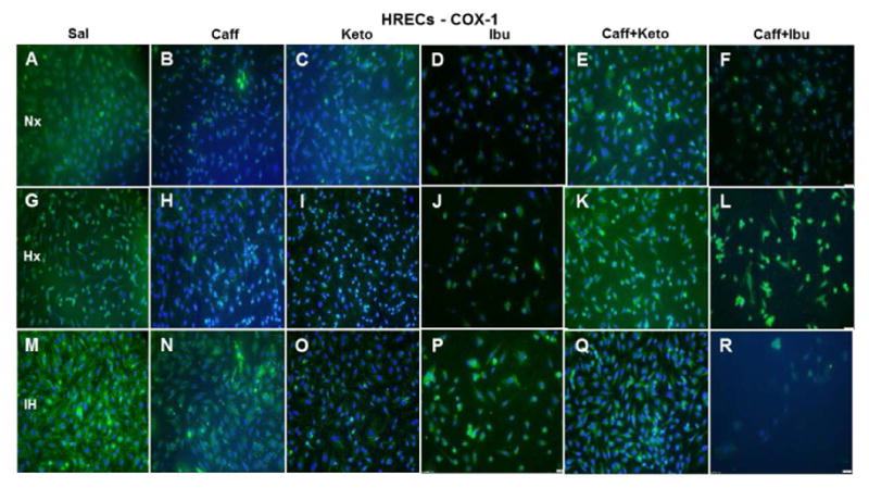
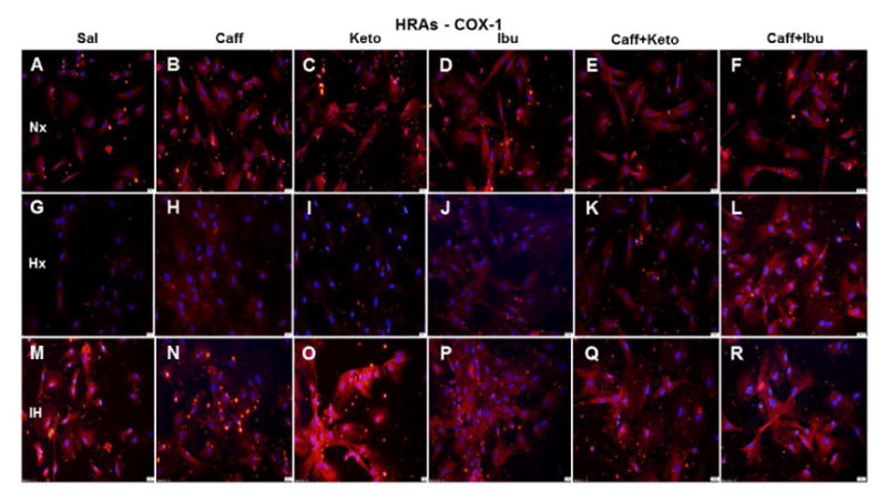
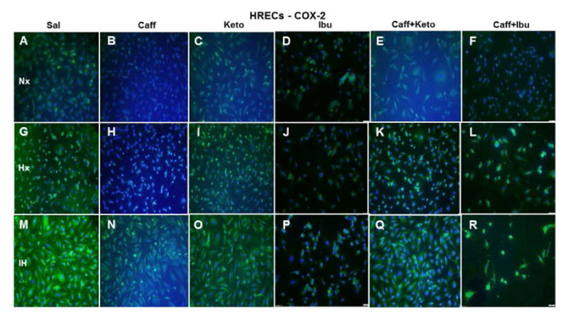
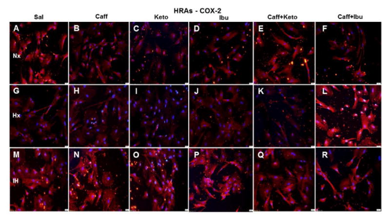
Topical ocular ketorolac improves the outcomes of severe retinopathy of prematurity and when administered with systemic caffeine, decreases the severity of oxygen-induced retinopathy. We tested the hypothesis that co-cultures of human retinal endothelial cells (HRECs) and human retinal astrocytes (HRAs) on 3-dimensional (3-D) hydrogel scaffolds is a more representative biomimetic paradigm of the blood-retinal-barrier (BRB) than 2-D cultures, and should be utilized for preclinical drug discovery and development. Mono- and co-cultures of HRECs and HRAs were treated with standard doses of ketorolac, ibuprofen, and/or caffeine, and exposed to hyperoxia, intermittent hypoxia (IH), or normoxia on 2-D surfaces or 3-D biodegradable hydrogel scaffolds (AlgiMatrix or Geltrex). Media and cells were collected at 72h post treatment for arachidonic acid metabolites. Cells cultured on 3-D scaffolds exhibited less oxidative stress and variability in drug responses. HRAs enhanced the responses of HRECs to drugs and changes in oxygen environment. PGE2 and PGI2 were the predominant prostanoids produced in response to IH, reflecting COX-2 immunoreactivity. We conclude that HRECs and HRAs co-cultured on 3-D scaffolds may recapitulate drug responses of the dynamic BRB and therefore should be implemented for preclinical ocular drug discovery and development.

Keywords: AlgiMatrix; Biodegradable 3-D scaffolds; Caffeine; Co-cultures; Geltrex; Human retinal astrocytes; Human retinal endothelial cells; Intermittent hypoxia; Non-steroidal anti-inflammatory drugs.


Conflict of interest statement

Conflict of interest: The authors declare that they have not conflicts of interest related to this manuscript.

Figures

Figure 1
Figure 1
8-iso-PGF levels in media from cells cultured on 2-D surfaces. Panel A represents data from HRECs, panel B from HRAs and panel C from co-cultures of HRECs+HRAs. Data are presented as mean±SEM (n=8 samples/groups). Comparison to saline Nx is represented by *p<0.05; **p<0.01; comparison to saline Hx is represented by #p<0.05; ##p<0.01; comparison to saline IH is represented by p<0.05; p<0.01; and comparison among the treated groups is represented by §p<0.05; §§p<0.01.
Figure 2
Figure 2
8-iso-PGF levels in media from 3-D AlgiMatrix biodegradable hydrogel scaffolds (panels A & B), and 3-D Geltrex biodegradable hydrogel scaffolds (panels C & D) in HRECs (panels A and C and HREC+HRA co-cultures (panels B & D). Data are presented as mean±SEM (n=8 samples/groups). Data are presented as mean±SEM (n=8 samples/groups). Comparison to saline Nx is represented by *p<0.05; **p<0.01; comparison to saline Hx is represented by #p<0.05; ##p<0.01; comparison to saline IH is represented by p<0.05; p<0.01; and comparison among the treated groups is represented by §p<0.05; §§p<0.01.
Figure 3
Figure 3
PGE2 levels in media from cells cultured on 2-D surfaces. Panel A represents data from HRECs, panel B from HRAs and panel C from co-cultures of HRECs+HRAs. Data are presented as mean±SEM (n=8 samples/groups). Comparison to saline Nx is represented by *p<0.05; **p<0.01; comparison to saline Hx is represented by #p<0.05; ##p<0.01; comparison to saline IH is represented by p<0.05; p<0.01; and comparison among the treated groups is represented by §p<0.05; §§p<0.01.
Figure 4
Figure 4
PGE2 levels in media from 3-D AlgiMatrix biodegradable hydrogel scaffolds. Panel A represents data from HRECs, panel B from HRAs and panel C from co-cultures of HRECs+HRAs. Data are presented as mean±SEM (n=8 samples/groups). Data are presented as mean±SEM (n=8 samples/groups). Comparison to saline Nx is represented by *p<0.05; **p<0.01; comparison to saline Hx is represented by #p<0.05; ##p<0.01; comparison to saline IH is represented by p<0.05; p<0.01; and comparison among the treated groups is represented by §p<0.05; §§p<0.01.
Figure 5
Figure 5
PGE2 levels in media from 3-D Geltrex biodegradable hydrogel scaffolds. Panel A represents data from HRECs, panel B from HRAs and panel C from co-cultures of HRECs+HRAs. Data are presented as mean±SEM (n=8 samples/groups). Data are presented as mean±SEM (n=8 samples/groups). Comparison to saline Nx is represented by *p<0.05; **p<0.01; comparison to saline Hx is represented by #p<0.05; ##p<0.01; comparison to saline IH is represented by p<0.05; p<0.01; and comparison among the treated groups is represented by §p<0.05; §§p<0.01.
Figure 6
Figure 6
PGF levels in media from cells cultured on 2-D surfaces. Panel A represents data from HRECs, panel B from HRAs and panel C from co-cultures of HRECs+HRAs. Data are presented as mean±SEM (n=8 samples/groups). Comparison to saline Nx is represented by *p<0.05; **p<0.01; comparison to saline Hx is represented by #p<0.05; ##p<0.01; comparison to saline IH is represented by p<0.05; p<0.01; and comparison among the treated groups is represented by §p<0.05; §§p<0.01.
Figure 7
Figure 7
PGF levels in media from 3-D AlgiMatrix biodegradable hydrogel scaffolds. Panel A represents data from HRECs, panel B from HRAs and panel C from co-cultures of HRECs+HRAs. Data are presented as mean±SEM (n=8 samples/groups). Data are presented as mean±SEM (n=8 samples/groups). Comparison to saline Nx is represented by *p<0.05; **p<0.01; comparison to saline Hx is represented by #p<0.05; ##p<0.01; comparison to saline IH is represented by p<0.05; p<0.01; and comparison among the treated groups is represented by §p<0.05; §§p<0.01.
Figure 8
Figure 8
PGF levels in media from 3-D Geltrex biodegradable hydrogel scaffolds. Panel A represents data from HRECs, and panel B from co-cultures of HRECs+HRAs. Data are presented as mean±SEM (n=8 samples/groups). Data are presented as mean±SEM (n=8 samples/groups). Comparison to saline Nx is represented by *p<0.05; **p<0.01; comparison to saline Hx is represented by #p<0.05; ##p<0.01; comparison to saline IH is represented by p<0.05; p<0.01; and comparison among the treated groups is represented by §p<0.05; §§p<0.01.
Figure 9
Figure 9
6-ketoPGF levels in media from cells cultured on 2-D surfaces. Panel A represents data from HRECs, panel B from HRAs and panel C from co-cultures of HRECs+HRAs. Data are presented as mean±SEM (n=8 samples/groups). Comparison to saline Nx is represented by *p<0.05; **p<0.01; comparison to saline Hx is represented by #p<0.05; ##p<0.01; comparison to saline IH is represented by p<0.05; p<0.01; and comparison among the treated groups is represented by §p<0.05; §§p<0.01.
Figure 10
Figure 10
6-ketoPGF levels in media from 3-D AlgiMatrix biodegradable hydrogel scaffolds. Panel A represents data from HRECs, panel B from HRAs and panel C from co-cultures of HRECs+HRAs. Data are presented as mean±SEM (n=8 samples/groups). Data are presented as mean±SEM (n=8 samples/groups). Comparison to saline Nx is represented by *p<0.05; **p<0.01; comparison to saline Hx is represented by #p<0.05; ##p<0.01; comparison to saline IH is represented by p<0.05; p<0.01; and comparison among the treated groups is represented by §p<0.05; §§p<0.01.
Figure 11
Figure 11
6-ketoPGF levels in media from 3-D Geltrex biodegradable hydrogel scaffolds. Panel A represents data from HRECs, panel B from HRAs, and panel C from co-cultures of HRECs+HRAs. Data are presented as mean±SEM (n=8 samples/groups). Data are presented as mean±SEM (n=8 samples/groups). Comparison to saline Nx is represented by *p<0.05; **p<0.01; comparison to saline Hx is represented by #p<0.05; ##p<0.01; comparison to saline IH is represented by p<0.05; p<0.01; and comparison among the treated groups is represented by §p<0.05; §§p<0.01.
Figure 12
Figure 12
TxB2 levels in media from cells cultured on 2-D surfaces. Panel A represents data from HRECs, panel B from HRAs and panel C from co-cultures of HRECs+HRAs. Data are presented as mean±SEM (n=8 samples/groups). Comparison to saline Nx is represented by *p<0.05; **p<0.01; comparison to saline Hx is represented by #p<0.05; ##p<0.01; comparison to saline IH is represented by p<0.05; p<0.01; and comparison among the treated groups is represented by §p<0.05; §§p<0.01.
Figure 13
Figure 13
TxB2 levels in media from 3-D AlgiMatrix biodegradable hydrogel scaffolds. Panel A represents data from HRECs, panel B from HRAs and panel C from co-cultures of HRECs+HRAs. Data are presented as mean±SEM (n=8 samples/groups). Data are presented as mean±SEM (n=8 samples/groups). Comparison to saline Nx is represented by *p<0.05; **p<0.01; comparison to saline Hx is represented by #p<0.05; ##p<0.01; comparison to saline IH is represented by p<0.05; p<0.01; and comparison among the treated groups is represented by §p<0.05; §§p<0.01.
Figure 14
Figure 14
TxB2 levels in media from 3-D Geltrex biodegradable hydrogel scaffolds. Panel A represents data from HRECs, and panel B from co-cultures of HRECs+HRAs. Data are presented as mean±SEM (n=8 samples/groups). Data are presented as mean±SEM (n=8 samples/groups). Comparison to saline Nx is represented by *p<0.05; **p<0.01; comparison to saline Hx is represented by #p<0.05; ##p<0.01; comparison to saline IH is represented by p<0.05; p<0.01
Figure 15
Figure 15
Representative image of COX-1 immunoreactivity in HRECs. The green stain is COX-1 immunoreactivity and the blue color is DAPI staining of the nuclei. Panels A through F represents the Nx groups, panels G through L represents the Hx groups, and panels M through R represents the IH groups. Treatment groups are saline (Panels A, G, and M), caffeine (panels B, H, and N), ketorolac (panels C, I, and O), ibuprofen (panels D, J, and P), caffeine+ketorolac (panels E, K, and Q), and caffeine+ibuprofen (panels F, L, and R). Images are captured at 20X magnification. Scale bar is 100 μm.
Figure 16
Figure 16
Representative image of COX-1 immunoreactivity in HRAs. The red stain is COX-1 immunoreactivity and the blue color is DAPI staining of the nuclei. Groups are as described in Figure 15. Images are captured at 20X magnification. Scale bar is 100 μm.
Figure 17
Figure 17
Representative image of COX-2 immunoreactivity in HRECs. The green stain is COX-2 immunoreactivity and the blue color is DAPI staining of the nuclei. Groups are as described in Figure 15. Images are captured at 20X magnification. Scale bar is 100 μm.
Figure 18
Figure 18
Representative image of COX-2 immunoreactivity in HRAs. The red stain is COX-2 immunoreactivity and the blue color is DAPI staining of the nuclei. Groups are as described in Figure 15. Images are captured at 20X magnification. Scale bar is 100 μm.

References

    1. Gilbert C, Fielder A, Gordillo L, et al. Characteristics of infants with severe retinopathy of prematurity in countries with low, moderate, and high levels of development: implications for screening programs. Pediatrics. 2005;115:e518–e525. - PubMed
    1. Kong L, Fry M, Al-Samarraie M, Gilbert C, Steinkuller PG. An update on progress and the changing epidemiology of causes of childhood blindness worldwide. J AAPOS. 2012;16:501–507. - PubMed
    1. York JR, Landers S, Kirby RS, Arbogast PG, Penn JS. Arterial oxygen fluctuation and retinopathy of prematurity in very-low-birth-weight infants. J Perinatol. 2004;24:82–87. - PubMed
    1. Di Fiore JM, Bloom JN, Orge F, et al. A higher incidence of intermittent hypoxemic episodes is associated with severe retinopathy of prematurity. J Pediatr. 2010;157:69–73. - PMC - PubMed
    1. Di Fiore JM, Kaffashi F, Loparo K, et al. The relationship between patterns of intermittent hypoxia and retinopathy of prematurity in preterm infants. Pediatr Res. 2012;72:606–612. - PMC - PubMed

Publication types

MeSH terms

LinkOut - more resources