. 2017 Nov 17;85(12):e00301-17.
doi: 10.1128/IAI.00301-17. Print 2017 Dec.

H-IPSE Is a Pathogen-Secreted Host Nucleus-Infiltrating Protein (Infiltrin) Expressed Exclusively by the Schistosoma haematobium Egg Stage

Affiliations

H-IPSE Is a Pathogen-Secreted Host Nucleus-Infiltrating Protein (Infiltrin) Expressed Exclusively by the Schistosoma haematobium Egg Stage

Luke F Pennington et al. Infect Immun. .

Abstract

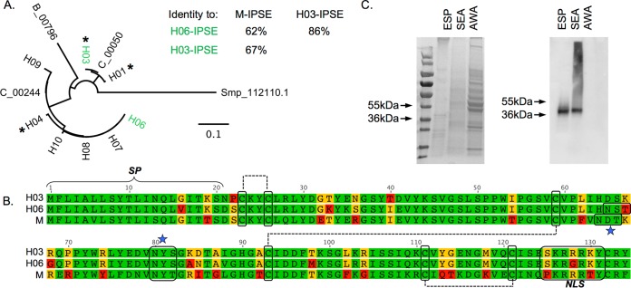
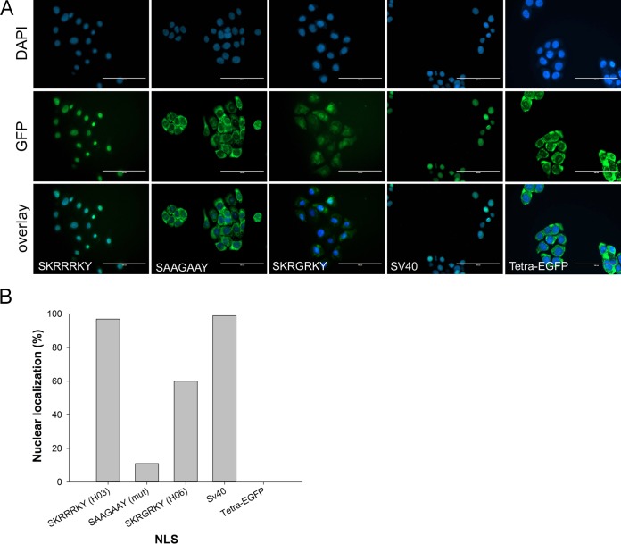
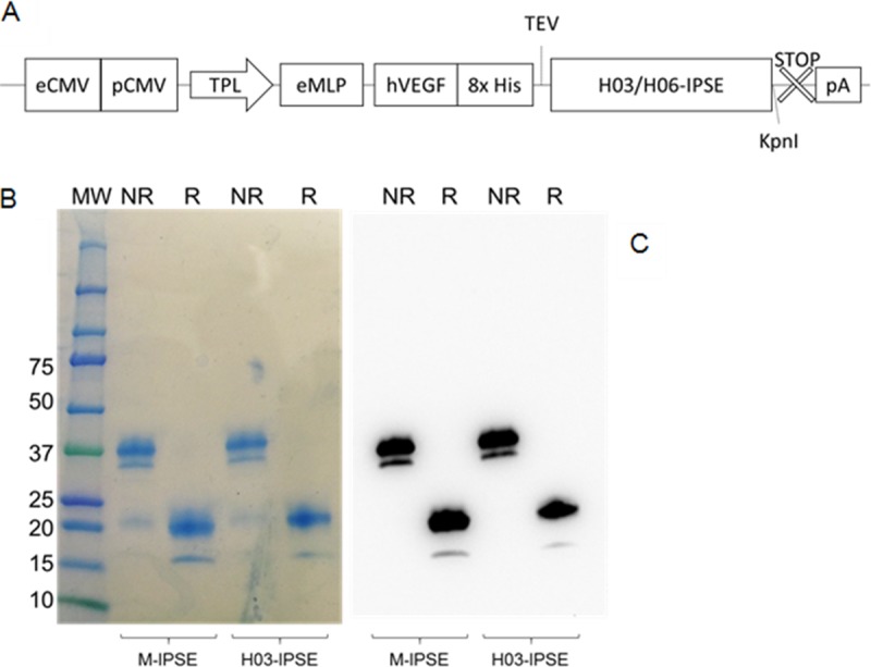
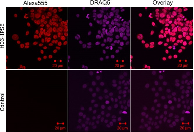
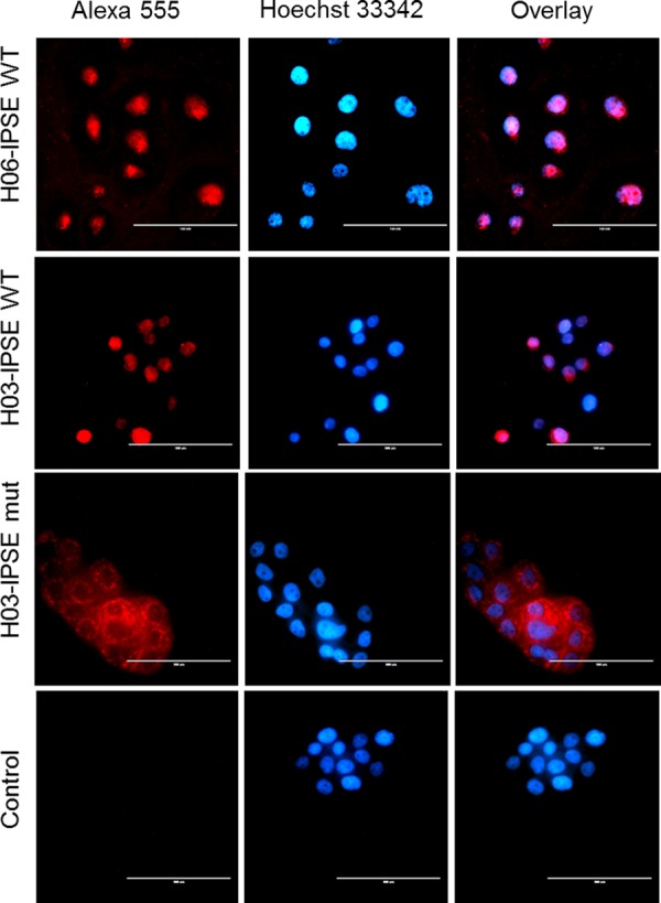
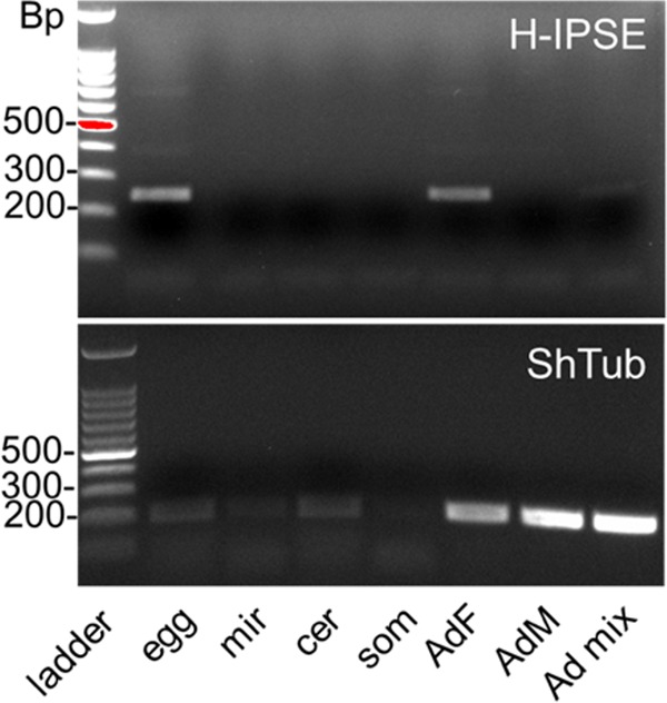
Urogenital schistosomiasis, caused by the parasitic trematode Schistosoma haematobium, affects over 112 million people worldwide. As with Schistosoma mansoni infections, the pathology of urogenital schistosomiasis is related mainly to the egg stage, which induces granulomatous inflammation of affected tissues. Schistosoma eggs and their secretions have been studied extensively for the related organism S. mansoni, which is more amenable to laboratory studies. Indeed, we have shown that IPSE/alpha-1 (here M-IPSE), a major protein secreted from S. mansoni eggs, can infiltrate host cells. Although the function of M-IPSE is unknown, its ability to translocate to the nuclei of host cells and bind DNA suggests a possible role in immune modulation of host cell tissues. Whether IPSE homologs are expressed in other schistosome species has not been investigated. Here, we describe the cloning of two paralog genes, H03-IPSE and H06-IPSE, which are orthologs of M-IPSE, from egg cDNA of S. haematobium Using PCR and immunodetection, we confirmed that the expression of these genes is restricted to the egg stage and female adult worms, while the H-IPSE protein is detectable only in mature eggs and not adults. We show that both H03-IPSE and H06-IPSE proteins can infiltrate HTB-9 bladder cells when added exogenously to culture medium. Monopartite C-terminal nuclear localization sequence (NLS) motifs conserved in H03-IPSE, SKRRRKY, and H06-IPSE SKRGRKY, are responsible for targeting the proteins to the nucleus of HTB-9 cells, as demonstrated by site-directed mutagenesis and green fluorescent protein (GFP) tagging. Thus, S. haematobium eggs express IPSE homologs that appear to perform similar functions in infiltrating host cells.

Keywords: Schistosoma haematobium; nuclear localization signal; schistosomiasis.


Figures

FIG 1
FIG 1
S. haematobium expresses multiple forms of H-IPSE. (A) Amino acid sequences of predicted H-IPSE paralogs, M-IPSE, and sequenced transcripts from egg cDNA were globally aligned by using a Blosum62 cost matrix, and a tree was built by using the neighbor-joining method in Geneious 7.1.4. The bar represents amino acid substitutions per site. H06 and H03 variants chosen for expression are highlighted in green, and their respective amino acid identities and identities to M-IPSE are shown. All variants identified through 3′-RACE cloning are indicated with asterisks. (B) Alignment of amino acid sequences of H03-IPSE (top rows) and H06-IPSE (middle row)s with the sequence of the homolog in S. mansoni (IPSE/alpha-1; here named M-IPSE) (bottom rows). These H-IPSE clones retain ∼63 to 68% amino acid identity and several features described previously for M-IPSE (24), including a 20-amino-acid classical signal sequence, seven cysteine residues involved in disulfide bonds, two N-linked glycosylation consensus motifs, and a predicted nuclear localization sequence. Residues shown in green are identical, residues in yellow share properties (e.g., hydrophobicity or polarity), and residues in red lack similarity. (C) SDS-PAGE gel (left) and Western blot with anti-H06-IPSE antiserum (right). Lanes contain parasite-derived adult worm antigen (AWA), egg secretory protein (ESP), or soluble egg antigen (SEA).
FIG 2
FIG 2
Effect of multiple amino acid substitutions on the NLS in H-IPSE. (A) The nucleotides encoding the H03/H06-IPSE nuclear localization sequence (SKRRRKY and SKRGRKY, respectively) were inserted into the pTetra-EGFP construct (2, 3). pTetra-EGFP encodes a tetrameric EGFP construct resulting in the expression of a fluorescent protein, which, due to its size (>100 kDa), is excluded from the nucleus in the absence of a functional NLS (tetra-EGFP) or imported into the nucleus in the presence of a functional NLS (canonical SV40 NLS and H03/H06-IPSE NLS). Nuclei were stained with DAPI, and green fluorescence was measured with the GFP light cube on an Evos fl microscope, 24 h after transfection. Bar, 100 μm. (B) Comparison of the effects of wild-type H06-IPSE and H03-IPSE and mutant H03-IPSE NLSs on the nuclear localization of the tetra-EGFP fusion protein. One hundred transfected HTB9 cells were evaluated under an Evos fl microscope for each transfection, and the percentage of cells displaying exclusively nuclear fluorescence, as opposed to cytosolic localization only or mixed cytosolic/nuclear localization, was recorded. The positive control was the SV40 canonical NLS, and the negative control was the unmodified tetra-EGFP vector (Tetra-EGFP).
FIG 3
FIG 3
Expression of M-IPSE and H-IPSE in HEK293-6A cells. (A) Schematic diagram of the pTT5 H03/06-IPSE expression cassette. eCMV, cytomegalovirus enhancer sequence; pCMV, cytomegalovirus promoter; TPL, tripartite leader sequence from adenovirus; eMLP, enhancer element from the adenovirus major late promoter; hVEGF, human vascular endothelial growth factor signal sequence; 8× His, octahistidine tag; TEV, tobacco etch virus protease cleavage site; STOP, stop codon; pA, β-globin polyadenylation signal. (B and C) Coomassie-stained 4-to-20% gradient SDS-PAGE gel (B) and Western blotting (C) of recombinant H03-IPSE (and M-IPSE, used for comparison) expressed in HEK293SF-3F6 cells, purified by IMAC from the serum-free culture supernatant, and run under nonreducing (NR) or reducing (R) conditions. MW, molecular weight marker (in thousands).
FIG 4
FIG 4
Recombinant H03-IPSE is taken up by HTB-9 host cells and translocates to the nucleus. HTB-9 cells, incubated for 24 h with 0.40 nM recombinant H03-IPSE, were stained with 5 μM DRAQ5 nuclear stain for 15 min at room temperature, followed by staining with a mouse anti-His antibody and Alexa Fluor 555-conjugated goat anti-mouse IgG(H+L) as a secondary antibody. The right column shows the overlay of the two channels. Uptake in HTB-9 cells was visualized by confocal microscopy. The primary anti-His antibody was omitted in the control lane.
FIG 5
FIG 5
Fluorescence microscopy of HTB-9 cells incubated with recombinant H03-IPSE (NLS, SKRRRKY), H06-IPSE (NLS, SKRGRKY), or the H03-IPSE mutant (NLS, SKAAAKY). HTB-9 cells were stained with Hoechst 33342 nuclear stain for 15 min at room temperature, followed by staining with a mouse anti-His antibody and Alexa Fluor 555-conjugated goat anti-mouse IgG(H+L) as a secondary antibody. The right column shows the overlay of the two channels. The primary anti-His antibody was omitted in the control lane. Bar, 100 μm.
FIG 6
FIG 6
Stage-specific expression of H-IPSE mRNA. Shown are RT-PCR results for H-IPSE obtained from cDNAs prepared by reverse transcription of DNase-treated RNAs isolated at various life stages of S. haematobium. Ladder, 100-bp DNA ladder; egg, S. haematobium egg cDNA; mir, miracidial cDNA; cer, cercarial cDNA; som, in vitro mechanically transformed schistosomulum cDNA; AdF, AdM, and Ad mix, mixed cDNAs from female, male, and mixed adult worms, respectively; ShTub, S. haematobium tubulin (control housekeeping gene).

References

    1. Gryseels B, Polman K, Clerinx J, Kestens L. 2006. Human schistosomiasis. Lancet 368:1106–1118. doi:10.1016/S0140-6736(06)69440-3. - DOI - PubMed
    1. Dewalick S, Bexkens ML, van Balkom BWM, Wu Y-P, Smit CH, Hokke CH, de Groot PG, Heck AJR, Tielens AGM, van Hellemond JJ. 2011. The proteome of the insoluble Schistosoma mansoni eggshell skeleton. Int J Parasitol 41:523–532. doi:10.1016/j.ijpara.2010.12.005. - DOI - PubMed
    1. Mathieson W, Wilson RA. 2010. A comparative proteomic study of the undeveloped and developed Schistosoma mansoni egg and its contents: the miracidium, hatch fluid and secretions. Int J Parasitol 40:617–628. doi:10.1016/j.ijpara.2009.10.014. - DOI - PubMed
    1. Cass CL, Johnson JR, Califf LL, Xu T, Hernandez HJ, Stadecker MJ, Yates JR, Williams DL. 2007. Proteomic analysis of Schistosoma mansoni egg secretions. Mol Biochem Parasitol 155:84–93. doi:10.1016/j.molbiopara.2007.06.002. - DOI - PMC - PubMed
    1. Fitzsimmons CM, Schramm G, Jones FM, Chalmers IW, Hoffmann KF, Grevelding CG, Wuhrer M, Hokke CH, Haas H, Doenhoff MJ, Dunne DW. 2005. Molecular characterization of omega-1: a hepatotoxic ribonuclease from Schistosoma mansoni eggs. Mol Biochem Parasitol 144:123–127. doi:10.1016/j.molbiopara.2005.08.003. - DOI - PubMed

Publication types

LinkOut - more resources