. 2018 Jan;8(1):74-93.
doi: 10.1158/2159-8290.CD-17-0682. Epub 2017 Sep 18.

Exploiting Drug Addiction Mechanisms to Select against MAPKi-Resistant Melanoma

Affiliations

Exploiting Drug Addiction Mechanisms to Select against MAPKi-Resistant Melanoma

Aayoung Hong et al. Cancer Discov. 2018 Jan.

Expression of concern in

Abstract

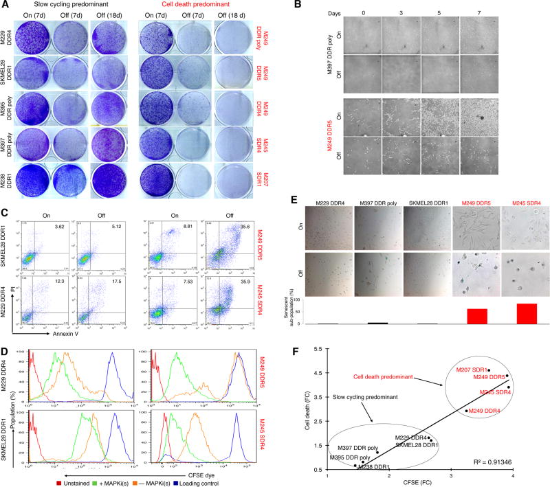
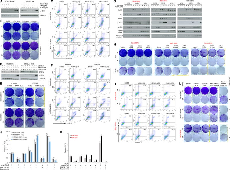
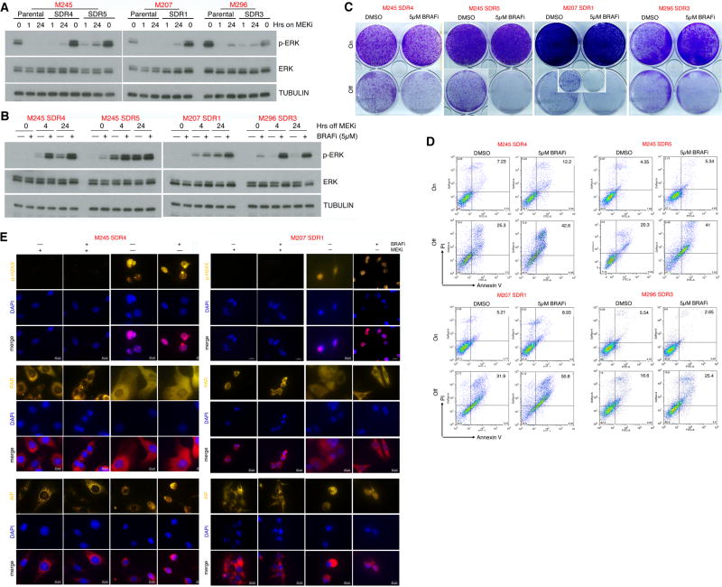
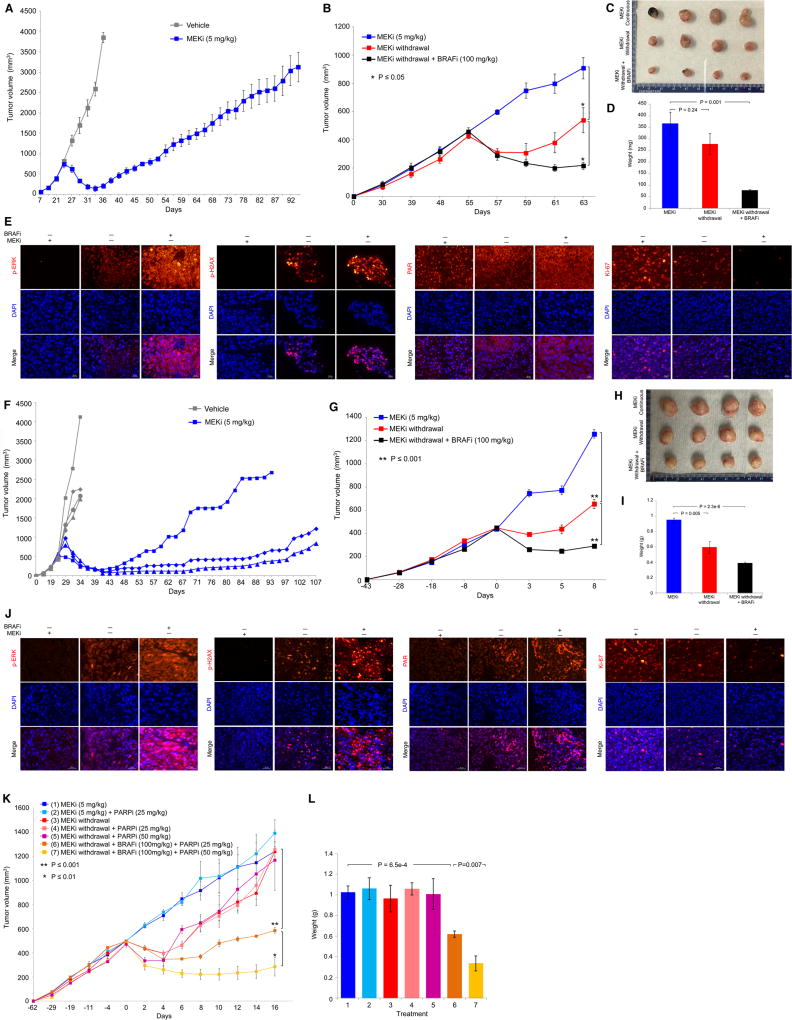
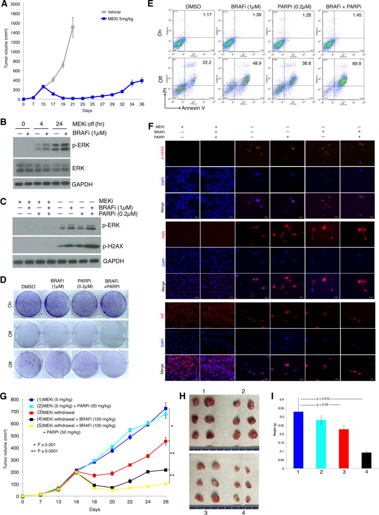
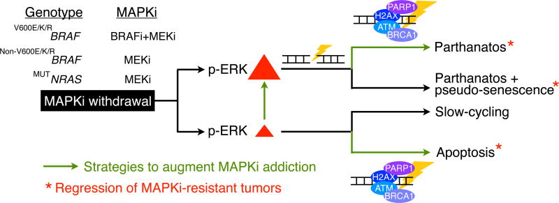
Melanoma resistant to MAPK inhibitors (MAPKi) displays loss of fitness upon experimental MAPKi withdrawal and, clinically, may be resensitized to MAPKi therapy after a drug holiday. Here, we uncovered and therapeutically exploited the mechanisms of MAPKi addiction in MAPKi-resistant BRAFMUT or NRASMUT melanoma. MAPKi-addiction phenotypes evident upon drug withdrawal spanned transient cell-cycle slowdown to cell-death responses, the latter of which required a robust phosphorylated ERK (pERK) rebound. Generally, drug withdrawal-induced pERK rebound upregulated p38-FRA1-JUNB-CDKN1A and downregulated proliferation, but only a robust pERK rebound resulted in DNA damage and parthanatos-related cell death. Importantly, pharmacologically impairing DNA damage repair during MAPKi withdrawal augmented MAPKi addiction across the board by converting a cell-cycle deceleration to a caspase-dependent cell-death response or by furthering parthanatos-related cell death. Specifically in MEKi-resistant NRASMUT or atypical BRAFMUT melanoma, treatment with a type I RAF inhibitor intensified pERK rebound elicited by MEKi withdrawal, thereby promoting a cell death-predominant MAPKi-addiction phenotype. Thus, MAPKi discontinuation upon disease progression should be coupled with specific strategies that augment MAPKi addiction.Significance: Discontinuing targeted therapy may select against drug-resistant tumor clones, but drug-addiction mechanisms are ill-defined. Using melanoma resistant to but withdrawn from MAPKi, we defined a synthetic lethality between supraphysiologic levels of pERK and DNA damage. Actively promoting this synthetic lethality could rationalize sequential/rotational regimens that address evolving vulnerabilities. Cancer Discov; 8(1); 74-93. ©2017 AACR.See related commentary by Stern, p. 20This article is highlighted in the In This Issue feature, p. 1.


Conflict of interest statement

Authors declare no conflicts of interest.

Figures

Figure 1
Figure 1
MAPKi-resistant melanoma display distinct drug-addiction phenotypes characterized by slow-cycling versus cell-death responses. (A) Clonogenic growth of double-drug resistant or DDR (MUTBRAF) or single-drug resistant or SDR (MUTNRAS) melanoma cell lines plated 24 hrs with BRAFi (vemurafenib)+MEKi (selumetinib) at 1 µM or MEKi (trametinib) at 0.1 µM followed by 7 d with (On) or 7 and 18 d without (Off) inhibitor withdrawal. (B) Temporal vital images of MAPKi-resistant or R-lines on or off BRAFi+MEKi. (C–E) Percentages of annexin V/PI-positive dead cells (C), CFSE dye dilution patterns (D), and levels of SA-βgal staining (E) in R-lines on or off MAPKi(s) for 6 d. Loading control (D) refers to the intensity of the CFSE dye initially loaded into the cells. (F) Correlation between fold-changes (FC) in CFSE dye dilution and % cell death off vs. on MAPKi(s).
Figure 2
Figure 2
Excessive ERK activation induces DNA damage and AIF-mediated death in the cell-death predominant MAPKi-addiction phenotype. (A) Heatmap showing the gene set variance analysis scores of differentially enriched gene signatures (median log fold-change (FC) ≥ 1.25 in the off-drug condition compared to the on-drug condition in either cell line; additionally, differential enrichment between the cell-death predominant and slow-cycling predominant R-lines must also be higher (by at least 25%) in the off-drug condition than in the on-drug condition). (B–C) Levels of AIF, PARP, and p-H2AX measured by Western blots (WB) (B) or immunofluorescence (IF) (C) in R-lines on or off MAPKi(s) for 1 and 3 d or 3 d, respectively. For b, mitochondrial (Mito), nuclear (Nucl), cytoplasmic (Cyto) cellular fractions or whole cell lysates (WCL). For c, nuclei visualized by DAPI; ruler, 20 µm. (D) DNA strand break measurements by the comet assay of R-lines on or off MAPKi(s) for 3 d. Tail length/moment FCs were quantified (n=5; mean ± SDs). (E) Temporal levels of p-ERK, p-H2AX and PARP in the nuclear fraction of R-cell lysates measured by WBs at indicate hrs off MAPKi(s). (F–G) Levels of indicated proteins by WBs in the nuclear (F, G) or WCL (F) fractions of R-lines, on and off MAPKi(s), transduced with control (V) or AIF (F) or H2AX (G) shRNA lentiviruses. (H–M) Percentages of annexin V/PI-positive dead cells (H, I), CFSE dye dilution patterns (J, K), and viable cell counts (L, M) in R-lines on or off MAPKi(s) for 6 d, transduced with control (Vector) or AIF (J, L) or H2AX (K, M) shRNA lentiviruses. For l, M, n=6; mean ± SDs; ***p <0.001 based on ANOVA. (N–O) Sub-cellular localization of PAR (N) or levels of PARG (O) by IF in R-lines. Nuclei visualized by DAPI; ruler, 20 µm. (P) PARG and p-ERK levels by WBs in the indicated fractions of M249 DDR5 or nuclear fractions of indicated additional R-lines. For WB, GAPDH, loading control.
Figure 3
Figure 3
Impairing DNA damage repair augments MAPKi-addiction. (A) Western blot (WB) analysis of p-H2AX levels in slow-cycling predominant R-lines on or off MAPKi(s), with or without ATMi and/or PARPi treatment for 3 d. (B–C) Clonogenic growth (B) or percentages of annexin V/PI-positive dead cells (C) in slow-cycling predominant R-lines on or off MAPKi(s) for 5 d, with or without ATMi and/or PARPi treatment. (D) WB analysis of BRCA1 and p-H2AX levels in in slow-cycling predominant R-lines, transduced with control (V) or BRCA1-specific shRNA lentiviruses, on or off MAPKi(s), with or without PARPi treatment for 3 d. (E–F) R-line cells from D were subjected to the same assays as in B and C, respectively. For WBs, TUBULIN, loading control. (G) Levels of indicated proteins by WBs in mitochondrial (MITO), nuclear (NUCL) subcellular fractions or whole-cell lysates (WCL) of slow-cycling versus cell-death predominant R-lines, on and off MAPKi(s), with or without ATMi and PARPi treatment for 3 d. HSP60, mitochondrial fraction control; Histone H3, nuclear fraction control. (H–I) Clonogenic growth (H) or percentages of annexin V/PI-positive dead cells (I) in cell-death predominant R-lines on or off MAPKi(s) for 6 d and 16 (only in H), with or without ATMi and/or PARPi treatment. (J–L) Levels of caspase-3 activity (J, K) or clonogenic growth (L) with indicated MAPKi(s) treatment, with or without ATMi and PARPi, with or without caspase-3 inhibitor, Z-DEVD-FMK (20 µM), in slow-cycling predominant R-lines (J) for 3, 6 d or cell-death predominant R-lines (K) for 3 d. For J, K, n=5; mean ± SDs; ***p <0.001 based on ANOVA. Staurosporine (1 µM) used to induce caspase-3 activity and death.
Figure 4
Figure 4
Paradoxical ERK activation by BRAFi potentiates drug addiction in MEKi-resistant MUTNRAS melanoma. (A–B) Western blot levels of p-ERK, ERK, and loading control TUBULIN in MUTNRAS parental and isogenic MEKi-resistant SDR-lines with indicated hrs on MEKi/trametinib (0.1 µM) treatment (A) or in SDR-lines with indicated hrs off MEKi/trametinib (0.1 µM) treatment, with or without BRAFi/vemurafenib (5 µM) treatment (B). (C–D) Clonogenic growth (C) and percentages of annexin V/PI-positive dead cells (D) in MUTNRAS SDR-lines on or off MEKi/trametinib (0.1 µM) for 6 d, with or without BRAFi/vemurafenib (5 µM) Inset for M207 SDR1 off MEKi for 16 d. (E) Levels and/or sub-cellular localization of p-H2AX, PAR, and AIF in cell-death predominant MEKi-addicted R-lines on or off MEKi for 3 d, with or without BRAFi (5 µM) treatment.
Figure 5
Figure 5
Excessive ERK and DNA damage induce regression of MEKi-resistant PDX tumors. (A) Tumor volumes (mean ± SEM) of a MUTNRAS PDX transplanted in NSG mice in response to daily gavage with the vehicle (n = 3) or trametinib (5 mg/kg; n = 5). Asterisk indicates the resistant tumor, R1, which was serially transplanted for experiment in B. (B) R1 trametinib-resistant MUTNRAS PDX tumors were grown in NSG mice for 55 days with daily trametinib (5 mg/kg) gavage until initiating indicated daily treatments or regimens (n = 4 in each group). Tumor volumes are shown as means ± SEM. P values, Student’s t-test. MEKi, trametinib (5 mg/kg); BRAFi, vemurafenib (100 mg/kg). (C) Pictures of tumors from three experimental groups in B at day 63. (D) Tumor weights (means ± SEM; p value, unpaired t-test) of three experimental groups in B. (E) Levels of indicated proteins in representative tissue sections of tumors in C. Ruler, 20 µm. (F–J) As in A to E except experiments used a distinct PDX model harboring S365LBRAF and individual tumor growth curves were plotted separately in F. The MEKi-resistant tumor (R3) which arose first was fragmented, serially passaged and used in G. (K–L) R1 trametinib-resistant MUTNRAS PDX tumors were serially passaged in NSG mice with daily trametinib (5 mg/kg) gavage until segregation into seven groups (1, 2, 3, 5, n=3 per group; 4, 6, 7, n=5 per group). Tumor volumes and weights are shown as means ± SEM. P values, Student’s t-test.
Figure 6
Figure 6
BRAF and PARP inhibitors augment MEKi addiction of MUTNras murine melanoma in an immune competent host. (A) Tumor volumes (mean ± SEM) of TpLN61R murine melanoma cells transplanted in C57BL/6 mice in response to daily gavage with the vehicle (n = 6) or trametinib (5 mg/kg; n = 6). One resistant tumor on day 36 was dissociated and cultured as a MEKi-resistant cell line (NILR2R). (B) Western blot levels of p-ERK, ERK, and the loading control GAPDH in the MEKi-resistant MUTNras SDR line, NILR2R, with indicated hrs off MEKi/trametinib (0.1 µM) treatment, with or without BRAFi/vemurafenib (1 µM) treatment. (C) Analysis of NILR2R protein lysates by Western blots of p-ERK and p-H2AX levels on or off MAPKi(s), with or without BRAFi (1µM) and/or PARPi (0.2µM) treatment for 3 d. (D–E) Clonogenic growth (D; 8 d) and percentages of Annexin V/PI-positive dead cells (E; 5 d) in NILR2R, on or off MEKi/trametinib (0.1 µM), with or without BRAFi/vemurafenib (1 µM) and/or PARPi (0.2µM) treatments. For D, cultures were seeded at 30K cells per well, except for the third row where cultures were seeded at 150K cells per well. (F) Levels and/or subcellular localization of p-H2AX, PAR, and AIF in NILR2R, on or off MEKi for 3 d, with or without BRAFi and/or PARPi treatment. Ruler, 20 µm. (G) Trametinib-resistant NILR2R cells were transplanted subcutaneously in C57BL/6 mice with daily trametinib (5 mg/kg) gavage until segregation into five groups (n=6 per group). Tumor volumes are shown as means ± SEM. P values, Student’s t-test. (H) Pictures of tumors from the first four experimental groups (mice sacrificed due to tumor ulceration) in G at day 26. (I) Tumor weights (means ± SEM; p value, unpaired two-way t-test) of the first four experimental groups in G.
Figure 7
Figure 7
Strategies to select against MAPKi-resistant melanoma. Schematic showing MAPKi-addiction phenotypes being driven by p-ERK rebound levels and potential therapeutic strategies (enhancing p-ERK or impairing DNA damage repair) that promote tumor cell-death (apoptosis or parthanatos) or regression.

Comment in

References

    1. Hugo W, Shi H, Sun L, et al. Non-genomic and Immune Evolution of Melanoma Acquiring MAPKi Resistance. Cell. 2015;162:1271–85. - PMC - PubMed
    1. Moriceau G, Hugo W, Hong A, et al. Tunable-Combinatorial Mechanisms of Acquired Resistance Limit the Efficacy of BRAF/MEK Cotargeting but Result in Melanoma Drug Addiction. Cancer Cell. 2015;27:240–56. - PMC - PubMed
    1. Nazarian R, Shi H, Wang Q, et al. Melanomas acquire resistance to B-RAF(V600E) inhibition by RTK or N-RAS upregulation. Nature. 2010;468:973–7. - PMC - PubMed
    1. Shi H, Hugo W, Kong X, et al. Acquired Resistance and Clonal Evolution in Melanoma during BRAF Inhibitor Therapy. Cancer Discov. 2014;4:80–93. - PMC - PubMed
    1. Shi H, Moriceau G, Kong X, et al. Preexisting MEK1 exon 3 mutations in V600E/KBRAF melanomas do not confer resistance to BRAF inhibitors. Cancer Discov. 2012;2:414–24. - PMC - PubMed

Publication types

Substances