. 2017 Oct 3;114(40):E8448-E8457.
doi: 10.1073/pnas.1711449114. Epub 2017 Sep 18.

Direct engagement of the PI3K pathway by mutant KIT dominates oncogenic signaling in gastrointestinal stromal tumor

Affiliations

Direct engagement of the PI3K pathway by mutant KIT dominates oncogenic signaling in gastrointestinal stromal tumor

Benedikt Bosbach et al. Proc Natl Acad Sci U S A. .

Abstract

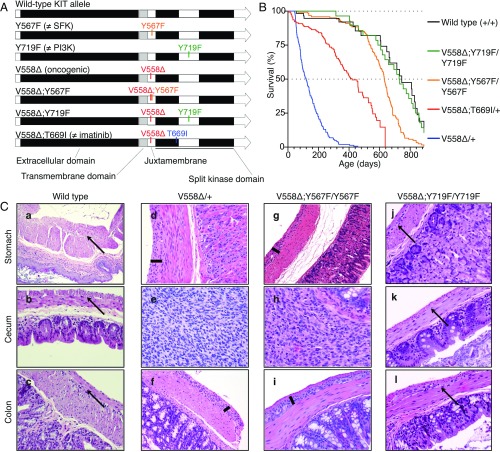
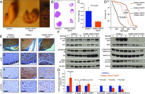
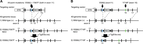
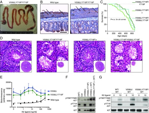
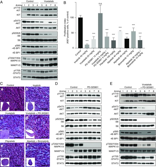
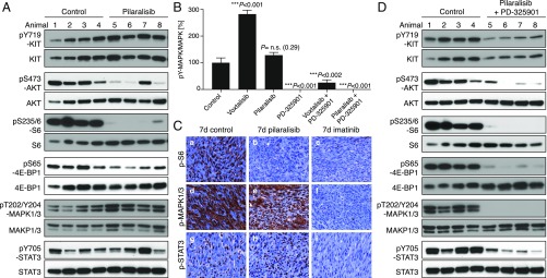
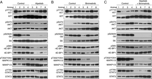
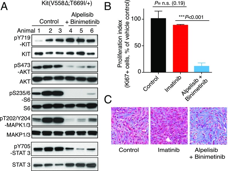
Gastrointestinal stromal tumors (GISTs) predominantly harbor activating mutations in the receptor tyrosine kinase KIT. To genetically dissect in vivo the requirement of different signal transduction pathways emanating from KIT for tumorigenesis, the oncogenic KitV558Δ mutation was combined with point mutations abrogating specific phosphorylation sites on KIT. Compared with single-mutant KitV558Δ/+ mice, double-mutant KitV558Δ;Y567F/Y567F knock-in mice lacking the SRC family kinase-binding site on KIT (pY567) exhibited attenuated MAPK signaling and tumor growth. Surprisingly, abrogation of the PI3K-binding site (pY719) in KitV558Δ;Y719F/Y719F mice prevented GIST development, although the interstitial cells of Cajal (ICC), the cells of origin of GIST, were normal. Pharmacologic inhibition of the PI3K pathway in tumor-bearing KitV558Δ/+ mice with the dual PI3K/mTOR inhibitor voxtalisib, the pan-PI3K inhibitor pilaralisib, and the PI3K-alpha-restricted inhibitor alpelisib each diminished tumor proliferation. The addition of the MEK inhibitor PD-325901 or binimetinib further decreased downstream KIT signaling. Moreover, combining PI3K and MEK inhibition was effective against imatinib-resistant KitV558Δ;T669I/+ tumors.

Keywords: GIST; Kit; PI3K; mouse.


Conflict of interest statement

The authors declare no conflict of interest.

Figures

Fig. 1.
Fig. 1.
Construction, survival, and histology of KitV558Δ/+, KitV558Δ;Y567F/Y567F, and KitV558Δ;Y719F/Y719F knock-in mice. (A) Schematic representation of targeted amino acids in the KIT protein. V558∆, oncogenic mutation due to deletion of valine 558; V558∆;T669I, double mutant containing the V558∆ and imatinib-resistant T669I mutations; V558∆;Y567F, double mutant containing the V558∆ and Y567F mutations; V558∆;Y719F, double mutant containing the V558∆ and Y719F mutations. Y567F, the docking site for the SFK tyrosine 567, is replaced by phenylalanine; Y719F, the docking site for the PI3K tyrosine 719, is replaced by phenylalanine. (B) Kaplan–Meier survival plot showing increased survival of KitV558Δ;Y567F/Y567F mice compared with fully signaling-competent KitV558Δ/+ mice. Survival of KitV558Δ;Y719F/Y719F mice was indistinguishable from that of WT mice (P = 0.791). Survival of KitV558Δ;Y567F/Y567F mice was significantly different from that of KitV558Δ;Y719F/Y719F and WT mice (P = 0.0003 and 0.0004, respectively). All survival curve comparisons are based on the log-rank/Mantel–Cox method. Median survival: KitV558Δ/+, 3 mo; KitV558Δ;T669I/+, 14 mo; KitV558Δ;Y567F/Y567F, 21 mo; KitV558Δ;Y719F/Y719F, 28 mo; WT, 29 mo. Values for KitV558Δ/+, KitV558Δ;T669I/+, and WT mice are from ref. . n ≥ 43 each; ticks indicate censored subjects. (C) Representative H&E-stained sections of FFPE samples of stomach, cecum, and colon from 3-mo-old mice. Arrows in ac and jl indicate the plane of normal myenteric plexus ICCs between the circular and longitudinal smooth muscle layers in WT and KitV558Δ;Y719F/Y719F mice. Bars in d, f, g, and i show hyperplastic extensions of this plane in gastric/colonic sections of KitV558Δ/+ and KitV558Δ;Y567F/Y567F mice. The tumor lesions in the cecum of KitV558Δ/+ (e) and KitV558Δ;Y567F/Y567F (h) mice exhibit typical GIST morphology with spindle-shaped/epithelioid tumor cells. The histology of KitV558Δ;Y719F/Y719F mice resembles that of WT mice. (Scale bar: 100 µm.) (Magnification, 10× in a, 20× in bl). n ≥3 animals each.
Fig. 2.
Fig. 2.
Tumors from KitV558Δ;Y567F/Y567F mice are smaller and have lower MAPK and SFK pathway activation than tumors from KitV558Δ/+ mice. (A) Representative gross cecal GISTs from 3-mo-old mice. (B) H&E staining of cross-sections of cecal GISTs from six mice. Note the unilateral tumor growth in relation to the cecal lumen in both genotypes and the compressed luminal diameter in KitV558Δ/+ mice. (C) Tumor weights. n = 3–6. Error bars indicate mean ± SD. (D) Kaplan–Meier survival plot showing reduced survival of KitV558Δ;Y567F/+ and KitV558Δ;Y567F/V558Δ;Y567F mice compared with KitV558Δ;Y567F/Y567F mice. The median survival of KitV558Δ;Y567F/+ and KitV558Δ;Y567F/V558Δ;Y567F mice was 15 (n = 16) and 7 (n = 46) months, respectively, and the median survival of KitV558Δ;Y567F/Y567F was 21 mo (n = 121), as shown in Fig. 1. (E) Comparison of KIT expression and downstream signal transduction in GISTs of 3- to 4-mo-old mice by immunohistochemistry. (Scale bars: 500 µm in ac, 200 µm in di, and 50 µm in jl.) (F and G) Immunoblot analysis (G) and its quantification (F). n.s., nonsignificant. Scale is in arbitrary units (intensity of gray shading).
Fig. S1.
Fig. S1.
(A) Targeting strategy for the simultaneous knock-in of V558Δ and Y567F into exon 11 of the 129/Sv Kit locus. (B) An analogous strategy with an extended 3′ homology arm used for the KitV558Δ;Y719F allele.
Fig. 3.
Fig. 3.
Absence of tumorigenesis and impaired spermatogenesis in KitV558Δ;Y719F/Y719F mice, and functional kinase activity in mutant KITV558∆;Y719F mice. (A) Gross pathology of the gastrointestinal tract of a 3-mo-old KitV558Δ;Y719F/Y719F mouse that appeared normal without cecal GIST, megaileum, or megacolon in comparison with KitV558Δ/+ and KitV558Δ;Y567F/Y567F mice (10, 11) (Fig. 2A). (B) Immunohistochemical KIT staining of ICCs in cross-sections of the flat-mounted cecum from 7-mo-old WT and KitV558Δ;Y719F/Y719F mice (n = 14; 3–29 mo old). The typical presence and distribution of individual ICCs in the plane of the myenteric plexus (ICC-MY, arrow) between the circular (CM) and longitudinal (LM) muscle layers, as well as intramuscular ICC (ICC-IM; arrowhead), are preserved in KitV558Δ;Y719F/Y719F mice. Note the increased tissue surface in the right section due to the tangential cut of the sample, but the absence of ICC hyperplasia. n = 3. (Scale bar: 100 µm.) (C) Kaplan–Meier survival plot showing similar survival of KitV558Δ;Y719F/Y719F, KitV558Δ;Y719F/+, and KitV558Δ;Y719F/V558Δ;Y719F mice. The median survival of KitV558Δ;Y719F/Y719F was 28 mo, as shown in Fig. 1. (D) Representative H&E-stained sections of testis showing normal morphology and spermatogenesis in 6-mo-old WT and KitV558Δ;Y719F/+ mice and empty tubules in testis of 6-mo-old KitV558Δ;Y719F/Y719F and 3-mo-old KitV558Δ;Y719F/V558Δ;Y719F mice. n = 3 each. (Scale bar: 50 µm.) (E) Kinase activity of WT KIT, KITV558∆, and KITV558∆;Y719F mutants using the PathHunter eXpress receptor tyrosine kinase functional assay. Quantitative detection of β-gal activity (bioluminescence) is reported as arbitrary units. (F) Western blots from 293T cells transfected with control empty vector (Ctrl vector), WT Kit, KitV558Δ, or KitV558Δ;Y719F/Y719F or cotransfected with KitV558Δ and WT or KitV558Δ;Y719F/Y719F and WT. (G) Immunoblot analysis of bone marrow-derived mast cell extracts from WT, KitV558Δ/+, and KitV558Δ;Y719F/+ mice. Antibodies specific for pKIT-Y567/Y569, pKIT-Y719, and KIT were used.
Fig. 4.
Fig. 4.
PI3K and MAPK inhibition with the dual PI3K/mTOR inhibitor voxtalisib, the pan PI3K inhibitor pilaralisib, and the MEK inhibitor PD-325901 in KitV558Δ/+ mice. (A) Immunoblot analysis of tumor extracts from KitV558Δ mice (n = 4/group) treated for 6 h with vehicle (Control) or voxtalisib (30 mg/kg). (B) Proliferation index measured by counting Ki-67–positive cells in GISTs from KitV558Δ mice treated as indicated. (C) Trichrome staining of GISTs from KitV558Δ mice treated for 7 d as indicated. Low-magnification overview of tumor section on bottom left corner. n ≥ 4. The number of stained cells was assessed in at least three 250 × 250-µm nonoverlapping fields per tumor. Doses are specified in Materials and Methods. Note that due to toxicity, the dose of voxtalisib had to be reduced from 30 (mg/kg) to 20 (mg/kg) for the 7-d single treatment, and to 5 (mg/kg) for the 7-d combination treatment with PD-325901. (D and E) Immunoblot analysis of tumor extracts from KitV558Δ/+ mice (n = 4/group) treated for 6 h with vehicle (Control) or 5 mg/kg (lanes 5 and 6) or 15 mg/kg (lanes 7 and 8) PD-325901 (D) or a combination of voxtalisib 30 mg/kg and 5 mg/kg PD-325901 (E).
Fig. 5.
Fig. 5.
Inhibition of PI3K resulted in a significant reduction in GIST proliferation. KitV558Δ/+ mice (n = 4/group) were treated for 4 h (A) with pilaralisib (100 mg/kg) alone, (B) with PD-325901 (15 mg/kg) alone, and (D) with both pilaralisib (100 mg/kg) and PD-325901 (15 mg/kg) and analyzed by immunoblotting. (B) Comparison of the level of activation of phospho-MAPK1/3 in GIST from mice treated with voxtalisib and pilaralisib alone or in combination with PD-325901. The level of activation of phospho-MAPK1/3 was determined by densitometry analysis of the respective bands from Western blots and measured as the ratio of phospho/total for each protein compared with control. The scale is arbitrary units (intensity of gray shading). (C) Representative results of IHC analysis of GIST after long-term (7 d) treatment of KitV558Δ/+ mice with vehicle (control), pilaralisib, or imatinib. As a positive control for IHC, imatinib down-regulated all three signaling readouts—p-MAPK, pS6, and p-STAT3—as reported previously (11). Antibodies used are specific for pS6 (ac), p-MAPK1/3 (df), and p-STAT3 (gi) on 5-µm sections of GISTs. Tumor sections from the different treatment groups (n ≥3 animals each) were placed next to each other on the same microscope slide to enable cross-comparison of staining intensities (40× objective).
Fig. 6.
Fig. 6.
PI3K-alpha inhibition by alpelisib in KitV558Δ/+ mice. Immunoblot analysis of tumor extracts from KitV558Δ/+ mice (n = 4/group) treated for 4 h with alpelisib 20 mg/kg (A), binimetinib 3.5 mg/kg (B), or both alpelisib 20 mg/kg and binimetinib 3.5 mg/kg (C) compared with vehicle (control).
Fig. 7.
Fig. 7.
GISTs from imatinib-resistant KitV558Δ;T669I/+ mice respond to combined PI3K and MAPK inhibition. KitV558Δ;T669I/+ mice (n = 3/group) were treated for 7 d with vehicle (Control), imatinib, or alpelisib (40 mg/kg) and binimetinib (10 mg/kg) and analyzed by immunoblotting (A), proliferation index (B), and Trichrome staining (C).

Similar articles

Cited by

References

    1. Hirota S, et al. Gain-of-function mutations of c-kit in human gastrointestinal stromal tumors. Science. 1998;279:577–580. - PubMed
    1. Lorincz A, et al. Progenitors of interstitial cells of Cajal in the postnatal murine stomach. Gastroenterology. 2008;134:1083–1093. - PMC - PubMed
    1. Huizinga JD, et al. W/kit gene required for interstitial cells of Cajal and for intestinal pacemaker activity. Nature. 1995;373:347–349. - PubMed
    1. Maeda H, et al. Requirement of c-kit for development of intestinal pacemaker system. Development. 1992;116:369–375. - PubMed
    1. Dematteo RP, et al. Adjuvant imatinib mesylate after resection of localised, primary gastrointestinal stromal tumour: A randomised, double-blind, placebo-controlled trial. Lancet. 2009;373:1097–1104. - PMC - PubMed

Publication types

MeSH terms

Substances