. 2017 Oct 3;114(40):E8468-E8477.
doi: 10.1073/pnas.1706546114. Epub 2017 Sep 18.

Defective decidualization during and after severe preeclampsia reveals a possible maternal contribution to the etiology

Affiliations

Defective decidualization during and after severe preeclampsia reveals a possible maternal contribution to the etiology

Tamara Garrido-Gomez et al. Proc Natl Acad Sci U S A. .

Abstract

In preeclampsia (PE), cytotrophoblast (CTB) invasion of the uterus and spiral arteries is often shallow. Thus, the placenta's role has been a focus. In this study, we tested the hypothesis that decidual defects are an important determinant of the placental phenotype. We isolated human endometrial stromal cells from nonpregnant donors with a previous pregnancy that was complicated by severe PE (sPE). Compared with control cells, they failed to decidualize in vitro as demonstrated by morphological criteria and the analysis of stage-specific antigens (i.e., IGFBP1, PRL). These results were bolstered by global transcriptional profiling data that showed they were transcriptionally inert. Additionally, we used laser microdissection to isolate the decidua from tissue sections of the maternal-fetal interface in sPE. Global transcriptional profiling revealed defects in gene expression. Also, decidual cells from patients with sPE, which dedifferentiated in vitro, failed to redecidualize in culture. Conditioned medium from these cells failed to support CTB invasion. To mimic aspects of the uterine environment in normal pregnancy, we added PRL and IGFBP1, which enhanced invasion. These data suggested that failed decidualization is an important contributor to down-regulated CTB invasion in sPE. Future studies will be aimed at determining whether this discovery has translational potential with regard to assessing a woman's risk of developing this pregnancy complication.

Keywords: cytotrophoblast; decidua; human endometrial stromal cells; preeclampsia; transcriptomics.


Conflict of interest statement

The authors declare no conflict of interest.

Figures

Fig. 1.
Fig. 1.
In vitro decidualization of hESCs was impaired in patients with previous pregnancies that were complicated by sPE. hESCs were obtained from nonpregnant women, patients with former sPE, or subjects who had a normal (norm) pregnancy outcome. The cells were decidualized by culturing for 5 d in the presence of cAMP and MPA. Nondecidualized cultures were maintained in the same medium without additives. (A) Localization of F-actin by rhodamine-phalloidin staining of hESCs from women with uncomplicated pregnancies showed the expected cytoskeletal reorganization and shape changes that were consistent with transformation from a fibroblast to a decidual phenotype. (B) hESCs from women who had sPE failed to undergo these changes. (C and D) PRL and (F and G) IGFBP1 secretion. (E and H) Summary of the ELISA data. Low levels of both molecules were detected in the CM of hESCs from the two donor groups before decidualization (solid bars). PRL and IGFBP1 levels greatly increased in most cultures of decidualized hESCs from donors who had normal pregnancy outcomes (gray hashed bars). In contrast, hESCs from patients with former sPE failed to show the decidualization-related increases in secretion of these molecules (black hashed bars; **P < 0.01 and ***P < 0.005; n.s., nonsignificant). (Scale bar: 100 μm.)
Fig. 2.
Fig. 2.
Global transcriptional profiling confirmed failed decidualization of hESCs obtained from nonpregnant women with a previous pregnancy complicated by sPE. (A) Schematic drawing of the study design. hESCs were isolated from endometrial biopsies and decidualized in vitro. The donors were nonpregnant women with previous normal pregnancy outcomes or patients with former sPE. (B) Summary of the LIMMA paired comparisons showing the number of DEGs by FDR <0.05 and at least twofold between the groups. (C) Before decidualization, there were five DEGs between hESCs from the normal pregnancy outcome group vs. patients with former sPE. (D) Heat map showing the 50 most highly DEGs (total = 74; SI Appendix, Table S3) that were modulated during decidualization of hESCs from donors who had normal pregnancy outcomes. (E) Heat map showing the 50 most highly DEGs (total = 129; SI Appendix, Table S4) that were misexpressed following decidualization of hESCs from donors with a former sPE pregnancy compared with those with normal pregnancies (*mRNA expression patterns validated by qRT-PCR; Δ, fold change).
Fig. 3.
Fig. 3.
Global transcriptional profiling of the DB and the DP revealed the DEGs in sPE vs. control pregnancies. (A) Schematic drawing of the study design. Laser microdissection enabled isolation of portions of the DB from the basal plate and DP, which was adjacent to the fetal membranes. (B) Summary of the LIMMA paired comparisons showing the number of DEGs between equivalent decidual compartments in sPE vs. nPTB (FDR < 0.05 and fold change ≥ 2). (C) Heat map showing the 50 most highly DEGs (total = 79; SI Appendix, Table S6) in the DB of nPTB vs. sPE patients. (D) Heat map showing the 50 most highly DEGs (total = 227; SI Appendix, Table S7) in the DP of nPTB vs. sPE patients.
Fig. 4.
Fig. 4.
sPE is associated with a striking down-regulation of PRL and IGFBP1 expression in the decidua (DEC). Tissue sections of the maternal/fetal interface that contained portions of the DB or the DP were coimmunostained with an antibody against CK7, which enabled visualization of CTBs, and decidual markers PRL (A and B) and IGFBP1 (C and D). (E and F) Adjacent sections were stained with anti-VIM, which labeled DEC cells. Nuclei were visualized with DAPI. In nPTB samples, anti-PRL and anti-IGFBP1 gave strong signals in the DB and DP (CK7-negative, VIM-positive). In contrast, the VIM-positive DEC cells in sPE samples failed to react with anti-PRL and anti-IGFBP1 or stained weakly for these markers. (G and H) The immunolocalization results shown in AD were semiquantified in ImageJ. Representative areas (n = 3 per sample) of the DB and DP from nPTB and sPE cases were subjected to ImageJ analyses. The results (intensity/area [0.3 mm2]) were expressed relative to the data from the control/nPTB DB samples. Data are expressed as the mean ± SEM (*P ≤ 0.05, **P ≤ 0.01, and ***P ≤ 0.001). Three or four representative areas of each sample were analyzed (sPE, n = 5 cases; nPTB, n = 4 cases). Am, amnion; iCTBs, invasive CTBs; schCTBs, smooth chorion CTBs. (Scale bars: 100 μm.)
Fig. 5.
Fig. 5.
Freshly isolated stromal cells from decidual biopsy specimens of patients with sPE displayed decidualization defects in culture. Cells were isolated from the DB or DP and analyzed at p0. Donors were women whose pregnancies were complicated by nPTB (n = 4) or sPE (n = 5). (A and B) The F-actin cytoskeleton of the cells was visualized by rhodamine-phalloidin staining, and nuclei were imaged with DAPI. In nPTB pregnancies, cells from either decidual compartment had a polygonal shape with a complex, well-developed network of actin filaments. (B) In contrast, the cells from sPE pregnancies were flattened with a much less well-developed actin cytoskeleton. Immunolocalization of PRL (C and D), IGFBP1 (E and F), and VIM (G and H). (C, E, and G) VIM-positive cells from the nPTB donors gave strong signals for the decidualization markers that were evaluated. (D, F, and H) In contrast, few of the VIM-positive cells from patients with sPE reacted with anti-PRL or anti-IGFBP1. (I and J) Secretion of PRL and IGFBP1 mirrored the immunostaining results. Data are the mean ± SEM of each sample, which was analyzed in triplicate (*P < 0.05, **P < 0.01, and ***P < 0.001). (Scale bars: 100 μm.)
Fig. 6.
Fig. 6.
Cultured hESCs from decidual biopsies of patients with sPE failed to redecidualize in vitro. Cells were isolated from the DB or DP and analyzed at p3–p5, i.e., after they lost expression of the decidualization markers PRL and IGFBP1. Donors were women whose pregnancies were complicated by nPTB (n = 3) or sPE (n = 4). Redecidualization was induced by treatment with cAMP (0.5 μM) and MPA (1 μM). CM and cells were analyzed after 5 d. (A and B) Visualization of the F-actin cytoskeleton via rhodamine-phalloidin staining. Nuclei were imaged with DAPI. When hESCs from donors who gave birth with nPTB were redecidualized, they enlarged, becoming polygonal/round with a complex actin cytoskeleton. In contrast, hESCs from donors with sPE failed to undergo the expected shape changes, often exhibiting disorganized actin cytoskeletons. (C and D) PRL and IGFBP1 secretion, measured by ELISA, were consistent with failed decidualization of hESCs from patients with sPE. Data are the mean ± SEM of each sample, which was analyzed in triplicate (*P < 0.05 and **P < 0.01; n.s., not significant). (Scale bar: 100 μm.)
Fig. 7.
Fig. 7.
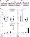
CM from decidual cells of patients with sPE inhibited CTB invasion in vitro. (A) Diagram of the experimental design. Decidual cells were isolated from the DB or DP and cultured overnight, and the CM was then isolated. The donors were women whose pregnancies were complicated by nPTB (n = 3) or by sPE (n = 4). CTBs were isolated from second-trimester placentas (15–17 wk, n = 4; 18–20 wk, n = 3; 21–23 wk, n = 3). They were cultured (72 h) on Matrigel-coated Transwell filters in medium conditioned by the nPTB or sPE decidual cells. CTBs and cellular processes that reached the undersides of the filters were counted. (B) Compared with the equivalent nPTB samples, CM from the cells of sPE donors significantly inhibited CTB invasion regardless of whether they were isolated from the DB or DP. (C) The addition of PRL and IGFBP1 (10 ng/mL each) to fresh medium increased CTB invasion to the levels that were observed when the cells were incubated in CM from nPTB cultures. Data are expressed as the mean ± SEM of duplicate wells (**P < 0.01 and ***P < 0.001; n.s., not significant).

Comment in

References

    1. Winn VD, Gormley M, Fisher SJ. The impact of preeclampsia on gene expression at the maternal-fetal interface. Pregnancy Hypertens. 2011;1:100–108. - PMC - PubMed
    1. Fisher SJ. Why is placentation abnormal in preeclampsia? Am J Obstet Gynecol. 2015;213(4) suppl:S115–S122. - PMC - PubMed
    1. Roberts JM, Cooper DW. Pathogenesis and genetics of pre-eclampsia. Lancet. 2001;357:53–56. - PubMed
    1. American College of Obstetricians and Gynecologists; Task Force on Hypertension in Pregnancy Hypertension in pregnancy. Report of the American College of Obstetricians and Gynecologists’ Task Force on Hypertension in Pregnancy. Obstet Gynecol. 2013;122:1122–1131. - PubMed
    1. Brosens IA, Robertson WB, Dixon HG. The role of the spiral arteries in the pathogenesis of preeclampsia. Obstet Gynecol Annu. 1972;1:177–191. - PubMed

Publication types