. 2017 Oct 3;114(40):E8508-E8517.
doi: 10.1073/pnas.1712592114. Epub 2017 Sep 18.

Identification of sialic acid-binding function for the Middle East respiratory syndrome coronavirus spike glycoprotein

Affiliations

Identification of sialic acid-binding function for the Middle East respiratory syndrome coronavirus spike glycoprotein

Wentao Li et al. Proc Natl Acad Sci U S A. .

Abstract

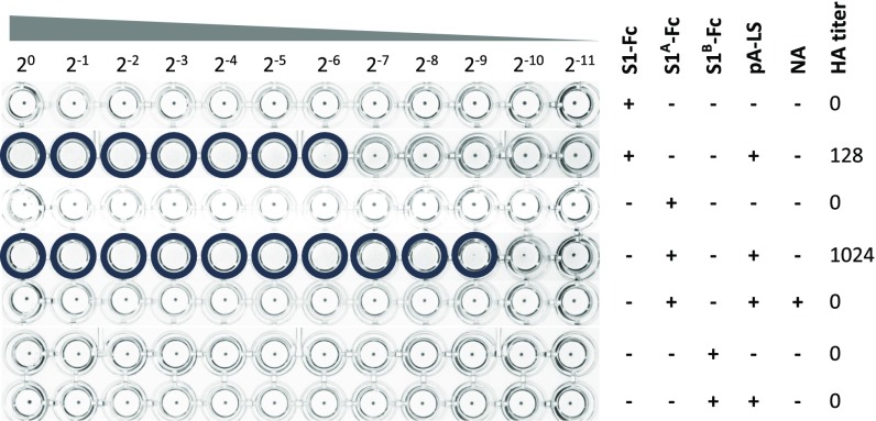
Middle East respiratory syndrome coronavirus (MERS-CoV) targets the epithelial cells of the respiratory tract both in humans and in its natural host, the dromedary camel. Virion attachment to host cells is mediated by 20-nm-long homotrimers of spike envelope protein S. The N-terminal subunit of each S protomer, called S1, folds into four distinct domains designated S1A through S1D Binding of MERS-CoV to the cell surface entry receptor dipeptidyl peptidase 4 (DPP4) occurs via S1B We now demonstrate that in addition to DPP4, MERS-CoV binds to sialic acid (Sia). Initially demonstrated by hemagglutination assay with human erythrocytes and intact virus, MERS-CoV Sia-binding activity was assigned to S subdomain S1A When multivalently displayed on nanoparticles, S1 or S1A bound to human erythrocytes and to human mucin in a strictly Sia-dependent fashion. Glycan array analysis revealed a preference for α2,3-linked Sias over α2,6-linked Sias, which correlates with the differential distribution of α2,3-linked Sias and the predominant sites of MERS-CoV replication in the upper and lower respiratory tracts of camels and humans, respectively. Binding is hampered by Sia modifications such as 5-N-glycolylation and (7,)9-O-acetylation. Depletion of cell surface Sia by neuraminidase treatment inhibited MERS-CoV entry of Calu-3 human airway cells, thus providing direct evidence that virus-Sia interactions may aid in virion attachment. The combined observations lead us to propose that high-specificity, low-affinity attachment of MERS-CoV to sialoglycans during the preattachment or early attachment phase may form another determinant governing the host range and tissue tropism of this zoonotic pathogen.

Keywords: MERS-CoV; attachment; receptor; sialic acid; spike.


Conflict of interest statement

The authors declare no conflict of interest.

Figures

Fig. 1.
Fig. 1.
MERS-CoV particles display a hemagglutination phenotype. MERS-CoV (EMC strain), SARS-CoV (HKU39849 strain), and IAV (PR8 strain) stocks were serially diluted twofold (starting dilution of 107 TCID50 per milliliter) and then incubated with washed human erythrocytes. Hemagglutination was scored after 2 h of incubation at 4 °C. Mixing of erythrocytes with blocking buffer was used as a negative control (mock). Wells positive for hemagglutination are encircled. Hemagglutination assays were performed at least two times.
Fig. 2.
Fig. 2.
(A) Design of self-assembling nanoparticles with Ig-binding potential. Surface representation of the 60-meric LS nanoparticle of A. aeolicus (N-terminal residues are labeled in red) and ribbon presentation of Ig-binding domain B of pA of S. aureus, including a schematic of LS, pA-LS, and pA-LS in complex with chimeric CoV S1 fused to Fc-part of human IgG1 (blue-purple). (B) Electron microscopic analysis of LS nanoparticles. Representative electron microscopic photographs of pA-LS (mean diameter of 15.21 ± 1.00) and pA-LS complexed with MERS-CoV S1-Fc (molar ratio of 1:1.2) are shown. (Scale bar, 50 nm.) (C) Schematic representation of the MERS-CoV spike protein sequence (drawn to scale). S1 and S2 subunits of the 1,353-aa-long MERS-CoV spike protein are indicated, as well as the four domains (A–D) within S1 and their respective boundaries: A (blue), B (green), C (yellow), and D (orange). The positions of the transmembrane domain (black bar; predicted by the TMHMM server) and of the predicted N-glycosylation sites (Ψ; predicted by the NetNGlyc server) are indicated.
Fig. 3.
Fig. 3.
(A) MERS-CoV S1-Fc hemagglutination phenotype is dependent on multivalent presentation on nanoparticles and on Sia-containing receptors on the erythrocyte surface. Human erythrocytes were mock-treated (PBS) or NA-treated, and incubated with a twofold serial dilution of MERS-CoV S1-Fc, pA-LS, or a combination. Hemagglutination was scored after 2 h of incubation at 4 °C. Wells positive for hemagglutination are encircled. Experiments were performed at least two times. (B) Defining the optimal pA-LS/S1-Fc ratio for hemagglutination. A fixed amount of MERS-CoV S1-Fc (2.5 μg) was preincubated with varying amounts of pA-LS (4, 2, 1, 0.5, 0.25, or 0.125 μg) serially diluted twofold and then incubated with washed human erythrocytes. Hemagglutination was scored after 2 h of incubation at 4 °C, and an optimal pA-LS/S1-Fc ratio was determined. Conditions lacking either pA-LS or MERS-CoV S1-Fc protein were used as a negative control. Wells positive for hemagglutination are encircled. Experiments were performed at least two times.
Fig. 4.
Fig. 4.
MERS-CoV S1-mediated Sia-binding activity resides in S1A. MERS-CoV S1-, S1A-, and S1B-Fc proteins alone or complexed with pA-LS were subjected to hemagglutination assay using human erythrocytes, after which HA titers were determined. MERS-CoV S1A-Fc hemagglutination activity was additionally tested with NA-treated human erythrocytes. Wells positive for hemagglutination are encircled. Hemagglutination assays were performed at least two times.
Fig. 5.
Fig. 5.
(A) MERS-CoV S1A-loaded nanoparticles preferentially bind α2,3-linked sialoglycans. Glycan array screen of MERS-CoV S1A-Fc–loaded pA-LS nanoparticles (Upper), IAV HA-Fc–loaded pA-LS nanoparticles (Middle), or pA-LS nanoparticles alone (Lower). MERS-CoV S1A-Fc nanoparticles show specific binding toward several α2,3-linked Neu5Ac glycans (salmon, 11–77) and some α2,6-linked Neu5Ac glycans (blue, 78–130). IAV HA-Fc nanoparticles show specific binding to α2,6-linked Neu5Ac glycans. Neither of the viral lectins showed binding to glycans lacking terminal Sia residues (gray, 1–10) or to Neu5Gc sialoglycans (purple, 131–135). No binding was observed for pA-LS nanoparticles alone. Bars display the average fluorescence units of six measurements after removal of the highest and lowest signals. Error bars represent the corresponding SEM. The list of glycans imprinted on the array and binding scores are provided in SI Appendix, Table S4. Note that the panel for IAV HA is on a different scale due to the relatively low avidity of MERS-CoV S1A. (B) List of glycans bound by MERS-CoV S1A–loaded nanoparticles above the arbitrary cutoff value of 2,000 relative fluorescence units. Indicated are the glycan schematic structure, average relative fluorescence units (RFU), and glycan number. When present on the glycan array, the α2,3- or α2,6-linked Neu5Ac and Neu5Gc derivatives of each hit are shown for comparison.
Fig. 6.
Fig. 6.
(A) Sia-binding activity of MERS-CoV S1A nanoparticles is inhibited by the acetyl moiety of 9-O-acetylated Sias. BSM-coated ELISA plates were mock treated, de–9-O-acetylated using BCoV HE, or desialylated using NA before incubation with twofold serially diluted (pA-LS–complexed) viral proteins. Fc-tagged IAV HA and HCoV-OC43 S1 were taken along as control proteins for HE and NA treatment. BSM-bound pA-LS nanoparticles were detected by ELISA with HRP-conjugated StrepMAB antibody as described above. BSM pretreated with NA served as a negative control. The experiment was performed in triplicate and repeated at least two times. A representative experiment is shown. Error bars represent the corresponding SEM. (B) Binding of MERS-CoV S1A nanoparticles to human mucin. Equimolar amounts of Fc-tagged MERS-CoV S1A, IAV HA, and pAPN were coupled to pA-LS nanoparticles, serially diluted twofold, and incubated on plates coated with human mucin that were mock-pretreated or pretreated with NA. IAV HA-Fc and pAPN-Fc were taken along as controls. Mucin-bound pA-LS nanoparticles were detected as described in A. The experiment was performed in triplicate and repeated at least two times. A representative experiment is shown. Error bars represent the corresponding SEM. (C) Staining with M. amurensis lectin II (specific for α2,3-linked Sias) of camel and human nose and lung tissues. (Magnification: 400×.)
Fig. 7.
Fig. 7.
(A) Sia depletion of Calu-3 cells inhibits MERS-CoV infection. Vero and Calu-3 cells were treated with or without NA and inoculated with MERS-CoV or IAV for 1 h at 37 °C. Cells were subsequently washed once, and fresh medium was added. Following incubation at 37 °C for 8 h, cells were fixed and the percentage of infected cells was determined by immunostaining and counting. Data are presented as mean ± SE and statistically analyzed with a t test. (B) Immunofluorescence staining on MERS-CoV–infected Vero and Calu-3 cells with and without NA pretreatment with the MERS-CoV N protein visualized in green. (Magnification: 100×.) (C) Surface expression of DPP4 on Vero and Calu-3 cells measured by flow cytometry. (D) Binding of nanoparticle-displayed MERS-CoV spike domain S1A to Vero and Calu-3 cells with and without NA pretreatment measured by flow cytometry. FACS data are presented as the geometric mean fluorescence intensity (gMFI) ratio between samples and isotype controls at a 95% confidence interval. The dotted line indicates the background level, equal to the isotype control. The data were analyzed with a nonparametric Mann–Whitney test (*P < 0.05; **P < 0.01; ***P < 0.001). All experiments were performed at least in triplicate and repeated three times. n.s., not significant.

References

    1. Zaki AM, van Boheemen S, Bestebroer TM, Osterhaus ADME, Fouchier RAM. Isolation of a novel coronavirus from a man with pneumonia in Saudi Arabia. N Engl J Med. 2012;367:1814–1820. - PubMed
    1. de Groot RJ, et al. Middle East respiratory syndrome coronavirus (MERS-CoV): Announcement of the Coronavirus Study Group. J Virol. 2013;87:7790–7792. - PMC - PubMed
    1. van Boheemen S, et al. Genomic characterization of a newly discovered coronavirus associated with acute respiratory distress syndrome in humans. MBio. 2012;3:1–9. - PMC - PubMed
    1. Lau SKP, et al. Genetic characterization of Betacoronavirus lineage C viruses in bats reveals marked sequence divergence in the spike protein of pipistrellus bat coronavirus HKU5 in Japanese pipistrelle: Implications for the origin of the novel Middle East respiratory syndrome coronavirus. J Virol. 2013;87:8638–8650. - PMC - PubMed
    1. Corman VM, et al. Characterization of a novel betacoronavirus related to MERS-CoV in European hedgehogs. J Virol. 2013;88:717–724. - PMC - PubMed

Publication types

MeSH terms