. 2017 Oct 3;114(40):E8498-E8507.
doi: 10.1073/pnas.1705972114. Epub 2017 Sep 18.

Leaderless secreted peptide signaling molecule alters global gene expression and increases virulence of a human bacterial pathogen

Affiliations

Leaderless secreted peptide signaling molecule alters global gene expression and increases virulence of a human bacterial pathogen

Hackwon Do et al. Proc Natl Acad Sci U S A. .

Abstract

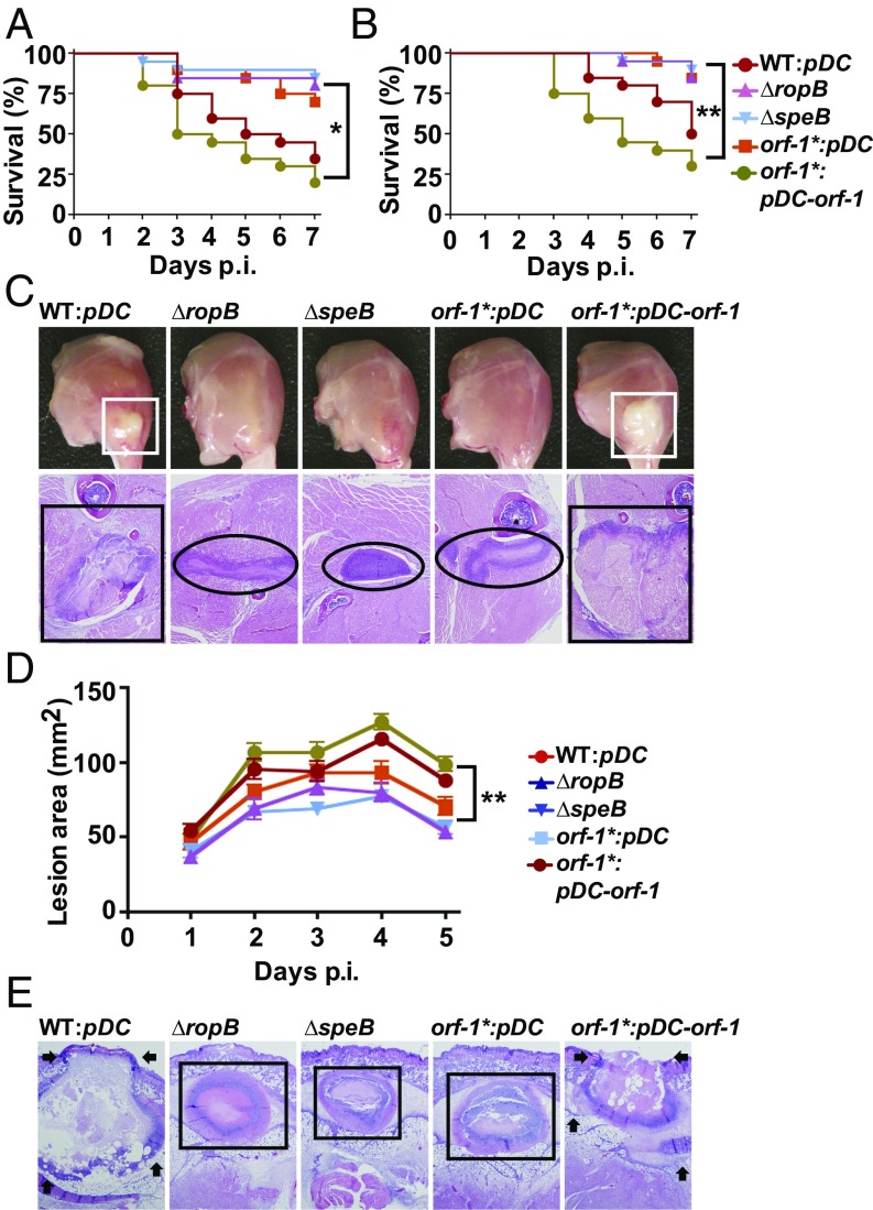
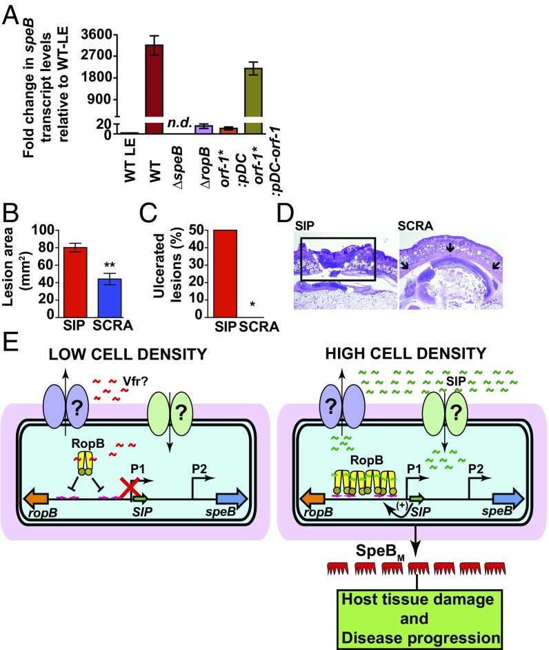
Successful pathogens use complex signaling mechanisms to monitor their environment and reprogram global gene expression during specific stages of infection. Group A Streptococcus (GAS) is a major human pathogen that causes significant disease burden worldwide. A secreted cysteine protease known as streptococcal pyrogenic exotoxin B (SpeB) is a key virulence factor that is produced abundantly during infection and is critical for GAS pathogenesis. Although identified nearly a century ago, the molecular basis for growth phase control of speB gene expression remains unknown. We have discovered that GAS uses a previously unknown peptide-mediated intercellular signaling system to control SpeB production, alter global gene expression, and enhance virulence. GAS produces an eight-amino acid leaderless peptide [SpeB-inducing peptide (SIP)] during high cell density and uses the secreted peptide for cell-to-cell signaling to induce population-wide speB expression. The SIP signaling pathway includes peptide secretion, reimportation into the cytosol, and interaction with the intracellular global gene regulator Regulator of Protease B (RopB), resulting in SIP-dependent modulation of DNA binding and regulatory activity of RopB. Notably, SIP signaling causes differential expression of ∼14% of GAS core genes. Several genes that encode toxins and other virulence genes that enhance pathogen dissemination and infection are significantly up-regulated. Using three mouse infection models, we show that the SIP signaling pathway is active during infection and contributes significantly to GAS pathogenesis at multiple host anatomic sites. Together, our results delineate the molecular mechanisms involved in a previously undescribed virulence regulatory pathway of an important human pathogen and suggest new therapeutic strategies.

Keywords: SpeB; Streptococcus pyogenes; leaderless peptide; quorum sensing; virulence regulation.


Conflict of interest statement

The authors declare no conflict of interest.

Figures

Fig. 1.
Fig. 1.
The ropB-speB intergenic region has the genetic element encoding the activation peptide signal for RopB-dependent speB expression. (A) Organization of the ropB and speB gene region in GAS. The ropB and speB genes are divergently transcribed. The angled arrows above the line indicate two transcription start sites for speB, designated P1 and P2. The angled arrow below the line indicates the transcription start site for ropB (PropB). The intergenic region with three predicted ORFs, designated orf-1 (orange), orf-2 (green), and orf-3 (blue), is shown as horizontal arrows. (B) Characterization of the sequences in the ropB-speB intergenic region for their role in speB expression. High-copy-number plasmids containing different fragments of the intergenic region were introduced individually into WT GAS, and the resulting strains were characterized for premature induction of speB expression. Cells were grown to the late exponential growth phase (A600 ∼ 1.0), and speB transcript levels were assessed by qRT-PCR. Numbers at either end of the constructs indicate the nucleotide positions relative to the first nucleotide of the speB start codon. Fold changes in speB transcript levels in the trans-complemented strains relative to reference GAS growth are shown. WT GAS (WT: empty vector) grown to late exponential growth was used as the reference. (C) Nucleotide sequence characteristics of the ropB-speB intergenic region. The numbers above the nucleotides indicate positions relative to the first nucleotide of the speB start codon. Nucleotides corresponding to transcription start sites P1 and P2 are highlighted in orange. Nucleotide sequences of orf-1, orf-2, orf-3, and speB are italicized and colored in red. An inferred ribosomal-binding site (RBS) located upstream of orf-1 is boxed and labeled. (D) Analysis of speB transcript levels in the indicated strains as determined by qRT-PCR. N.D., not detected. (E) Western immunoblot analysis of secreted SpeB in filtered growth media from indicated strains. Growth media samples were probed with anti-SpeB polyclonal rabbit antibody and chemiluminescence. The masses of molecular weight markers in kilodaltons (kDa) are shown. The mature form of purified recombinant SpeB (SpeBM; 25 kDa) was used as a marker. (F) Milk plate clearing assay to assess SpeB protease activity in indicated strains. Protease activity was determined by the presence of a clear zone around the bacterial growth.
Fig. 2.
Fig. 2.
Synthetic peptides containing the amino acid sequences of SIP activate speB expression. (A) The orf1 gene, encoding SIP, is expressed during the stationary phase of GAS growth. Total RNA extracted from WT GAS grown to either the late exponential (LE; A600 ∼ 1.0) or stationary (STAT) phase of growth and orf-1* mutant grown to the STAT phase of growth were analyzed by Northern blot. (B, Inset) Amino acid sequences of the synthetic peptides (SIP-1–SIP-7) used in the experiment. (B) SCRA peptide of an identical length and amino acid composition as SIP-1 but differing in the order of sequence was used as a negative control. The orf-1* mutant strain was grown in chemically defined medium (CDM) to the early STAT phase (A600 ∼ 1.7) and supplemented with either 100 nM indicated synthetic peptide or the carrier for the synthetic peptides (DMSO). After 60 min of incubation, transcript levels of speB were assessed by qRT-PCR. The orf-1* mutant strain supplemented with DMSO was used as a reference, and fold changes in speB transcript levels relative to the reference are shown. (C) Western immunoblot analysis of secreted SpeB in filtered growth media from the indicated samples. Cell growth and synthetic peptide supplementation were performed as described in A. Growth media collected were probed with anti-SpeB polyclonal rabbit antibody and detected by chemiluminescence. The masses of molecular weight markers in kilodaltons (kDa) are marked. (D) Milk plate clearing assay to assess the ability of various SIPs to induce SpeB protease activity in the orf-1* mutant. (E) Addition of SIP-1 and SIP-5 decouples the growth phase dependency of speB expression in WT GAS. The WT GAS was grown in CDM to the mid-exponential growth phase (A600 ∼ 0.6), and cells were incubated with 100 nm of each synthetic peptide for 60 min. Transcript levels of speB were assessed by qRT-PCR, and the fold change in speB expression relative to DMSO-supplemented growth is shown.
Fig. 3.
Fig. 3.
Eep protease, Opp, and Dpp do not participate in SIP biosynthesis. (A) Alignment of amino acid sequences of characterized propeptides specific for each founding member of RRNPP family regulators. The propeptides of small hydrophobic peptide 3 (SHP3) from Streptococcus pyogenes, phosphatase regulator A (PhrA) from Bacillus subtilis, peptide signal for neutral protease regulator (NprX) from B. cereus, peptide controlling conjugative transfer of plasmids (cCF10) from E. faecalis, peptide activating PlcR (PapR) from B. cereus, arbitrium communication peptide (AimP) from phage Phi3T, and SIP from S. pyogenes are shown. The positively charged residues characteristic of bacterial peptide signals are shown in red, and the amino acid sequence corresponding to each mature peptide is boxed and highlighted in pink. Transcript levels of the speB (B) and SpeB protease activity of SpeB (C) were assessed in the indicated strains by qRT-PCR and milk plate clearing assay, respectively. (D) Genetic inactivation of sip results in loss of regulatory activity in the secreted component of GAS growth. A qRT-PCR analysis of speB transcript level in WT GAS grown in cell‐free culture supernatants obtained from the indicated strains is shown. Secretome preparation and secretome swap assay were performed as described in SI Appendix, Supplemental Materials and Methods. Triplicate biological replicates were grown on two different occasions and analyzed in duplicate. The data were graphed as the mean ± SD. ME, mid-exponential phase of growth; ME SEC, total secretome prepared from mid‐exponential growth phase; orf-1* STAT SEC, total secretome prepared from the stationary growth phase of the orf-1* mutant; WT STAT SEC, total secretome prepared from the stationary growth phase of WT GAS. (E, Inset) Amino acid sequence of the synthetic peptide SIP-1 with fluorescein modification at its amino terminus (FITC–SIP-1) used in the experiment. (E) The orf-1* mutant strain was grown in chemically defined medium (CDM) to the early stationary phase (STAT, A600 ∼ 1.7) and supplemented with either the indicated synthetic peptide or the carrier for the synthetic peptides (DMSO). Unmodified SIP-1 was added at a final concentration of 1 μM, whereas varying concentrations of FITC–SIP-1 were used. After 60 min of incubation at 37 °C, cells were washed three times with sterile PBS, suspended in PBS, and lysed. Fluorescence measurements were obtained with clarified cell lysates using excitation and emission wavelengths of 480 nm and 520 nm, respectively. The unsupplemented orf-1* mutant strain was used as a reference, and changes in relative fluorescence units (RFU) relative to the reference are shown. (F) Confocal microscopy images of the orf-1* mutant strain either unsupplemented or supplemented with the indicated synthetic peptide. Synthetic peptide addition to the orf-1* mutant strain was performed as described in E. For each sample, bright-field, fluorescence-field, and merged images are shown. (Bottom) Magnified view of the FITC–SIP-1–supplemented growth. [Scale bars: 63.4 μm × 63.4 μm (y axis × x axis) at 100× magnification.]
Fig. 4.
Fig. 4.
SIP directly interacts with RopB and controls gene regulation by inducing allosteric changes in RopB. (A) Analysis of the binding between purified RopB and fluoresceinated SIP by an FP assay. (B) Ability of SIP or SCRA peptide to compete with the FITC-labeled SIP–RopB complex for binding. A preformed RopB (350 nM)-labeled SIP (10 nM) complex was titrated with the indicated unlabeled peptides. (C) Schematics of the location of RopB-binding sites within the P1 promoter. The transcription start site of the P1 promoter is shown as bent arrows, whereas the two RopB-binding sites with the inverted repeats are marked as arrows. Alignment of the nine-base-long nucleotide sequences of the RopB-binding half-sites from site 1 and site 2 are aligned from a 5′→3′ direction, and the identical bases among the half-sites are shaded in gray. (D) Nucleotide sequence of the RopB-binding site used in the binding studies is shown. The pseudoinverted repeat within the RopB-binding site is marked by arrows. Analysis of the binding between the FITC-labeled oligoduplex containing the putative RopB-binding site and apo-RopB (E) or SIP-bound RopB (F) by FP assay is shown. (G) Size exclusion chromatography analysis of purified RopB with or without the presence of either synthetic SIP or SCRA peptide. Molecular masses were calculated based on the calibration curve using molecular weight standards. (H) Mass spectrometry analyses of the apo- or SIP-bound RopB complex purified as described in E for the presence of SIP. (I) Increasing concentrations of the apo- or peptide-bound form of RopB (+, 1.5 μg: ++, 3 μg) were analyzed by Blu-native PAGE. The oligomeric forms of RopB, assessed based on the molecular weight marker [M; in kilodaltons (kDa)], are labeled.
Fig. 5.
Fig. 5.
SIP-mediated regulation of virulence genes is critical for GAS pathogenesis in mouse models of infection. (A) Twenty outbred CD-1 mice were inoculated i.p. with each indicated strain. Kaplan–Meier survival curves with P values derived by the log-rank test are shown. p.i., postinfection. (B) Twenty outbred CD-1 mice per strain were injected i.m. with each indicated strain. Kaplan–Meier survival curves with P values derived by the log rank-test are shown. (C) Gross (Top) and microscopic (Bottom) analyses of hind-limb lesions from mice infected with each indicated strain. (Top) Larger lesions with extensive tissue damage in SpeB-expressing strains are boxed (white boxes). (Bottom) Areas of disseminated lesions in the infected tissues are boxed (black box), whereas confined, less destructive lesions are circled. (D) Fifteen immunocompetent hairless mice were infected s.c. with each indicated strain, and the lesion area produced by each strain was determined. The lesion area was measured and graphed (mean ± SEM). The P value was derived by two-way ANOVA. (E) Histopathologic analysis of lesions from mice infected s.c. with each indicated strain. Areas of disseminated lesions and ulcerations on the skin surfaces caused by SpeB-producing strains are marked by arrows, whereas confined, less destructive lesions caused by SpeB-deficient strains are boxed. [Scale bars: C and E, 2.2 mm × 1.7 mm (y axis × x axis) at 4× magnification.]
Fig. 6.
Fig. 6.
SIP signaling controls speB expression during infection. (A) Analysis of the speB transcript level in the s.c. lesions from mice infected with the indicated strains. Samples were collected 24 h postinfection (P.I.) from the lesions of four mice per strain and analyzed in triplicate. Data were graphed as mean SD, with P values derived from a two-sample t test. Ten immunocompetent hairless mice per group were infected s.c. with the orf-1* mutant coinjected with either 10 μg of synthetic SIP or SCRA peptide. LE, late exponential GAS growth in laboratory medium. The lesion area (B) and ulceration (C) caused by each peptide at 24 h P.I. were determined. The lesion area was measured and graphed (mean ± SEM). The P value was derived by the Mann–Whitney test. *P < 0.05; **P < 0.001. (D) Histopathologic analysis of lesions from mice coinjected s.c. with SIP or SCRA peptide. SIP-induced ulcerated lesions that extend beyond the field of view are boxed. Coinjection with SCRA peptide caused small abscesses that are confined to the inoculation site (indicated by arrows). [Scale bars: 3.3 mm × 4.4 mm (y axis × x axis) at 2× magnification.] (E) Proposed model for the mechanism of intercellular communication and GAS virulence regulation. (Left) At low cell density, the secretion signal sequence of Vfr binds to RopB and negatively influences RopB-dependent transcription activation from the P1 promoter, possibly by disrupting RopB–DNA interactions. (Right) At high cell density, SIP is produced, secreted, and reimported into the cytosol. The high-affinity RopB–DNA interactions and RopB polymerization aided by SIP binding lead to up-regulation of sip expression, which results in robust induction of SIP production by a positive-feedback mechanism. In addition to up-regulation of virulence genes, the SIP signaling circuit down-regulates the expression of categories of genes involved in GAS growth and host cell attachment. Finally, the SIP-dependent up-regulation of speB leads to abundant secretion of mature SpeB (SpeBM), which facilitates host tissue damage and disease dissemination by cleavage of various host and GAS proteins.

References

    1. Parker CT, Sperandio V. Cell-to-cell signalling during pathogenesis. Cell Microbiol. 2009;11:363–369. - PMC - PubMed
    1. Hughes DT, Sperandio V. Inter-kingdom signalling: Communication between bacteria and their hosts. Nat Rev Microbiol. 2008;6:111–120. - PMC - PubMed
    1. Sanson M, et al. Phosphorylation events in the multiple gene regulator of group A Streptococcus significantly influence global gene expression and virulence. Infect Immun. 2015;83:2382–2395. - PMC - PubMed
    1. Dubbs JM, Mongkolsuk S. Peroxide-sensing transcriptional regulators in bacteria. J Bacteriol. 2012;194:5495–5503. - PMC - PubMed
    1. Hood MI, Skaar EP. Nutritional immunity: Transition metals at the pathogen-host interface. Nat Rev Microbiol. 2012;10:525–537. - PMC - PubMed

Publication types

MeSH terms

LinkOut - more resources