. 2017 Dec 4;216(12):3959-3969.
doi: 10.1083/jcb.201704145. Epub 2017 Sep 18.

Prepatterning by RhoGEFs governs Rho GTPase spatiotemporal dynamics during wound repair

Affiliations

Prepatterning by RhoGEFs governs Rho GTPase spatiotemporal dynamics during wound repair

Mitsutoshi Nakamura et al. J Cell Biol. .

Abstract

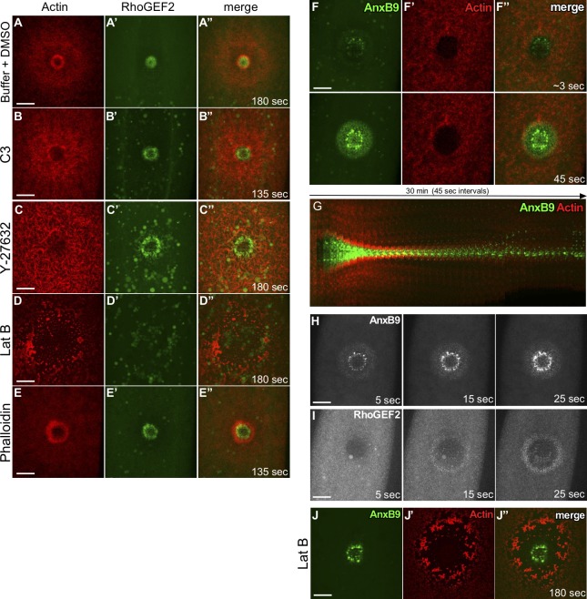
Like tissues, single cells are subjected to continual stresses and damage. As such, cells have a robust wound repair mechanism comprised of dynamic membrane resealing and cortical cytoskeletal remodeling. One group of proteins, the Rho family of small guanosine triphosphatases (GTPases), is critical for this actin and myosin cytoskeletal response in which they form distinct dynamic spatial and temporal patterns/arrays surrounding the wound. A key mechanistic question, then, is how these GTPase arrays are formed. Here, we show that in the Drosophila melanogaster cell wound repair model Rho GTPase arrays form in response to prepatterning by Rho guanine nucleotide exchange factors (RhoGEFs), a family of proteins involved in the activation of small GTPases. Furthermore, we show that Annexin B9, a member of a class of proteins associated with the membrane resealing, is involved in an early, Rho family-independent, actin stabilization that is integral to the formation of one RhoGEF array. Thus, Annexin proteins may link membrane resealing to cytoskeletal remodeling processes in single cell wound repair.


Figures

Figure 1.
Figure 1.
RhoGEF2, Pbl, RhoGEF3, and Tum exhibit discrete localization patterns and are required for cell wound repair. (A) Schematic diagram summarizing the localization patterns of actin, Rho1, Rac1, and Cdc42 at cell wounds. (B–H) Confocal xy projection images from Drosophila NC4–6 staged embryos coexpressing an actin marker (sGMCA or sChMCA) and fluorescently tagged Rho family GTPases: ChFP-Rho1 (B–B″), ChFP-Cdc42 (C–C″), and GFP-Rac1 (D–D″). The actin ring and halo regions are indicated in (B′). (E–H) Confocal xy projection images from Drosophila NC4–6 staged embryos coexpressing sChMCA and GFP-tagged RhoGEFs or Tum: sfGFP-Tum (E–E″), sfGFP-RhoGEF2 (F–F″), Pbl-eGFP (G–G″), and sfGFP-RhoGEF3 (H–H″). (I–K) Confocal xy projection images from Drosophila NC4–6 staged embryos coexpressing two fluorescently tagged RhoGEFs: Pbl-eGFP and RFP-RhoGEF2 (I–I’’), sfGFP-RhoGEF3 and RFP-RhoGEF2 (J–J’’), and Pbl-eGFP and sfGFP-RhoGEF3 (K–K’’). (L–P) Actin dynamics (sChMCA or sGMCA) during cell wound repair in control (GAL4 driver 7063 alone) (L), RhoGEF2RNAi(1) (M), pblRNAi(1) (N), RhoGEF3RNAi(1) (O), and Tum-i+antibodies (Abs) (P). (L’–P’) xy Kymograph across the wound area depicted in (L–P), respectively (yellow dashed lines, wound edges; yellow arrowheads, actin ring; red arrowheads, actin accumulation inside wounds). (Q–T) Quantification of the wound area over time for control, RhoGEF2RNAi(1), and RhoGEF2RNAi(2) (n = 10 for each; Q); control, pblRNAi(1), and pblRNAi(2) (n = 10 for each; R); control, RhoGEF3RNAi(1), and RhoGEF3RNAi(2) (n = 10 for each; S); and GAL4 control, 9E10 control, tumRNAi, Tum Abs, and tumRNAi+Abs (n = 10 for controls and n = 15 for tumRNAi and Tum Abs; T). Time after wounding is indicated. Bars, 20 µm. Error bars represent ± SEM.
Figure 2.
Figure 2.
RhoGEF2, Pbl, and RhoGEF3 interact with Rho1, Cdc42, and Rac1, respectively, during cell wound repair. (A–I″) Confocal xy projection images at 180 s after wounding from Drosophila NC4–6 staged embryos coexpressing sfGFP-RhoGEF2 (A–C″), Pbl-eGFP (D–F″), or sfGFP-RhoGEF3 (G–I″) with ChFP-Rho1 (A–A″, D–D″, and G–G″), ChFP-Rac1 (B–B″, E–E″, and H–H″), or ChFP-Cdc42 (C–C″, F–F″, and I–I″). (J–R) Smoothened fluorescence (Fluor.) intensity (arbitrary units) profiles derived from averaged fluorescence intensity values over a 10-pixel width across the wound area in the embryo shown (A″–I″). Error bars represent the 95% confidence interval. (S) GST pulldown assays with 35S-labeled in vitro translated RhoGEF2, Pbl, and RhoGEF3. The GST-Rho1, Rac1, and Cdc42 proteins were loaded with GDP or GTP as indicated. Time after wounding is indicated. Bars, 20 µm.
Figure 3.
Figure 3.
Prepattern established by RhoGEFs is required for correct localization of Rho family GTPases. (A–D) Localization of ChFP-Rho1 colabeled with sGMCA in control (A–A″), RhoGEF2RNAi(1) (B–B″), pblRNAi(1) (C–C″), and RhoGEF3RNAi(1) (D–D″) mutant backgrounds. (E–H) Fluorescence intensity profiles across the wound area in (A″–D″). (I–L) Localization of GFP-Rac1 colabeled with sChMCA in control (I–I″), RhoGEF2RNAi(1) (J–J″), pblRNAi(1) (K–K″), and RhoGEF3RNAi(1) (L–L″) mutant backgrounds. (M–P) Smoothened fluorescence (Fluor.) intensity (arbitrary units) profiles derived from averaged fluorescence intensity values over a 10-pixel width across the wound area in the embryo shown (I″–L″). Error bars represent the 95% confidence interval. (Q–T) Localization of ChFP-Cdc42 colabeled with sGMCA in control (Q–Q″), RhoGEF2RNAi(1) (R–R″), pblRNAi(1) (S–S″), and RhoGEF3RNAi(1) (T–T″) mutant backgrounds. (U–X) Fluorescence intensity profiles across the wound area in (Q″–T″). Time after wounding is indicated. Bars, 20 µm.
Figure 4.
Figure 4.
RhoGEF2, but not AnxB9, accumulation at cell wounds is actin-dependent. (A–E) Confocal xy projection images from Drosophila NC4–6 staged embryos coexpressing sfGFP-RhoGEF2 and sChMCA injected with injection buffer + 50% DMSO (control; A–A″), C3 exoenzyme (Rho1 inhibitor; B–B″), Y-27632 (Rok inhibitor; C–C″), LatB (depolymerizes F-actin; D–D″), and phalloidin (stabilizes F-actin; E–E″). (F–F″) Two confocal xy projection images at ∼3-s and 45-s time points from Drosophila NC4–6 staged embryos coexpressing eGFP-AnxB9 and sK2MCA. The fastest acquirable time point is labeled ∼3 s (G) xy Kymograph across the wound area from Drosophila NC4–6 staged embryos coexpressing eGFP-AnxB9 and sK2MCA. (H and I) Confocal xy projection images from Drosophila NC4–6 staged embryos expressing eGFP-AnxB9 (H) or sfGFP-RhoGEF2 (I) at three time points (5, 10, and 15 s). (J–J″) Confocal xy projection images from Drosophila NC4–6 staged embryos coexpressing eGFP-AnxB9 and sK2MCA that have been injected with LatB. Time after wounding is indicated. Bars, 20 µm.
Figure 5.
Figure 5.
Actin stabilization mediated by AnxB9 is required for RhoGEF2 recruitment to cell wounds. (A–C) Actin dynamics during cell wound repair in control (GAL4 driver 7063 alone) (A), AnxB9RNAi(1) (B), and AnxB9RNAi(2) (C) NC4–6 staged embryos expressing an actin reporter (sGMCA). (A′–C′) xy Kymograph across the wound area in A–C, respectively. Arrowheads denote actin accumulation within the wound. (D) Quantification of the wound area over time (control, AnxB9RNAi(1), and AnxB9RNAi(2); n = 10 respectively). Error bars represent ± SEM (E–G″) Confocal xy projection images from Drosophila NC4–6 staged embryos coexpressing sfGFP-RhoGEF2 and sChMCA in an AnxB9RNAi background injected with buffer (E–E″) or phalloidin (F–G″). RhoGEF2 partially accumulates at the wounds (arrowheads; F). (H) Schematic diagram summarizing the localization patterns of actin, AnxB9, RhoGEF2, Pbl, RhoGEF3, Tum, Rho1, Rac1, and Cdc42 at cell wounds. (I) Schematic diagram summarizing the pathways among AnxB9, actin, RhoGEFs, Tum, and Rho family GTPases in response to cell wounds. Time after wounding is indicated. Bars, 20 µm.

Similar articles

Cited by

References

    1. Abreu-Blanco M.T., Verboon J.M., and Parkhurst S.M.. 2011a Cell wound repair in Drosophila occurs through three distinct phases of membrane and cytoskeletal remodeling. J. Cell Biol. 193:455–464. 10.1083/jcb.201011018 - DOI - PMC - PubMed
    1. Abreu-Blanco M.T., Verboon J.M., and Parkhurst S.M.. 2011b Single cell wound repair: Dealing with life’s little traumas. BioArchitecture. 1:114–121. 10.4161/bioa.1.3.17091 - DOI - PMC - PubMed
    1. Abreu-Blanco M.T., Verboon J.M., and Parkhurst S.M.. 2014. Coordination of Rho family GTPase activities to orchestrate cytoskeleton responses during cell wound repair. Curr. Biol. 24:144–155. 10.1016/j.cub.2013.11.048 - DOI - PMC - PubMed
    1. Bement W.M., Mandato C.A., and Kirsch M.N.. 1999. Wound-induced assembly and closure of an actomyosin purse string in Xenopus oocytes. Curr. Biol. 9:579–587. 10.1016/S0960-9822(99)80261-9 - DOI - PubMed
    1. Benaud C., Le Dez G., Mironov S., Galli F., Reboutier D., and Prigent C.. 2015. Annexin A2 is required for the early steps of cytokinesis. EMBO Rep. 16:481–489. 10.15252/embr.201440015 - DOI - PMC - PubMed

MeSH terms