Review
. 2017 Oct 1;199(7):2203-2213.
doi: 10.4049/jimmunol.1700744.

Emerging Concepts in TCR Specificity: Rationalizing and (Maybe) Predicting Outcomes

Affiliations
Review

Emerging Concepts in TCR Specificity: Rationalizing and (Maybe) Predicting Outcomes

Nishant K Singh et al. J Immunol. .

Abstract

T cell specificity emerges from a myriad of processes, ranging from the biological pathways that control T cell signaling to the structural and physical mechanisms that influence how TCRs bind peptides and MHC proteins. Of these processes, the binding specificity of the TCR is a key component. However, TCR specificity is enigmatic: TCRs are at once specific but also cross-reactive. Although long appreciated, this duality continues to puzzle immunologists and has implications for the development of TCR-based therapeutics. In this review, we discuss TCR specificity, emphasizing results that have emerged from structural and physical studies of TCR binding. We show how the TCR specificity/cross-reactivity duality can be rationalized from structural and biophysical principles. There is excellent agreement between predictions from these principles and classic predictions about the scope of TCR cross-reactivity. We demonstrate how these same principles can also explain amino acid preferences in immunogenic epitopes and highlight opportunities for structural considerations in predictive immunology.


Figures

Figure 1
Figure 1
High peptide specificity emerging from how a TCR interfaces with the MHC protein. A) In the structure of the A6 TCR bound to Tax11-19/HLA-A2, Thr98 and Asp99 of CDR3α form strong electrostatic interactions with Ar65 on the HLA-A2 α1 helix (29). B) In order to interact with Arg65, CDR3α must undergo a conformational change upon binding (31). In the absence of the loop conformational change, steric clashes would occur between the CDR3α backbone and Arg65. Upon making the conformational change, the backbone of CDR3α is tightly packed against position 4 of the peptide. If position 4 is anything other than glycine, steric clashes would exist, preventing the loop from adopting its needed conformation, as shown in bottom right for an alanine at position 4.
Figure 2
Figure 2
Structural and physiochemical constraints on which peptides a TCR can recognize permit a high degree of cross-reactivity but also high specificity. In the example shown here for a class I system, there are 512 billion possible 9-mer peptides. Constraining one primary anchor and limiting the second reduces this to 12 billion. Further restraints designed to mimic principles of TCR specificity progressively reduce the number of possible peptides. This is illustrated for a hypothetical case where “compatibility” with a TCR is introduced in step-wise fashion. Compatibility adds a requirement for a glycine at P4, a requirement for a hydrophobic leucine or isoleucine at P5, an aromatic tyrosine or phenylalanine at P7, removal of charges from the center, and exclusion of glycine and proline from all remaining positions. Under all these constraints, there are still 1.6 million compatible peptides. Two, seemingly unrelated peptides are shown in the lower right. Note that “compatibility” as defined indicates a peptide which permits a TCR to bind with an affinity strong enough to productively signal, implying that all compatible peptides need not be recognized with the same affinity.
Figure 3
Figure 3
Hot spots demonstrated by sequence landscapes in TCR-pMHC interfaces. A) Illustration of a structurally conserved hot spot within the interface between the 42F3 TCR and peptides presented by H2-Ld (47). The left panel shows the interface between 42F3 and the QL9 (QLSPFPFDL) mimotope FLSPFWFDI/Ld. The hot spot region is localized to the peptide bulge and engaged primarily by residues of CDR3β. The right panel shows the hot spot as found in eight different agonist 42F3-agonist/Ld structures. Peptide backbones at position 6 through 9 are colored by contact frequency with the TCR, with red indicating the greatest number of contacts. The side chains of the key amino acids at positions 7 and 8 are shown, as is the conformation of the CDR3β loop. Pro97β remains in position to interact with peptide position 6, whereas Asp95β adjusts its conformation to optimize charge complementarity with peptide position 7. B) In altering peptide specificity, the molecular evolution process acted upon a hot spot in the interface between A6 TCR and Tax11-19/HLA-A2 (see also Fig. 1) (38). By changing Thr98α to a lysine and Asp99α to a tyrosine, the complex electrostatic interactions between the TCR and the HLA-A2 α1 helix were disrupted, forcing Arg65 of HLA-A2 to adopt a new conformation, permitting Trp104 of CDR3α to sandwich between the arginine and the peptide backbone and forming a new hot spot in the interface the modified TCR forms with the MART-126-35/HLA-A2 ligand.
Figure 4
Figure 4
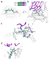
The MAGE-A3 and Titin epitopes are presented and recognized almost identically by the MAG-IC3 TCR (74). A) Near-identical presentation of the two epitopes in the HLA-A1 peptide binding groove. The inset shows the peptide sequences and the color scheme used for all panels. B) Overview of how the two ligands are engaged by the MAG-IC3 TCR. C) Details of identical, key interactions in the two interfaces. Charge complementarity with pGlu1 is optimized by the positioning of the carbonyl oxygen of Ala98 of CDR3α away from the glutamate side chain (not shown is a salt-bridge from Arg170 of HLA-A1 that helps fix the glutamate). Tyr32 of CDR1α hydrogen bonds with pAsp3. Phe101 of CDR3α “caps” the hydrophobic pPro4, and Arg56 of CDR2β hydrogen bonds with the pPro4 backbone. Not evident in the figure is how pIle5 packs between the TCR and the HLA-A2 α2 helix. D) The Titin and MAGE-A3 peptides have a valine and a glycine at P6, respectively. Substitution with larger amino acids would result in clashes with Ala69 and Thr73 of the HLA-A1 α1 helix, explaining why pathogen-derived peptides similar to the Titin and MAGE-A3 peptides would not be recognized. Dashed lines show distances between the side chains of Val6 of the Titin peptide and residues of HLA-A1 (note that in generating this figure, the CDR1α loop in the two structures was optimized to better fit the experimental electron density and amino acid geometry, yielding coordinates slightly altered from those deposited in the PDB).
Figure 5
Figure 5
Structural studies of alloreactivity illustrate core principles. A) The LC13 TCR cross-reacts between the syngeneic viral-peptide/HLA-B*0801 complex (left panel) and the allogeneic self-peptide/HLA-B*4405 complex (right panel). Despite considerable sequence differences, the peptides adopt very similar conformations in the ternary complexes, with key interactions between the TCR and the protruding aromatic P7 side chain maintained (86). The TCR also discriminates between closely-related B44 subtypes due to a single amino acid difference that prohibits the peptide from adopting a compatible conformation in B*4403 (right panel, circled detail). In B*4405, the aspartic acid at position 156 forms a hydrogen bond with pTyr3. In B*4403, position 156 is a leucine (yellow) and would clash with pTyr3 as shown. B) In the structure of the HCV1406-NS3/HLA-A2 complex, the conformation and chemistry of the NS3 peptide are similar to those of the MART-126-36 peptide, except for the residue at P1 (left panel). Structurally, the MART-126-35 peptide fits within the complex without any steric clashes or chemical incompatibility, save for the P1 residue, which would experience charge repulsion with Glu134 of CDR1α (right panel). Replacing pGlu1 with lysine resulted in a MART-126-35 variant that was recognized by the HCV1406 TCR (35).

References

    1. Mobley DL, Gilson MK. Predicting Binding Free Energies. Annual Review of Biophysics. 2017:46. - PMC - PubMed
    1. Erijman A, Rosenthal E, Shifman JM. How Structure Defines Affinity in Protein-Protein Interactions. PLOS ONE. 2014;9:e110085. - PMC - PubMed
    1. Vangone A, Bonvin AMJJ. Contacts-based prediction of binding affinity in protein–protein complexes. eLife. 2015;4:e07454. - PMC - PubMed
    1. Zhu C, Jiang N, Huang J, Zarnitsyna VI, Evavold BD. Insights from in situ analysis of TCR–pMHC recognition: response of an interaction network. Immunological Reviews. 2013;251:49–64. - PMC - PubMed
    1. Grasberger B, Minton AP, DeLisi C, Metzger H. Interaction between proteins localized in membranes. Proc Natl Acad Sci U S A. 1986;83:6258–6262. - PMC - PubMed

Publication types

MeSH terms