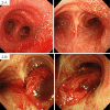
Bloody Bronchial Cast Formation Due to Alveolar Hemorrhage Associated with H1N1 Influenza Infection
- PMID: 28924110
- PMCID: PMC5675937
- DOI: 10.2169/internalmedicine.8137-16
Bloody Bronchial Cast Formation Due to Alveolar Hemorrhage Associated with H1N1 Influenza Infection
Abstract
A previously healthy 55-year-old man with H1N1 influenza A presented with severe respiratory failure and cardiac arrest. Following the return of spontaneous circulation, venovenous extracorporeal membrane oxygenation was required to maintain oxygenation. On day 2, bronchoscopy revealed a bloody bronchial cast obstructing the right main bronchus. A pathological examination revealed that it was composed of intrabronchial and intra-alveolar hemorrhagic tissue. Unfortunately, the patient died due to severe brain ischemia; a subsequent autopsy revealed marked alveolar hemorrhage. It is possible that anticoagulant therapy, alveolar collapse, and neuromuscular blocking agents provoked cast development in this case.
Keywords: ECMO; alveolar collapse; anticoagulation; complication; plastic bronchitis; white out.
Figures


References
-
- Deng J, Zheng Y, Li C, Ma Z, Wang H, Rubin BK. Plastic bronchitis in three children associated with 2009 influenza A(H1N1) virus infection. Chest 138: 1486-1488, 2010. - PubMed
-
- Eberlein MH, Drummond MB, Haponik EF. Plastic bronchitis: a management challenge. Am J Med Sci 335: 163-169, 2008. - PubMed
-
- Isobe M, Sasaki S, Hojo M, et al. . A bronchial cast caused by pulmonary hemorrhage due to a vitamin K deficiency. Pediatr Int 53: 133-134, 2011. - PubMed
-
- Coonar A. Blood clot cast of the bronchial tree. Eur J Cardiothorac Surg 28: 490, 2005. - PubMed
Publication types
MeSH terms
LinkOut - more resources
Full Text Sources
Other Literature Sources
Medical
Miscellaneous

