. 2017 Sep 18;7(1):11765.
doi: 10.1038/s41598-017-12292-9.

α-SNAP is expressed in mouse ovarian granulosa cells and plays a key role in folliculogenesis and female fertility

Affiliations

α-SNAP is expressed in mouse ovarian granulosa cells and plays a key role in folliculogenesis and female fertility

Alexis Arcos et al. Sci Rep. .

Erratum in

Abstract

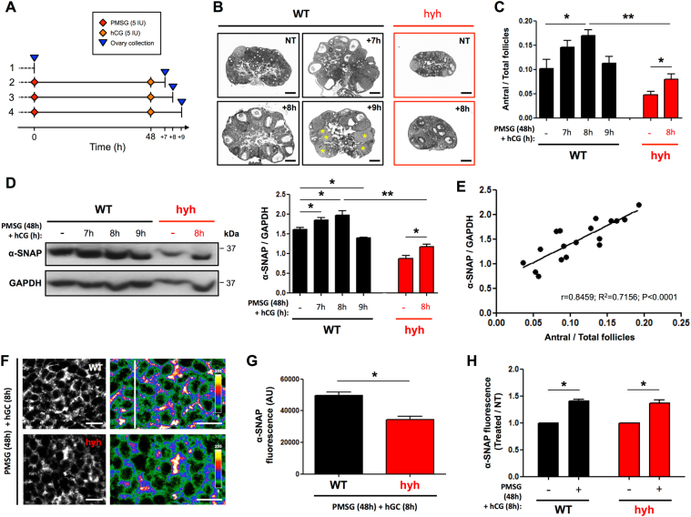
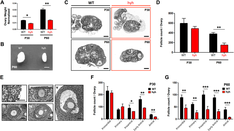
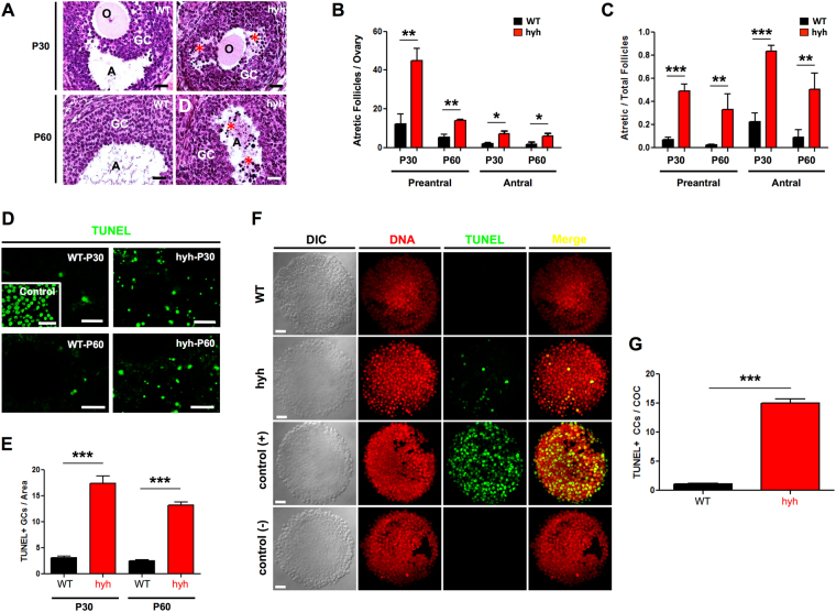
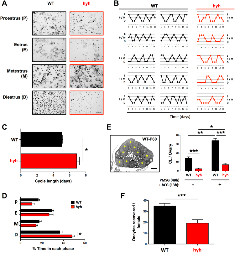
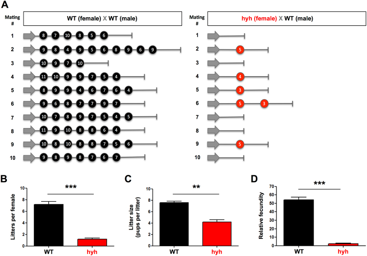
The balance between ovarian folliculogenesis and follicular atresia is critical for female fertility and is strictly regulated by a complex network of neuroendocrine and intra-ovarian signals. Despite the numerous functions executed by granulosa cells (GCs) in ovarian physiology, the role of multifunctional proteins able to simultaneously coordinate/modulate several cellular pathways is unclear. Soluble N-ethylmaleimide-sensitive factor (NSF) attachment protein (α-SNAP) is a multifunctional protein that participates in SNARE-mediated membrane fusion events. In addition, it regulates cell-to-cell adhesion, AMPK signaling, autophagy and apoptosis in different cell types. In this study we examined the expression pattern of α-SNAP in ovarian tissue and the consequences of α-SNAP (M105I) mutation (hyh mutation) in folliculogenesis and female fertility. Our results showed that α-SNAP protein is highly expressed in GCs and its expression is modulated by gonadotropin stimuli. On the other hand, α-SNAP-mutant mice show a reduction in α-SNAP protein levels. Moreover, increased apoptosis of GCs and follicular atresia, reduced ovulation rate, and a dramatic decline in fertility is observed in α-SNAP-mutant females. In conclusion, α-SNAP plays a critical role in the balance between follicular development and atresia. Consequently, a reduction in its expression/function (M105I mutation) causes early depletion of ovarian follicles and female subfertility.


Conflict of interest statement

The authors declare that the research was conducted in the absence of any commercial or financial relationships that could be construed as a potential conflict of interest.

Figures

Figure 1
Figure 1
α-SNAP expression in mouse ovarian tissue and granulosa cells (GCs) of wild type (WT) and α-SNAP mutant (hyh) females. (A) Western blot analysis of α-SNAP in postpubertal (postnatal day 60; P60) WT female mouse tissue extracts. GAPDH levels serve as loading control. Cropped blots (dotted lines) are displayed. Full-length blots including other tissue extracts are included in the Supplementary Figures file online (see Supplementary Figure S1). (B) Western blot analysis of α-SNAP in prepubertal (P7 and P14), peripubertal (P30), and postpubertal (P60 and P120) ovary extracts. β-tubulin levels serve as loading control. (C) Western blot analysis of α-SNAP in purified GCs and ovary remnants depleted of GCs (Re) at P30 and P60. N-cadherin (Ncad) was used as a marker of GC enrichment (see Supplementary Information) and Histone H3 was used as loading control. (AC) Bars represent mean ± SEM of densitometric analyses (n = 3 or 4 independent experiments). (D–F) α-SNAP immunolabeling in P60 WT (E) and mutant hyh (F) ovarian follicles. Omission of the primary antibody was used as a negative control (D). Note the expression of α-SNAP in GCs. The expression of α-SNAP in the oocyte (O) cannot be established because of a non-specific fluorescent signal detected in the oocyte-zona pellucida region. (D) Scale bars, 10 μm. (D’–F’) Magnifications of GC regions in ovarian follicles. Images were pseudocolored using the lookup table shown at the right of the figures to highlight the differences in fluorescence intensity between WT (E’) and mutant hyh (F’) samples. Scale bars, 10 μm. (G) Quantification of α-SNAP immunofluorescence intensity in WT (black bar) and hyh (red bar) GCs. Bars represent mean ± SEM of 4 independent experiments. (H) Western blot images and densitometric analysis of α-SNAP in WT and hyh ovaries at P30 and P60. GAPDH was used as loading control. Note the hypomorphism of α-SNAP in hyh samples at both developmental stages. Bars represent mean ± SEM of densitometric analyses (n = 3 independent experiments). *p < 0.05; **p < 0.01; ***p < 0.001 (ANOVA with Tukey’s post hoc test or Student’s t-test).
Figure 2
Figure 2
Expression of α-SNAP in ovarian tissue and granulosa cells (GCs) after gonadotropin stimulation. (A,B). Scheme of the protocol used for gonadotropin stimulation and ovarian tissue collection (A), and representative images of hematoxylin-eosin staining of ovary sections (B). Wild type (WT) females were divided into four groups: 1, control non-treated females (NT); 2–4, females received gonadotropin stimulation (one i.p. injection of PMSG (5 IU) + one i.p. injection of hCG (5 IU) 48 h after PMSG injection) and ovaries were collected 7 h (+7; group 2), 8 h (+8; group 3) and 9 h (+9; group 4) after hCG injection (see Material and Methods for details). Mutant females were included in protocol 1 (NT) and 3 (+8). Note the presence of abundant antral follicles in +7 and +8 WT ovaries; on the other hand, several corpora lutea are observed in +9 ovary (yellow asterisks). Scale bars, 200 μm. (C) Morphometric analysis of antral follicles in WT (black bars) and hyh (red bars) ovaries from control or non-treated females (−) and after PMSG + hCG treatment (7 h, 8 h, 9 h). (D) Western blot analysis of α-SNAP in ovary protein extracts obtained from WT and hyh non-treated females (−), and after PMSG + hCG treatment (7 h, 8 h, 9 h). GAPDH was used as loading control. Blots are representative of 3 independent experiments. Bars represent densitometric analysis of Western blots (mean ± SEM, n = 3). (E) Correlation (linear regression) analysis of normalized α-SNAP protein levels and the relative number of antral follicles. A strong positive linear correlation (r = 0.8459) and coefficient of determination (R2 = 0.7156) is observed (p < 0.0001). (F) α-SNAP immunolabeling in P60 WT and mutant hyh ovarian follicles 8 h after gonadotropin stimulation (PMSG + hCG). GC region in ovarian follicles are magnified and pseudocolored using the lookup table shown at the right of the figures to highlight the differences in fluorescence intensity between WT and mutant hyh samples. Scale bars, 10 μm. (G,H) Quantification of α-SNAP immunofluorescence intensity in WT (black bar) and hyh (red bar) GCs. Bars represent mean ± SEM of 4 independent experiments. (H) Relative α-SNAP immunofluorescence intensity in GCs from PMSG + hCG treated females compared with non-treated (control) females. Note that the increase in WT (black bars) is similar to that of hyh (red bars) GCs. *p < 0.05; **p < 0.01 (C,D, ANOVA with Tukey’s post hoc test; G,H, Student’s t-test).
Figure 3
Figure 3
Ovarian phenotype and ovulation capacity of α-SNAP mutant (hyh) females. (A) Ovary relative to body weight in WT (black bars) and hyh mutant (red bars) P30 and P60 females. (B) Representative image of P60 WT and hyh ovaries. Scale bar, 2mm. (C) Representative histological sections of WT and hyh mutant ovaries at P30 and P60. Scale bars, 200 μm. (D) Number of follicles per ovary in P30 and P60 WT and hyh females. (E) Representative images of ovarian follicles at different developmental stages: primordial (i), primary (ii), preantral (iii), early antral (iv), and antral (v) follicles. Scale bars, 25 μm (i–ii) and 50 μm (iii–v). (F,G) The number of follicles in each stage per ovary was analyzed in P30 (F) and P60 (G) WT and mutant hyh ovarian histological serial sections. Bars represent mean ± SEM of 4 independent experiments. *p < 0.05; **p < 0.01; ***p < 0.001 (Student’s t-test).
Figure 4
Figure 4
Follicular atresia and apoptosis of granulosa cells (GCs) in α-SNAP mutant (hyh) ovaries. (A) Sections of ovaries obtained from P30 and P60 WT and hyh mutant mice stained with hematoxylin-eosin. Magnifications of parts of the ovaries showing representative follicles. Atretic follicles characterized by the presence of condensed (pyknotic) nuclei (red asterisks) at the mural GC layers and in the follicular antrum (A) were abundant in hyh mutant ovaries. O, oocyte. Scale bars, 50 μm. (B) Number of preantral and antral atretic follicles per ovary in P30 and P60 WT and hyh females. Bars represent mean ± SEM of 4 independent experiments. (C) Quantification of the relative number of atretic follicles among preantral and antral follicles in WT and hyh mutant mice at P30 and P60. Follicles were scored as atretic if they demonstrate at least 5% pyknotic nuclei. Bars represent mean ± SEM of 4 independent experiments. (D) Representative images of TUNEL assays in histological sections of WT and hyh mutant ovaries at P30 and P60. For positive controls (control), histological sections were incubated with recombinant DNase I prior to labeling procedures. Scale bars, 50 μm. (E) Quantification of TUNEL + granulosa cells and bodies per area. Bars represent mean ± SEM of 3 independent experiments. (F) Representative images of TUNEL assays in COCs isolated from P60 WT and hyh ovaries 48 h after PMSG (10 IU) treatment. Propidium iodide was used for DNA detection (DNA). DIC, differential interference contrast microscopy. Scale bars, 20 μm. (G) Quantification of TUNEL + cumulus cells (CCs) per cumulus-oocyte-complex (COC). Twenty-nine WT and 33 hyh COCs obtained from 3 WT and 3 hyh females were analyzed. Bars represent mean ± SEM. ***p < 0.001 (Student’s t-test).
Figure 5
Figure 5
Estrous cycle and ovulation rate of α-SNAP mutant (hyh) females. (A) Estrous cycle phases in postpubertal WT and mutant hyh females. Representative images of the cytology of vaginal secretion. According to the relative abundance of nucleated epithelial cells, cornified epithelia cells and leucocytes, the cycle was divided in Proestrus (P), Estrus (E), Metestrus (M) and Diestrus (D). Note the abundance of cornified epithelial cells in E and leukocytes in D. (B–D) Daily analysis of vaginal secretion cytology during a 15 days period (n = 10 WT and 5 hyh). (B) Estrous cycles in hyh mutant females (red lines) are irregular compared with the cycles of WT females (black lines). (C,D) The average length of the estrous cycle was larger in hyh mutant females (C), and it was mainly due to a prolonged diestrus phase (D). Bars represent mean ± SEM (n = 10 WT and 5 hyh). (E) Quantification of corpora lutea (CL) in WT (black bars) and hyh (red bars) ovaries from non-treated (control) females (−) and 13 h after PMSG (48 h) + hCG treatment. A representative image of P60 WT ovary 13 h after gonadotropin stimulation is shown. Note the presence of abundant corpora lutea (yellow asterisks). Scale bar, 400 μm. Bars represent mean ± SEM of 4 independent experiments. (F) Quantification of MII oocytes collected from the oviductal ampulla of WT (black bars) and mutant hyh (red bars) females, 13 h after gonadotropin (PMSG × 48 h + hCG) stimulation. Bars represent mean ± SEM (n = 31 WT and 19 hyh). *p < 0.05; **p < 0.01; ***p < 0.001(Student’s t-test).
Figure 6
Figure 6
Reproductive performance of α-SNAP mutant (hyh) females. (A) Schematic representation of the reproductive profile of WT (n = 10) and hyh mutant (n = 10) females. Each circle represents a litter and the number inside the circle represents the litter size (pups born in that litter). (B–D) Quantification of the number of litters per female (B), litter size (C) and relative fecundity (D). Relative fecundity was obtained as: (litter size) × (number of litters) × (productive matings/100); the obtained value is a measure of the overall fecundity according to the Handbook of Genetically Standardized JAX Mice. Bars represent mean ± SEM (n = 10 WT and 10 hyh). **p < 0.01; ***p < 0.001 (Student’s t-test).

Similar articles

Cited by

References

    1. Matzuk MM, Lamb DJ. Genetic dissection of mammalian fertility pathways. Nat Cell Biol. 2002;4(Suppl):s41–49. doi: 10.1038/ncb-nm-fertilityS41. - DOI - PubMed
    1. McGee EA, Hsueh AJ. Initial and cyclic recruitment of ovarian follicles. Endocr Rev. 2000;21:200–214. doi: 10.1210/edrv.21.2.0394. - DOI - PubMed
    1. Moley KH, Schreiber JR. Ovarian follicular growth, ovulation and atresia. Endocrine, paracrine and autocrine regulation. Adv Exp Med Biol. 1995;377:103–119. doi: 10.1007/978-1-4899-0952-7_7. - DOI - PubMed
    1. Schwartz, N. B. In Neuroendocrinology in Physiology and Medicine (ed P. M. Conn, Freeman ME) 135–146 (Humana Press, 2000).
    1. Edson MA, Nagaraja AK, Matzuk MM. The mammalian ovary from genesis to revelation. Endocr Rev. 2009;30:624–712. doi: 10.1210/er.2009-0012. - DOI - PMC - PubMed

Publication types

MeSH terms

Substances