. 2017 Sep 18;7(1):11747.
doi: 10.1038/s41598-017-11648-5.

An alternate mode of oligomerization for E. coli SecA

Affiliations

An alternate mode of oligomerization for E. coli SecA

Aliakbar Khalili Yazdi et al. Sci Rep. .

Abstract

SecA is the ATPase of preprotein translocase. SecA is a dimer in solution and changes in its oligomeric state may function in preprotein translocation. The SecA-N68 construct, in which the C-terminal helical domains of SecA are deleted, was used to investigate the mechanism of SecA oligomerization. SecA-N68 is in equilibrium between monomers, dimers, and tetramers. Subunit interactions in the SecA-N68 tetramer are mediated entirely by unstructured regions at its N- and C-termini: when the termini are deleted to yield SecA-N68∆NC, the construct is completely monomeric. This monomeric construct yielded crystals diffracting to 2.6 Å that were used to solve the structure of SecA-N68, including the "preprotein crosslinking domain" (PPXD) that was missing from previous E. coli SecA structures. The SecA-N68 structure was combined with small angle X-ray scattering (SAXS) data to construct a model of the SecA-N68 tetramer that is consistent with the essential roles of the extreme N- and C-termini in oligomerization. This mode of oligomerization, which depends on binding of the extreme N-terminus to the DEAD motor domains, NBD1 and NBD2, was used to model a novel parallel and flexible SecA solution dimer that agrees well with SAXS data.


Conflict of interest statement

The authors declare that they have no competing interests.

Figures
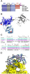
Figure 1
Figure 1
Structure of SecA-N68 (A) Domain structure of SecA and deletion constructs. Full length SecA consists of nucleotide binding domain 1 (NBD1); the preprotein cross-linking domain (PPXD), which is connected to NBD1 through a β-hairpin linker; NBD2; the C-terminal domains (CTD), which can be further sub-divided into the helical scaffold domain (HSD), residues 611 to 670 and 755 to 835 and helical wing domain (HWD), residues 671 to 754; finally there is the zinc binding domain, which consists of a 22 residue zinc-binding motif at the extreme C-terminus, connected to residue 835 by an unstructured linker. The unstructured N-terminus of SecA is illustrated by the short green line, and has the sequence MLIKLLTKVFGSRN; in some constructs, the N-terminal sequence included a hexahistidine tag with sequence MHHHHHHLTKVFGSRN. An unstructured C-terminal sequence (short orange line) was also present in SecA-N68, which, starting from residue 597, had the sequence RIFASDRVSGMMRK. (B) Structure of SecA-N68∆NC, with domains colour-coded as in Panel A. The structure also contained Mg2+ (green sphere) and ADP (sticks), bound between NBD1 and NBD2. (C) Structure-based sequence alignment of the PPXDs from the SecA molecules of 5 different organisms: M. tuberculosis (1NKT), T. maritima (3JUX), T. thermophilus (2IPC), B. subtilis (1TF5), and E. coli. The numbering and secondary structure (H for helix, B for strand) is for E. coli SecA. Residues that have a relatively small CA RMSD between the 5 structures are indicated with black dots. (D) Superposition of PPXD structures. The E. coli PPXD from the SecA-N68∆NC structure is shown as a grey ribbon and the other four structures (as in Panel C) are shown as CA-traces. The 5 superposed PPXD structures have been mapped onto the SecYEG-bound PPXD in the T. maritima SecA-SecYEG complex (3DIN).
Figure 2
Figure 2
Oligomerization of SecA-N68 is Mediated by Unstructured Termini (A) The oligomerization of H6-SecA-N68 and SecA-N68∆NC was characterized by gel filtration chromatography. H6-SecA-N68 is a SecA construct with an unstructured extreme N-terminal sequence MHHHHHHLTKVFGSRNDRTL (the wild-type sequence is MLIKLLTKVFGSRNDRTL) and also an unstructured C-terminal sequence; SecA-N68∆NC is the same construct, except that the unstructured N- and C-terminal sequences have been removed. H6-SecA-N68 was applied to the column at concentrations of 4 mg•mL−1 (solid black curve), 2 mg•mL−1 (dashed curve), 0.6 mg•mL−1 (dashed-dotted curve), and 0.35 mg•mL−1 (dotted curve); SecA-N68∆NC (grey curve) was applied at 10 mg•mL−1. The elution volume for molecular weight standards is shown on the upper axis. Panels B to E are SedFit analyses from sedimentation velocity AU of 4 different SecA-N68 constructs at a range of concentrations; all constructs were analyzed under identical buffer conditions consisting of 50 mM Tris-HCl pH 7.5, 100 mM KCl, 2 mM EDTA, 5 mM MgCl2 and 5 mM TCEP-HCl. (B) SecA-N68, with a wild-type N-terminal sequence, was analyzed at concentrations of 1.0 (dashed) and 5.0 (solid) mg•mL−1. (C) SecA-N68ΔNC, which lacks both unstructured termini, was analyzed at concentrations of 0.1 (dashed) and 4 (solid) mg•mL−1. (D) Analysis of SecA-N68ΔC, which carries only the wild-type unstructured N-terminus, at concentrations of 0.25 (dotted), 1 (dashed), and 2 (solid) mg•mL−1. (E) Analysis of SecA-N68ΔN, which carries only the unstructured C-terminus, at concentrations of 0.4 (dotted), 0.6 (dashed), and 4 (solid) mg•mL−1.
Figure 3
Figure 3
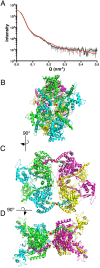
A SAXS-Based Model for the SecA-N68 Tetramer (A) The SecA-N68∆NC crystal structure was used with the program GLOBSYMM to find a tetramer with D2 symmetry that matches the solution SAXS data, which were recorded with SecA-N68 at 6.7 mg/mL. The experimental SAXS data are indicated by the grey curve, with the vertical bars showing the standard deviation of the replicate measurements. The red curve is the scattering from the SecA-N68 tetramer model (Panels B, C, and D), calculated using Crysol. The experimental radius of gyration was 48.2 Å, while the value for the hydrated tetramer model was 48.9 Å; the overall χ2 value for the fit of the model to the data was 2.24. The tetramer model is shown from three perspectives related by 90° rotations about a vertical axis (B to C) and horizontal axis (C to D). The N- and C-termini of SecA-N68∆NC are shown with red spheres. The additional unstructured residues at the N- and C-termini that mediate tetramer formation in SecA-N68 have been built into the structure to illustrate a potential mode of interaction. The extreme N-terminus is binding in the cleft between NBD1 and NBD2 to mediate dimer formation between the green and magenta protomers, and the yellow and cyan protomers. The extreme C-terminus is binding in a groove next to the hairpin connecting NBD1 to the PPXD, and mediates dimer formation between the green and cyan protomers, and yellow and magenta protomers. Together, the extreme N- and C-termini, illustrated with spheres at CA positions, mediate a dimer-of-dimers tetramer.
Figure 4
Figure 4
The SecA N-terminus is Essential for Function To test the functionality of SecA constructs, BL21.19(DE3) cells, in which expression of SecA is temperature-sensitive such that the cells do not grow at 42 °C, were transformed with plasmids that express SecA, SecA-N95, or the same constructs but with 14 residues deleted at the N-terminus, SecA∆N and SecA-N95∆N. The cells were grown in liquid culture for 12 hours and diluted to a common OD600 of 1, followed by 5 subsequent 10-fold dilutions; 5 µL of the resulting liquid cultures were spotted onto LB-agar and grown at either 28 °C or 42 °C for 30 hours in the absence or presence of IPTG (500 µM).
Figure 5
Figure 5
Sedimentation Analyses of SecA-N95 and SecA-N95ΔN Sedimentation velocity (Panels A and B) and equilibrium (Panels C and D) were used to evaluate the effect of N-terminal deletion on the dimerization of SecA-N95. For all experiments, the buffer used was 50 mM Tris-HCl, 100 mM KCl, 2 mM EDTA, 5 mM MgCl2, and pH 7.5. (A) Sedimentation velocity analysis of SecA-N95 at 31 µM (solid curve), 10.5 µM (dashed curve), and 2 µM (dotted curve). A sedimentation coefficient of 8.4 S is observed at concentrations of 10 µM and above and corresponds to the SecA-N95 dimer. (B) Sedimentation velocity analysis of SecA-N95ΔN at 26 µM (solid curve), 10 µM (dashed curve) and 2 µM (dotted curve). At 26 µM concentration, SecA-N95ΔN sediments with a major species at 7.4 S and a minor species at 4.6 S while at 2 µM it sediments with an overall coefficient of 5.6 S. In Panels A and B, the positions for sedimentation of the dimer at 8.4 S and monomer at 4.6 S are indicated. (C) Sedimentation equilibrium analysis of SecA-N95 was carried out in a 3-sector cell at rotor speeds of 7000, 10000, and 12000 rpm; the data from all nine curves were globally fit to a model of a single ideal species to yield a MW of 173.5 kDa (the theoretical dimer MW is 189.3 kD). Representative sedimentation curves for the equilibration at 10000 rpm are indicated; the other six curves are omitted for clarity. (D) Sedimentation equilibrium analysis of SecA-N95ΔN was carried out at rotor speeds of 10000, 12000, and 16000 rpm; the data from all nine curves were globally fit to a monomer-dimer equilibrium model, using a MW of 93.2 kDa, which yielded a dimer dissociation constant of 24.5 µM. Representative sedimentation curves for the equilibration at 12000 rpm are indicated; the other six curves are omitted for clarity. In Panels (C) and (D), the absorbance data are indicated by the circles and the fit to the data by the solid curves; the residuals are indicated above the data.
Figure 6
Figure 6
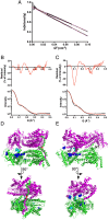
Parallel and Antiparallel Dimer Models (A) Guinier plot of the low angle SAXS data for full-length SecA (bottom curve) and SecA-N95 (top curve). For clarity, the curves have been normalized to the same I(0) value of 1000, and in both cases the standard deviation of the measurements are indicated by the vertical bars. Data points used for estimation of R G are indicated by circles for full-length SecA and squares for SecA-N95; least-squares fits to these data are shown by the red lines. The analysis yielded an R G of 42.3 ± 0.3 Å for full-length SecA and 38.6 ± 0.2 Å for SecA-N95. (B) Comparison of theoretical scattering from the parallel SAXS-derived dimer model of SecA-N95 with experimental SAXS data. The bottom panel shows the experimental data in grey, with standard deviations of the measurements as small black bars; the superimposed red curve is the theoretical scattering calculated using the FoXS server,. The top panel shows the residuals as a percentage of the intensity, on a linear scale. The R G of the model is 38.1 Å and the fit yields a χ value of 10.8. (C) Comparison of theoretical scattering from the antiparallel E. coli dimer based on the crystallographic dimer present in the original B. subtilis SecA structure and modelled using the E. coli SecA-N68 crystal structure and homology models of the C-terminal domains. The R G of the model is 39.8 Å and the fit yields a χ value of 17.6. (D) The parallel dimer with its long axis parallel to the page (top panel) and rotated 90° about a horizontal axis (bottom panel) to create a view down the two-fold rotation axis. The N-termini mediating dimer formation are indicated by magenta or green spheres at CA positions. The interactions between the N-termini and the opposite protomer are the same as those modelled for the interaction in the SecA-N68 tetramer, with adjustments of residues 15 through 18 to accommodate the somewhat different orientation of the protomers with respect to each other. Residue 15, the first residue present in the SecA-N68∆NC structure, is indicated with a red sphere. Sites of in vivo photo-activated cross-linking are indicated by residues highlighted with spheres and coloured cyan for one study and blue for a second study. (E) The antiparallel dimer viewed down its two-fold rotation axis (top panel) and rotated 90° about a vertical axis; in this case the N-terminal amino acids of E. coli SecA from residue 3 (indicated by “NT”) onwards, were modelled from the B. subtilis structure. Cross-linking sites are indicated as in Panel D.

Similar articles

Cited by

References

    1. Chatzi KE, Sardis MF, Economou A, Karamanou S. SecA-mediated targeting and translocation of secretory proteins. Biochim. Biophys. Acta. 2014;1843:1466–1474. doi: 10.1016/j.bbamcr.2014.02.014. - DOI - PubMed
    1. Collinson I, Corey RA, Allen WJ. Channel crossing: how are proteins shipped across the bacterial plasma membrane? Philos. Trans. R. Soc. Lond. B. Biol. Sci. 2015;370:20150025. doi: 10.1098/rstb.2015.0025. - DOI - PMC - PubMed
    1. Lycklama a Nijeholt JA, Driessen AJM. The bacterial Sec-translocase: structure and mechanism. Philos. Trans. R. Soc. Lond. B. Biol. Sci. 2012;367:1016–1028. doi: 10.1098/rstb.2011.0201. - DOI - PMC - PubMed
    1. Sardis MF, Economou A. SecA: a tale of two protomers. Mol. Microbiol. 2010;76:1070–1081. doi: 10.1111/j.1365-2958.2010.07176.x. - DOI - PubMed
    1. Li L, et al. Crystal structure of a substrate-engaged SecY protein-translocation channel. Nature. 2016;531:395–399. doi: 10.1038/nature17163. - DOI - PMC - PubMed

Publication types

MeSH terms

LinkOut - more resources