. 2017 Sep 18;7(1):11769.
doi: 10.1038/s41598-017-12328-0.

Residual Ammonium Persulfate in Nanoparticles Has Cytotoxic Effects on Cells through Epithelial-Mesenchymal Transition

Affiliations

Residual Ammonium Persulfate in Nanoparticles Has Cytotoxic Effects on Cells through Epithelial-Mesenchymal Transition

Chen Song et al. Sci Rep. .

Abstract

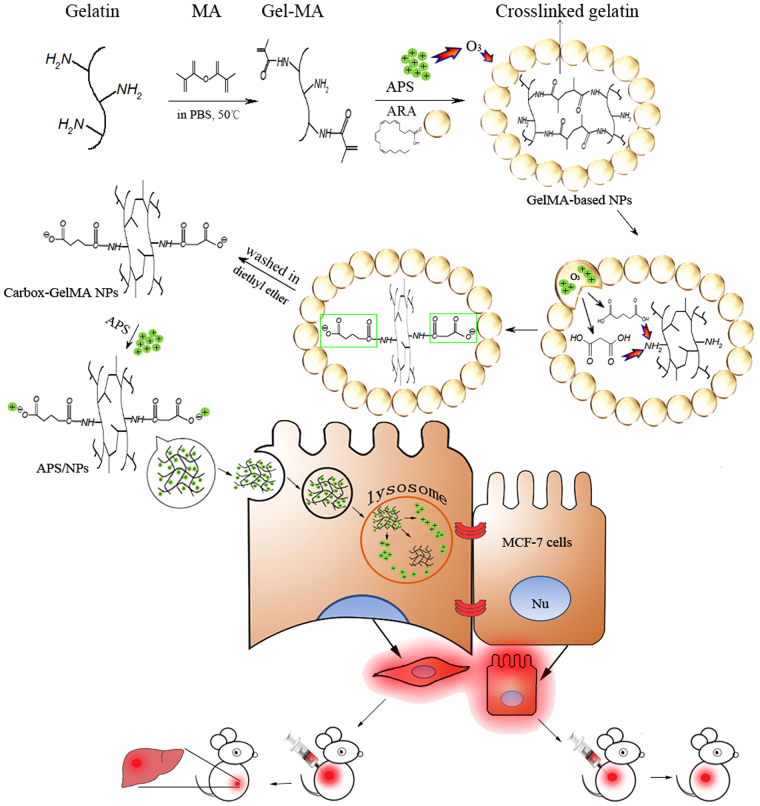
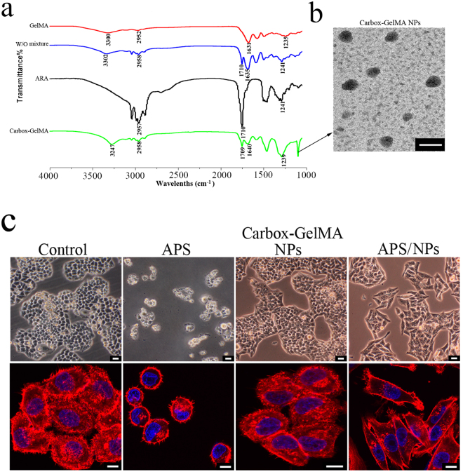
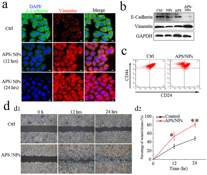
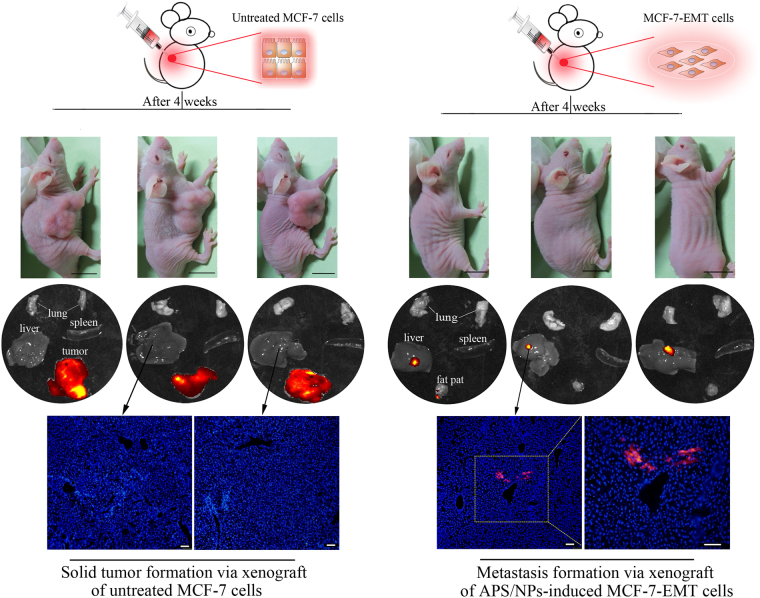
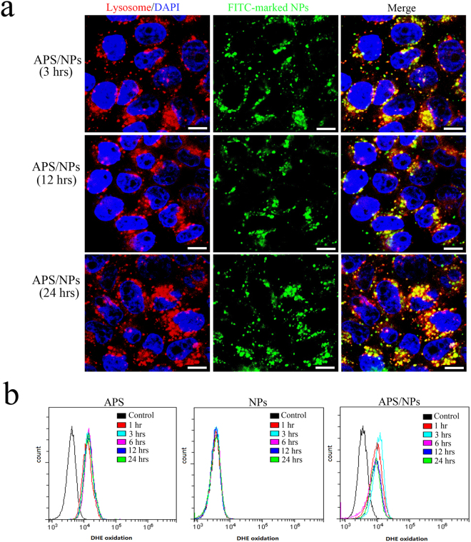
Ammonium persulfate (APS), a low molecular weight chemical compound with strong oxidizing properties, should to be totally removed during preparation of nanomaterials due to its cytotoxicity. APS exerts its oxidative stress effects mainly on cell membrane, but its intracellular influence remains unclear. Here, we designed a facile negatively-charged carboxylic gelatin-methyacrylate (carbox-GelMA) nanoparticle (NP) as a cargo-carrier through the catalytic and oxidizing action of APS in W/O system. The formed APS-loaded carbox-GelMA NPs (APS/NPs) were transported into the lysosome in MCF-7 breast cancer cells. The intracellular APS/NPs produced a high level of oxidative stress in lysosome and induced epithelial-mesenchymal transition (EMT). Consequently, the MCF-7 cells challenged with APS/NPs had a strong metastatic and invasive capability in vitro and in vivo. This study highlights that a facile APS-loaded nanocarrier has cyctotoxicity on cells through EMT. Unexpectedly, we found a novel pathway inducing EMT via lysosomal oxidative stress.


Conflict of interest statement

The authors declare that they have no competing interests.

Figures

Figure 1
Figure 1
The scheme describing the fabrication process of APS-loaded carbox-GelMA nanoparticles (APS/NPs) and their influence on EMT in breast cancer MCF-7 cells. ARA and GelMA were emulsively blending to form the water-in-oil (W/O) mixture, and then the mixture was catalyzed to form GelMA-based NPs by APS. Meanwhile, ARA was oxidized to produce malonic acid and glutaric acid by APS. The carboxyl groups in malonic acid and glutaric acid could react with the partial free amino group in GelMA to generate the negative charges. After the excessive oil layer was washed by diethyl ether, the carbox-GelMA NPs were produced. The negatively-charged carbox-GelMA NPs could carry the positively-charged APS through electrostatic interaction. The APS/NPs showed high effective induction for EMT in MCF-7 cells in vitro through lysosome pathway and endow MCF-7 cells the metastasic capability to liver in vivo.
Figure 2
Figure 2
Characterization of carbox-GelMA NPs and their influence for MCF-7 breast cancer cells. (a) The component analysis of ARA, GelMA, W/O GelMA/ARA mixture (W/O mixture) and carbox-GelMA NPs by the Fourier transform infrared spectroscopy (FTIR). (b) The ultrastructure of carbox-GelMA NPs under transmission electron microscopy (TEM). Scale bars: 200 nm. (c) The upper row: The MCF-7 cells shapes were observed under phase-contrast microscopy. Scale bars: 100 µm. The lower row: The F-actin staining MCF-7 cells were observed under confocal microscope. Scale bars: 20 µm.
Figure 3
Figure 3
APS/NPs could activate EMT, increase the migration ability and the stemness in breast cancer MCF-7 cell lines in vitro. (a) The expression of epithelial marker E-Cadherin (green) and mesenchymal marker vimentin (red) in MCF-7 cells was detected by immunoflurescence staining. The expression of E-cadherin was downregulated whereas the expression of vimentin was upregulated in the MCF-7 cells treated with APS/NPs for 12 hrs and 24 hrs, compared to the control group (Ctrl, untreated cells). Nuclei were stained with DAPI (blue). Scale bars: 10 μm. (b) The expressions of E-Cadherin protein and vimentin protein in MCF-7 cells were detected by western blotting. The MCF-7 cells were treated with pure carbox-GelMA nanoparticles (NPs), pure APS (APS) and APS/NPs for 24 hrs respectively, the untreated cells were taken as the control group. Low expression of E-cadherin protein whereas high expression of vimentin protein appeared simultaneously in APS/NPs group. (c) FACS analysis of cell-surface markers, CD44 and CD24, in MCF-7-EMT cells. The MCF-7 cells being treated with APS/NPs for 72 hrs were named MCF-7-EMT cells (see text part). More CD44high/CD24lowcells were detected in MCF-7-EMT cells, compared with the untreated cells. (d) Wound-healing assay of cell migration capability. The MCF-7-EMT cells exhibited significantly higher migration rates, compared with the untreated cells.
Figure 4
Figure 4
The APS/NPs-induced MCF-7-EMT cells owned strong metastatic ability in vivo. The DiI+ untreated MCF-7 cells and the DiI+ APS/NPs-induced MCF-7-EMT cells were orthotopically transplanted into the BALB/c nude mice respectively. 4 weeks later, solid tumors were obviously found in the untreated MCF-7 cells-transplantation mice, while no evident solid tumors were found in the MCF-7-EMT cells-transplanted mice. The fluorescent signals of the major organs, including the lungs, livers, spleens and tumor tissues (or fat pats) from the mice in 2 groups were detected. The DiI+ signals (red) were gathered in tumor tissues in the untreated MCF-7 cells-transplanted mice, while the DiI+ signals were detected in the livers of the MCF-7-EMT cells-transplanted mice. The fluorescence analysis of liver tissue sections further revealed that the DiI-marked cells had migrated into the liver tissues in the MCF-7-EMT cells-transplanted group. Blue: DAPI-stained nucleus. Black scale bars: 1 cm; White scale bars: 100 µm.
Figure 5
Figure 5
The oxidant stress in lysosome induced by APS could activate EMT in MCF-7 cells. (a) Fluorescence co-localization of lysosome (red) and FITC-labeled NPs (green) after the MCF-7 cells were treated with APS/NPs for 3 hrs, 12 hrs, and 24 hrs respectively. Yellow: the overlap views of lysosome with NPs. Blue: DAPI-stained nucleus. The scale bars: 10 µm. (b) FACS analysis for intracellular ROS levels in MCF-7 cells after the cells were treated with APS, NPs, and APS/NPs respectively for different times.
Figure 6
Figure 6
The heatmap of differently expressed genes among the APS-treated MCF-7 cells, the APS/NPs-induced MCF-7-EMT cells, and the untreated MCF-7 cells. (a) The heatmap of oxidative stress-related genes in the APS-treated MCF-7 cells (APS groups), the APS/NPs-induced MCF-7-EMT cells (APS/NPs groups), and the untreated MCF-7 cells (control groups). §Means the differently expressed genes between the ANP/NPs groups and the control groups. Φ means the differently expressed genes between the APS groups and the control groups. (b) The heatmap of cell motility-related and lysosome-related genes among the APS-treated MCF-7 cells (APS groups), the APS/NPs-induced MCF-7-EMT cells (APS/NPs groups), and the untreated MCF-7 cells (control groups). Green area means downregulated expression and red area means upregulated expression.

Similar articles

Cited by

References

    1. Luengo Y, Nardecchia S, Morales MP, Serrano MC. Different cell responses induced by exposure to maghemite nanoparticles. Nanoscale. 2013;5:11428–11437. doi: 10.1039/c3nr02148c. - DOI - PubMed
    1. Schwensen JF, et al. Occupational contact dermatitis in hairdressers: an analysis of patch test data from the Danish contact dermatitis group, 2002–2011. Contact Dermatitis. 2014;70:233–237. doi: 10.1111/cod.12176. - DOI - PubMed
    1. Hoekstra M, van der Heide S, Coenraads PJ, Schuttelaar ML. Anaphylaxis and severe systemic reactions caused by skin contact with persulfates in hair-bleaching products. Contact Dermatitis. 2012;66:317–322. doi: 10.1111/j.1600-0536.2012.02047.x. - DOI - PubMed
    1. Munoz X, et al. Occupational asthma due to persulfate salts: diagnosis and follow-up. Chest. 2003;123:2124–2129. doi: 10.1378/chest.123.6.2124. - DOI - PubMed
    1. Moscato G, Pala G, Perfetti L, Frascaroli M, Pignatti P. Clinical and inflammatory features of occupational asthma caused by persulphate salts in comparison with asthma associated with occupational rhinitis. Allergy. 2010;65:784–790. doi: 10.1111/j.1398-9995.2009.02288.x. - DOI - PubMed

Publication types