. 2017 Sep 18;8(1):571.
doi: 10.1038/s41467-017-00628-y.

The autophagy initiator ULK1 sensitizes AMPK to allosteric drugs

Affiliations

The autophagy initiator ULK1 sensitizes AMPK to allosteric drugs

Toby A Dite et al. Nat Commun. .

Abstract

AMP-activated protein kinase (AMPK) is a metabolic stress-sensing enzyme responsible for maintaining cellular energy homeostasis. Activation of AMPK by salicylate and the thienopyridone A-769662 is critically dependent on phosphorylation of Ser108 in the β1 regulatory subunit. Here, we show a possible role for Ser108 phosphorylation in cell cycle regulation and promotion of pro-survival pathways in response to energy stress. We identify the autophagy initiator Unc-51-like kinase 1 (ULK1) as a β1-Ser108 kinase in cells. Cellular β1-Ser108 phosphorylation by ULK1 was dependent on AMPK β-subunit myristoylation, metabolic stress associated with elevated AMP/ATP ratio, and the intrinsic energy sensing capacity of AMPK; features consistent with an AMP-induced myristoyl switch mechanism. We further demonstrate cellular AMPK signaling independent of activation loop Thr172 phosphorylation, providing potential insight into physiological roles for Ser108 phosphorylation. These findings uncover new mechanisms by which AMPK could potentially maintain cellular energy homeostasis independently of Thr172 phosphorylation.AMPK is involved in sensing of metabolic stress. The authors show that the autophagy initiator ULK1 phosphorylates β1-Ser108 on the regulatory β1-subunit, sensitizing AMPK to allosteric drugs, and activates signaling pathways that appear independent of Thr172 phosphorylation in the kinase activation loop.


Conflict of interest statement

The authors declare no competing financial interests.

Figures

Fig. 1
Fig. 1
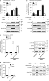
A-769662 activation of cellular AMPK signaling is dependent on β1-pSer108. a Reconstitution of basal AMPK signaling in AMPK β1/2 double knockout (β1/2-dKO) iMEFs by lentiviral transduction of AMPK β1 WT or S108A mutant. b Immunoblots for pACC from β1/2-dKO iMEFs-expressing β1 WT or S108A mutant, stimulated with 20 μM A-769662 for 90 min. n = 3. Error bars, mean pACC fold change relative to basal ± s.e.m. Statistical analysis was performed using unpaired two-tailed Student’s t-test. **P < 0.01 indicates significant increase in pACC compared to basal
Fig. 2
Fig. 2
Quantitative global and phosphoproteomic analysis uncovers cellular roles for β1-pSer108. a Representative immunoblots for β1/2-dKO iMEFs-expressing β1 mutants S108A or S108E, stimulated with 2 mM phenformin for 1 h. b Workflow showing the stable isotope dimethyl labeling-based quantitative proteomic and phosphoproteomic approach. c Heatmap showing significantly perturbed cellular phosphoproteins and corresponding phosphosites. Red indicates increased, and green decreased, phosphorylation in β1-S108E compared to β1-S108A-expressing cells. Gray indicates missing phosphopeptide in that replicate. d Immunoblot/densitometry analysis confirming increased PAK2-Ser141 phosphorylation in β1-S108E-expressing cells. n = 3, representative immunoblot is shown. Error bars, mean PAK2-Ser141 phosphorylation (arbitrary units) ± s.e.m. Statistical analysis was performed using unpaired two-tailed Student’s t-test. *P < 0.05 indicates significant increase in PAK2-pSer141 in S108E-expressing cells compared to S108A-expressing cells. e ‘‘Cell cycle, connective tissue development and function, cellular movement’’ is one of the top networks associated with changes in phosphoproteome between S108A and S108E-expressing iMEFs, as identified by Ingenuity Pathway Analysis software
Fig. 3
Fig. 3
β1-Ser108 trans-phosphorylation occurs via an AMPK independent mechanism. a Rationale for employing kinase inactive (KI) AMPK to examine cellular Ser108 phosphorylation. β1-Ser108 phosphorylation (blue) can potentially be performed by an upstream kinase in both WT and KI α1(D141A) AMPK mutant. LKB1/CaMKK2-mediated phosphorylation of α-Thr172 (red) activates WT AMPK (orange) leading to background Ser108 cis-autophosphorylation. This is excluded using KI AMPK, which can be phosphorylated on Thr172 but remains inactive. b Immunoblot for β1-pSer108 in KI-α1β1γ1 purified from HEK293T cells treated with AMPK-activating agents/conditions: glucose free (glucose-free DMEM + 10% serum, 4 h), AICAR (2 mM, 1 h), H2O2 (1 mM, 45 min), A-769662 (300 μM, 1 h), phenformin (2 mM, 1 h), ionomycin (2.5 μM, 15 min) and amino-acid free (EBSS medium, 4 h). n = 3, representative immunoblots shown. C: Bacterial expressed, CaMKK2-treated α1β1γ1 standard. c Reconstitution of basal AMPK signaling in AMPK α1/2 double knockout (α1/2-dKO) iMEFs by lentiviral transduction of AMPK α1 WT, but not the KI mutant. d Immunoblot for β1-pSer108 from α1/2-dKO iMEFs-expressing KI-α1, stimulated with 2 mM phenformin for 1 h. n = 3, representative immunoblots shown
Fig. 4
Fig. 4
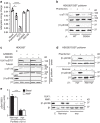
ULK1 phosphorylates β1-Ser108 in vitro. a Sequence alignment of the ULK consensus motif/favorable substitutions with human AMPK β1- and β2-residues 104–112 (Ser108 in lower case). x denotes positions with no demonstrated preference. Preferred/favored consensus β-residues are in bold. Sequence of the synthetic peptide S108tide is shown in red. b Dose curve of S108tide phosphorylation by ULK1 and ULK2 (plotted to left y-axis), and ULK1/FIP200/Atg13 complex (plotted to right y-axis). n = 3. Error bars, mean activity ± s.e.m. c Immunoblots for β1-pSer108 and α-pThr172 in bacterial-expressed KI-α1β1γ1 phosphorylated with ULK1 (left) or ULK2 (right) for 30 min. n = 3, representative immunoblots shown. C: CaMKK2-treated α1β1γ1 control. d Activity of ULK1-phosphorylated α1(C176S)β1γ1 in the presence of 20 μM A-769662 or 10 mM salicylate. n = 3, representative immunoblots of pSer108 in assayed AMPK preparations are shown. Error bars, mean fold AMPK activation relative to ULK1-untreated ± s.e.m. Statistical analyses were performed using one way ANOVA with post hoc Dunnett’s multiple comparison test. **P < 0.01, ****P < 0.0001 indicate significant increase in AMPK activation compared to ULK1-untreated
Fig. 5
Fig. 5
ULK1 phosphorylation of Ser108 is specific for the AMPK β1 isoform. a Immunoblots for β1-pSer108, β2-pSer108, and α-pThr172 in bacterial-expressed KI-α1β1γ1 (left) or KI-α1β2γ1 (right) phosphorylated with ULK1 or CaMKK2 for 30 min. Controls: CaMKK2-treated WT α1β1γ1 (left) or α1β2γ1 (right). b Immunoblots for β-pSer108 in KI-α1β1γ1 (left) or KI-α1β2γ1 (right) purified from HEK293T cells stimulated with 1 mM H2O2 for 45 min. Controls: p-β1 and p-β2, CaMKK2-treated WT α1β1γ1 and α1β2γ1, respectively. In both panels n = 3, representative immunoblots shown
Fig. 6
Fig. 6
ULK phosphorylates β1-Ser108 in cells. Statistical analyses were performed using one-way ANOVA with post hoc Dunnett’s multiple comparison test, unless indicated. Immunoblots for β1-pSer108 and α-pThr172 in KI-α1β1γ1 purified from HEK293T cells incubated with a 1 mM H2O2 and 10 μM 6965 for 45 min, or b 2 mM phenformin and 10 μM 6965 for 1 h. n = 3, representative immunoblots shown. Error bars, mean % phosphorylation relative to H2O2- or phenformin-treated ± s.e.m. **P < 0.01 indicates significant increase, and ## P < 0.01, ### P < 0.001 and #### P< 0.0001 indicate significant decrease, compared to H2O2- or phenformin-treated. c Immunoblots for β1-pSer108 and α-pThr172 in lysates from HEK293T cells, WT or ulk1/2-dKO iMEFs incubated in 25 mM glucose DMEM + 10% serum. n = 3 individual cultures per cell line, representative immunoblots shown. Error bars, mean fold increase in phosphorylation relative to HEK293T cells ± s.e.m. ****P < 0.0001 indicates significant increase in phosphorylation compared to HEK293T cells. d Immunoblots for β1-pSer108 and α-pThr172 in KI-α1 AMPK purified from WT or ulk1/2-dKO iMEFs stimulated with 2 mM phenformin for 1 h. n = 3, representative immunoblots shown. Error bars, mean fold increase in phosphorylation relative to basal ± s.e.m. Statistical analysis was performed using unpaired two-tailed Student’s t-test. *P < 0.05 indicates significant decrease compared to WT iMEFs
Fig. 7
Fig. 7
An AMP-myristoyl switch triggers ULK1 phosphorylation of β1-Ser108. a Adenine nucleotides extracted from HEK293T cells incubated with phenformin (2 mM, 1 h), H2O2 (1 mM, 45 min), AZD8055 (1 μM, 1 h), or INK128 (1 μM, 1 h) were quantitated by mass spectrometry. Adenylate energy charge was calculated as described in Online Methods. n = 3. Error bars, mean adenylate energy charge ± s.e.m. Statistical analyses were performed using one-way ANOVA with post hoc Dunnett’s multiple comparison test. ****P > 0.001, *P < 0.05 indicate significant decrease in mean adenylate energy charge compared to basal. b Immunoblots for β1-pSer108 and α-pThr172 in KI-α1β1γ1 or KI-α1β1γ1(D245A) purified from HEK293T cells stimulated with 2 mM phenformin for 1 h. n = 3, representative immunoblots shown. c Immunoblots for ULK1-pSer757, and β1-pSer108 and α-pThr172 in KI-α1β1γ1 purified from HEK293T cells incubated with 1 μM mTOR inhibitors AZD8055 or INK128 for 1 h. n = 3, representative immunoblots shown. C: Bacterial expressed, CaMKK2-treated α1β1γ1 standard. d Immunoblots for β1-pSer108 in KI-α1β1γ1 (myr) or KI-α1β1(G2A)γ1 (non-myr) purified from HEK293T cells incubated with 2 mM phenformin for 1 h (upper) or glucose free medium for 4 h (lower). n = 3, representative immunoblots shown. e Immunoblot for β1-pSer108 in bacterial-expressed, non-myristoylated (non-myr) or myristoylated (myr) KI-α1β1γ1 phosphorylated with ULK1 for 30 min in the presence of 100 μM AMP. n = 3, representative immunoblots shown. C: CaMKK2-treated α1β1γ1 standard. Error bars, mean increase in β1-pSer108 relative to ULK1-untreated ± s.e.m. Statistical analysis was performed using unpaired two-tailed Student’s t-test. ***P < 0.001 indicates significant decrease in β1-pSer108 relative to non-myristoylated AMPK
Fig. 8
Fig. 8
Cellular AMPK signaling occurs independently of α1-pThr172. Statistical analyses were performed using one-way ANOVA with post hoc Dunnett’s multiple comparison test. a Immunoblots for α2-pThr172 and pACC from α1/2-dKO iMEFs-expressing α2 WT or T172A mutant, stimulated with 2 mM phenformin and/or 100 μM A-769662 for 1 h. n = 3, representative immunoblots shown. Error bars, mean pThr172 and pACC (fold change vs. untreated WT α2-expressing cells) ± s.e.m. ***P < 0.001, **P > 0.01, *P < 0.05 indicate significant increase compared to basal WT α2 cells. #### P > 0.0001, ## P < 0.01 indicate significant increase compared to basal α2(T172A) cells. b Immunoblots for β1-pSer108 and ULK1-pSer555 from α1/2-dKO iMEFs-expressing α2 T172A mutant, stimulated with 2 mM phenformin and 100 μM A-769662 for 1 h. n = 3, representative immunoblots shown. Error bars, mean Ser108 and Ser555 phosphorylation (arbitrary units) ± s.e.m. ***P < 0.001, *P < 0.05 indicate significant increase compared to basal. # P < 0.05 indicates significant increase compared to phenformin treated
Fig. 9
Fig. 9
An integrated model for ULK1 regulation of β1-AMPK signaling. The initial ULK stimulus (e.g., Ser555 phosphorylation) is provided by AMPK, activated itself in response to energy stress and elevated AMP:ATP. Once activated, ULK1 suppresses Thr172 phosphorylation and AMPK sensitivity to AMP via negative feedback (red arrows). ULK1 simultaneously phosphorylates Ser108, sensitizing AMPK to drugs/metabolites acting at the ADaM site. This would promote AMP-independent AMPK signaling and maintain ULK1 activity via positive feedback (green arrows). In our cell model, activity of α2(T172A) AMPK is not elevated in response to AMP and requires additional stimulation with A-769662 to achieve ULK1 phosphorylation

Similar articles

Cited by

References

    1. Hardie DG. AMPK - sensing energy while talking to other signaling pathways. Cell Metab. 2014;20:939–952. doi: 10.1016/j.cmet.2014.09.013. - DOI - PMC - PubMed
    1. Steinberg GR, Kemp BE. AMPK in health and disease. Physiol. Rev. 2009;89:1025–1078. doi: 10.1152/physrev.00011.2008. - DOI - PubMed
    1. Oakhill JS, et al. β-Subunit myristoylation is the gatekeeper for initiating metabolic stress sensing by AMP-activated protein kinase (AMPK) Proc. Natl Acad. Sci. USA. 2010;107:19237–19241. doi: 10.1073/pnas.1009705107. - DOI - PMC - PubMed
    1. Houde VP, et al. Investigation of LKB1 Ser431 phosphorylation and Cys433 farnesylation using mouse knockin analysis reveals an unexpected role of prenylation in regulating AMPK activity. Biochem. J. 2014;15:41–56. doi: 10.1042/BJ20131324. - DOI - PMC - PubMed
    1. Xiao B, et al. Structure of mammalian AMPK and its regulation by ADP. Nature. 2011;472:230–233. doi: 10.1038/nature09932. - DOI - PMC - PubMed

Publication types

MeSH terms

Substances