

Aggressive Posterior Retinopathy of Prematurity in a Premature Male Infant
- PMID: 28924435
- PMCID: PMC5597914
- DOI: 10.1159/000478694
Aggressive Posterior Retinopathy of Prematurity in a Premature Male Infant
Abstract
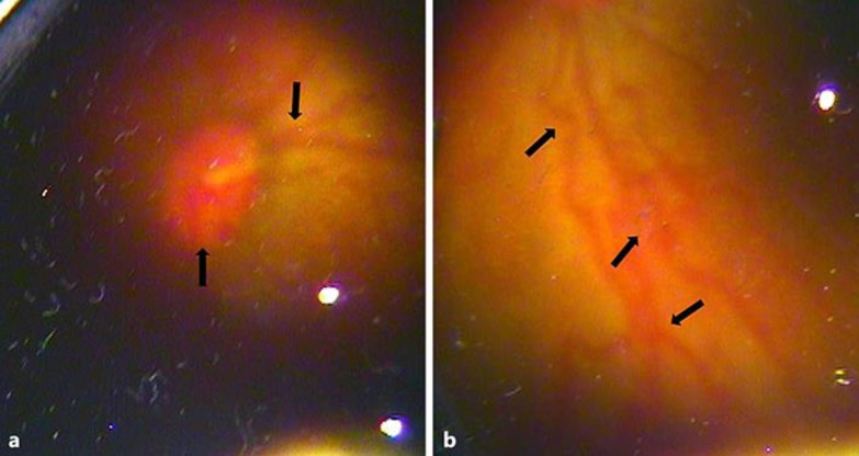
A premature male infant was born at 30 weeks' gestation with a birth weight of 1,700 g in a rural hospital. He was diagnosed with respiratory distress syndrome and received continuous positive airway pressure treatment for 26 days. At 26 days after birth, the patient was transferred to our hospital for further evaluation and management. A comprehensive eye examination revealed a stage 3 retinopathy of prematurity (ROP) involving zone 2 in both eyes. The patient was recommended to a provincial-level eye hospital for emergency laser therapy. Five months after birth, the feedback from the eye hospital showed that the patient had a high risk of blindness in both eyes. Our case report shows that delaying first screening examination increases the possibility of developing aggressive posterior ROP in infants with ROP. Doctors in rural hospitals should be aware of this possibility and trained for early screening and treatment in high-risk infants.
Keywords: Blindness; Premature infant; Retinopathy of prematurity.
Figures

References
-
- International Committee for the Classification of Retinopathy of Prematurity The international classification of retinopathy of prematurity revisited. Arch Ophthalmol. 2005;123:991–999. - PubMed
-
- Drenser KA, Trese MT, Capone A., Jr Aggressive posterior retinopathy of prematurity. Retina. 2010;30(suppl 4):S37–S40. - PubMed
-
- Fortes Filho JB, Eckert GU, Procianoy L, Barros CK, Procianoy RS. Incidence and risk factors for retinopathy of prematurity in very low and in extremely low birth weight infants in a unit-based approach in southern Brazil. Eye (Lond) 2009;23:25–30. - PubMed
Publication types
LinkOut - more resources
Full Text Sources
Other Literature Sources
