. 2017:1663:219-230.
doi: 10.1007/978-1-4939-7265-4_18.

Correlative Fluorescence Super-Resolution Localization Microscopy and Platinum Replica EM on Unroofed Cells

Affiliations

Correlative Fluorescence Super-Resolution Localization Microscopy and Platinum Replica EM on Unroofed Cells

Kem A Sochacki et al. Methods Mol Biol. 2017.

Abstract

Platinum replicas of unroofed mammalian cells can be imaged with a transmission electron microscope (TEM) to produce high contrast, high resolution images of the structure of the cytoplasmic side of a plasma membrane. A complementary approach, super-resolution fluorescence localization microscopy, can be used to localize labeled molecules with better than 20 nm precision in cells. Here, we describe a correlative method that couples these two techniques and produces images where localization microscopy data can be used to highlight specific proteins of interest within the structural context of the platinum replica TEM image. This combined method is uniquely suited to investigate the nanometer-scale structural organization of the plasma membrane and its associated organelles and proteins.

Keywords: CLEM; Correlative microscopy; Electron microscopy; Fluorescence; Localization microscopy; Nanoscopy; PREM; Platinum replica; Super-resolution; Unroofed cells.


Figures

Figure 1)
Figure 1)
The imaged region is marked with a diamond objective marker. (a) The diamond objective marker is shown in the microscope turret with our custome-made spacer attached at the bottom. (b) A 4 mm etched circle is shown on the bottom of the coverslip.
Figure 2)
Figure 2)
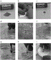
Lifting the replica onto a grid. (a) The coverslip fragment is shown after platinum shadowing and carbon coating. (b-c) The coverslip is cut to approximately the size of the loop that will be used to lift the replica. (d) The coverslip is placed on the top surface of 5% hydrofluoric acid. (e) The glass coverslip falls away leaving the platinum/carbon replica floating on top of the surface. (f) The hydrofluoric acid is replaced with water by serial dilutions with a plastic pipette. (g-h) The replica is lifted from below and (i) placed over the top of the TEM grid.
Figure 3)
Figure 3)
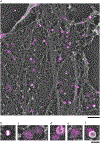
Correlative data from GFP-Clathrin light chain expressing cells labeled with GFP-nanobody conjugated to Alexa Fluor 647. (a) Large zoomed out view of unroofed HeLa cell after correlation. Fluorescence signal is shown in magenta. Scale bar is 1 μm. (b) Gold nanoparticle from (a). (c-e) Examples of clathrin correlation from (a). (f) example of vesicle that moved during critical point drying due to a weak attachment to the membrane. Scale bar in b-f is 200 nm.

References

    1. Moor H, Muhlethaler K (1963) Fine structure in frozen-etched yeast cells. The Journal of cell biology 17 (3):609–628 - PMC - PubMed
    1. Lung B (1968) Whole-mount electron microscopy of chromatin and membranes in bull and human sperm heads. Journal of ultrastructure research 22 (5):485–493 - PubMed
    1. Heuser J, Keen J (1988) Deep-etch visualization of proteins involved in clathrin assembly. The Journal of cell biology 107 (3):877–886 - PMC - PubMed
    1. Svitkina T (2015) Imaging Cytoskeleton Components by Electron Microscopy. Methods in molecular biology (Clifton, NJ) 1365:99–118 - PMC - PubMed
    1. Heuser J (2000) The production of ‘cell cortices’ for light and electron microscopy. Traffic 1 (7):545–552 - PubMed

LinkOut - more resources