. 2017 Oct;33(5):483-492.
doi: 10.1007/s12264-017-0179-1. Epub 2017 Sep 18.

Age-Dependent Alpha-Synuclein Accumulation and Phosphorylation in the Enteric Nervous System in a Transgenic Mouse Model of Parkinson's Disease

Affiliations

Age-Dependent Alpha-Synuclein Accumulation and Phosphorylation in the Enteric Nervous System in a Transgenic Mouse Model of Parkinson's Disease

Chong-Bin Zhong et al. Neurosci Bull. 2017 Oct.

Abstract

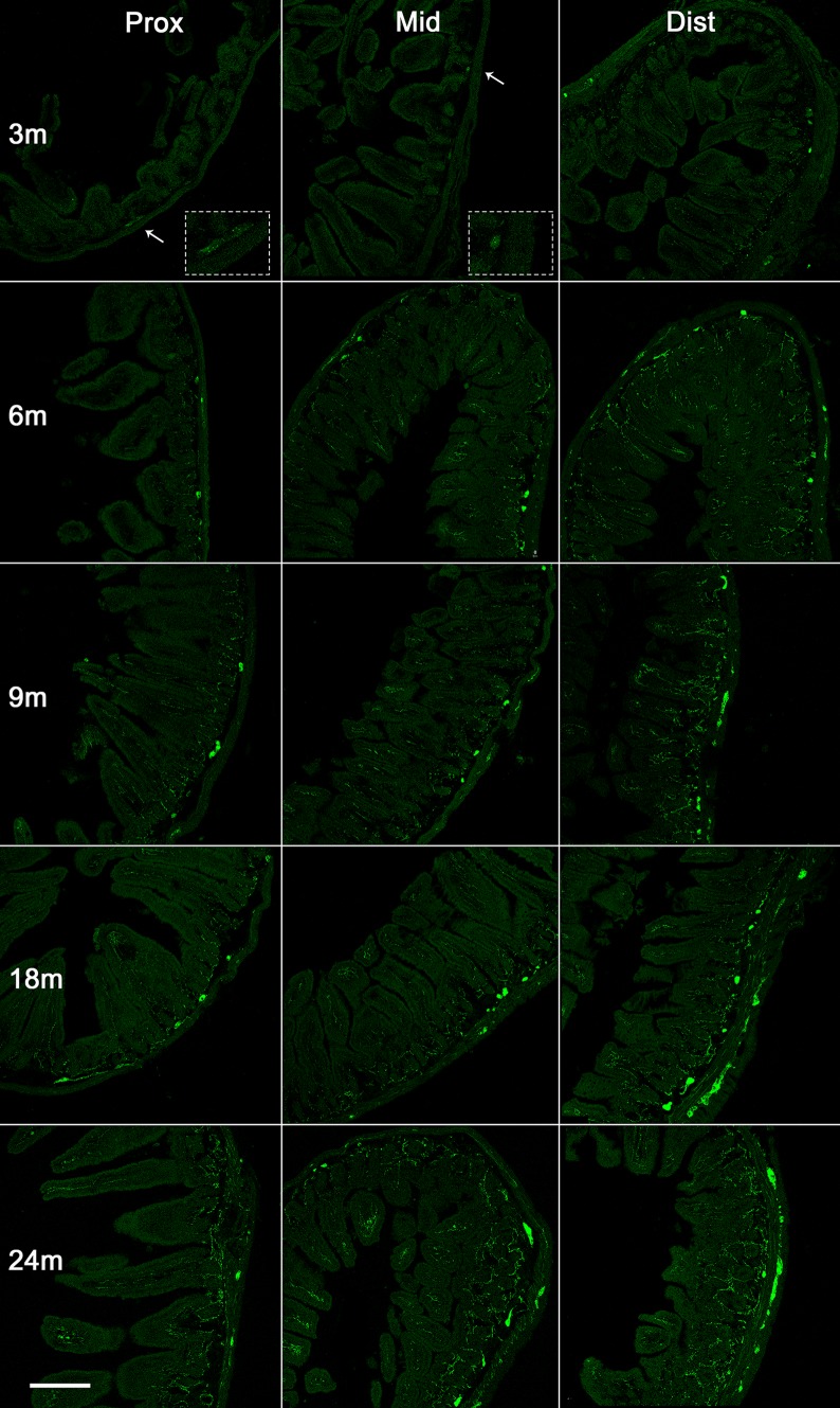
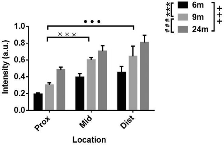
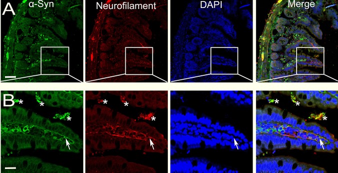
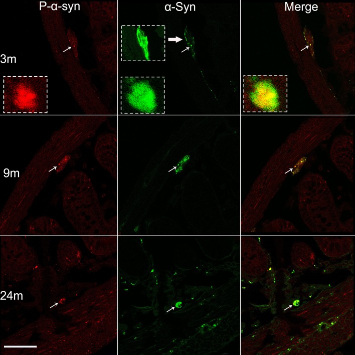
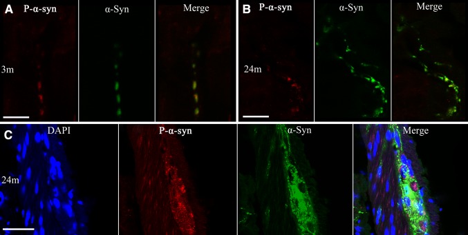
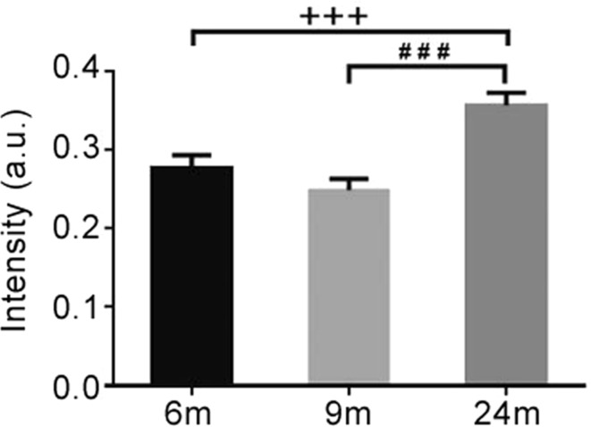
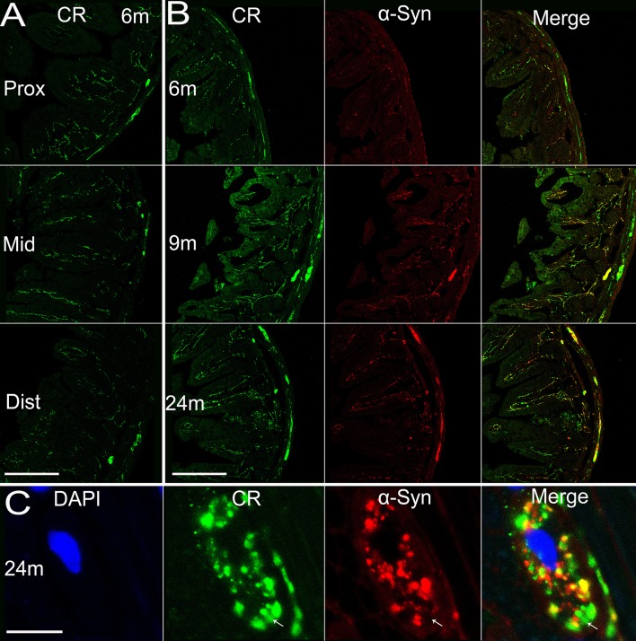
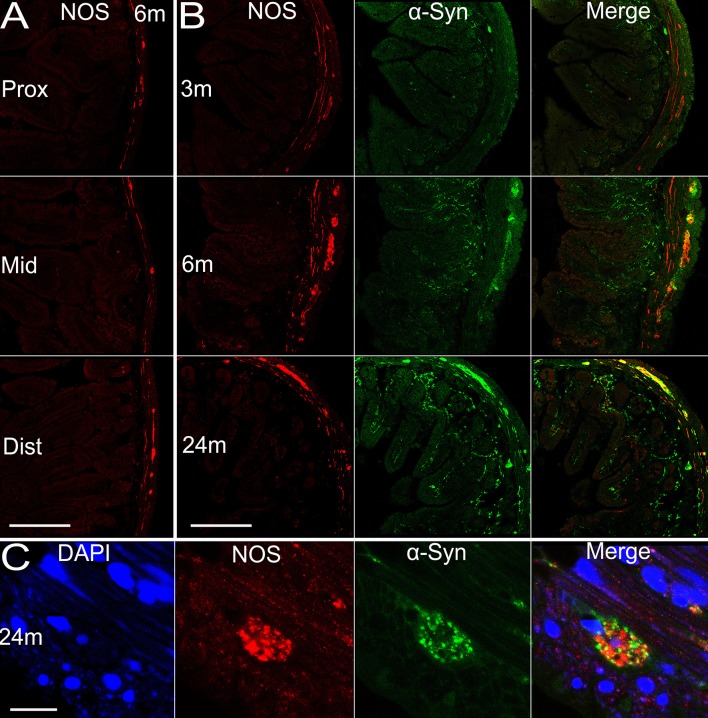
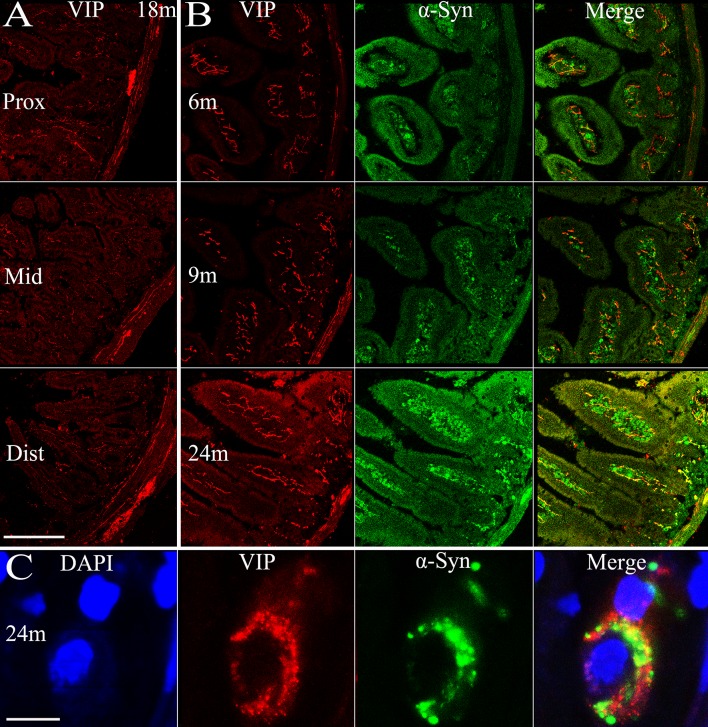
The enteric nervous system (ENS) controls the function of the gastrointestinal tract and has been implicated in various diseases, including Parkinson's disease (PD). PD is a neurodegenerative disease with Lewy bodies (LBs) and Lewy neurites (LNs) as the main pathological features. In addition to the typical motor symptoms in PD, attention has been drawn to non-motor symptoms, such as constipation, implying dysfunction of the ENS. In the present study, we characterized the age-dependent morphological alterations and aggregation of α-synuclein (α-syn), the primary protein component in LBs and LNs, in the ENS in an α-syn transgenic mouse model. We found that the expression and accumulation of α-syn increased gradually in neurons of Meissner's and Auerbach's plexuses of the gastrointestinal tract with age (from 1 week to 2 years). In addition, α-syn was increasingly phosphorylated at the serine 129 residue, reflecting pathological alterations of the protein over time. Furthermore, α-syn was present in different subtypes of neurons expressing vasoactive intestinal polypeptide, neuronal nitric oxide synthase, or calretinin. The results indicated that BAC-α-Syn-GFP transgenic mice provide a unique model in which to study the relationship between ENS and PD pathogenesis.

Keywords: Enteric system; Parkinson’s disease; Phosphorylation; Protein aggregation; α-synuclein.


Figures

Fig. 1
Fig. 1
Expression of α-syn in the small intestine of transgenic mice with age. Representative images of α-syn-GFP expression in the small intestine proximal to the stomach (left), the middle of the small intestine (middle), and the distal intestine (right) at 3, 6, 9, 18, and 24 months (m). Arrows, high-power images in insets; scale bar 250 μm.
Fig. 2
Fig. 2
Semi-quantitative analysis of α-syn fluorescence (GFP) intensity in the small intestine of transgenic mice at different ages. Two-way ANOVA (F = 32.041, P < 0.001, in age; F = 24.110, P < 0.001, in location), followed by LSD post hoc test (***P < 0.005, 6 vs 9 months; ### P < 0.005, 9 vs 24 months; +++ P < 0.005, 6 vs 24 months; ••• P < 0.005, Prox vs Dist; ××× P < 0.005, Prox vs Mid.); error bars, standard error of the mean.
Fig. 3
Fig. 3
Overlap of α-syn and neurofilament in the distal segment of the small intestine. α-Syn- and neurofilament-positive profiles largely overlap in the cell bodies of Auerbach’s and Meissner’s plexuses, and also in nerve terminals extending into the microvilli (arrows in B). *Non-specific labels of residual contents in the lumen; scale bars, 75 μm in A and 25 μm in B.
Fig. 4
Fig. 4
Age-dependent phosphorylation of α-syn in Meissner’s plexus of the small intestine. Profiles of phospho-α-syn (P-α-syn, left), pan-α-syn (middle), and their co-localization (right), in 3, 9, and 24 month-old mice. The photomicrographs acquired under a confocal microscope show increased phosphorylation of α-syn with age (left). Subsets of pan-α-syn-positive profiles (middle) are overlapped with phospho-α-syn-positive profiles (right) (arrows and the insets). Scale bar 50 μm.
Fig. 5
Fig. 5
Co-localization of α-syn and phosphorylated α-Syn (P-α-syn) in transgenic mice at indicated ages. AB Photomicrographs acquired under a confocal microscope show increased phosphorylation of α-syn with age (A, 3 months; B 24 months) in the mucous layer and in Auerbach’s plexus of the distal segment of the small intestine. Subsets of pan-α-syn-positive are overlapped with phospho-α-syn-positive profiles (AC). Scale bars 2.5 μm (A), 10 μm (B), and 50 μm (C).
Fig. 6
Fig. 6
Semi-quantitative analysis of fluorescence intensity of phospho-α-syn immunoreactivity in the small intestine (distal segment) of transgenic mice at different ages. One-way ANOVA (F = 12.029, P = 0.001) followed by LSD post hoc test (### P < 0.005, 9 vs 24 months; +++ P < 0.005, 6 vs 24 months); error bars SEM.
Fig. 7
Fig. 7
Expression of calretinin in the small intestine of transgenic mice with age and the co-localization of calretinin and α-syn. A Expression of calretinin in different sections (proximal, middle, and distal segments) of the small intestine in transgenic mice. B α-Syn and calretinin expressed in transgenic mice and their co-localization at 6, 9, and 24 months. C Co-localization of calretinin and α-syn, and the nucleus (DAPI) of an enteric neuron in Auerbach’s plexus at 24 months. Arrows, area where calretinin is expressed while α-syn not. Scale bars 200 μm in A, B; 10 μm in C. CR, calretinin.
Fig. 8
Fig. 8
Expression of nNOS in the small intestine of transgenic mice with age and the co-localization of nNOS and α-syn. A Expression of nNOS in different sections (proximal, middle, and distal segments) of the intestine in transgenic mice. B α-Syn and nNOS expressed in transgenic mice and the co-localization at 3, 6, and 24 months. C Co-localization of nNOS and α-syn, and the nucleus of an enteric neuron at 24 months. Scale bars 200 μm in A, B; 10 μm in C.
Fig. 9
Fig. 9
Expression of VIP in the small intestine of transgenic mice with age and the co-localization of VIP and α-syn. A Expression of VIP in different sections (proximal, middle, and distal segments) of the small intestine in transgenic mice. B α-Syn and VIP expressed in transgenic mice and their co-localization at 6, 9, and 24 months. C Co-localization of VIP and α-syn, and the nucleus (DAPI) of an enteric neuron at 24 months. Scale bars 200 μm in A, B; 10 μm in C.

References

    1. Eschbach J, Danzer KM. Alpha-synuclein in Parkinson’s disease: pathogenic function and translation into animal models. Neurodegener Dis. 2014;14:1–17. doi: 10.1159/000354615. - DOI - PubMed
    1. Cersosimo MG, Benarroch EE. Pathological correlates of gastrointestinal dysfunction in Parkinson’s disease. Neurobiol Dis. 2012;46:559–564. doi: 10.1016/j.nbd.2011.10.014. - DOI - PubMed
    1. Hilton D, Stephens M, Kirk L, Edwards P, Potter R, Zajicek J, et al. Accumulation of alpha-synuclein in the bowel of patients in the pre-clinical phase of Parkinson’s disease. Acta Neuropathol. 2014;127:235–241. doi: 10.1007/s00401-013-1214-6. - DOI - PubMed
    1. Stirpe P, Hoffman M, Badiali D, Colosimo C. Constipation: an emerging risk factor for Parkinson’s disease? Eur J Neurol. 2016;23:1606–1613. doi: 10.1111/ene.13082. - DOI - PubMed
    1. Cao M, Gu ZQ, Li Y, Zhang H, Dan XJ, Cen SS, et al. Olfactory dysfunction in Parkinson’s disease patients with the LRRK2 G2385R variant. Neurosci Bull. 2016;32:572–576. doi: 10.1007/s12264-016-0070-5. - DOI - PMC - PubMed