. 2018 Jan;66(1):126-144.
doi: 10.1002/glia.23232. Epub 2017 Sep 19.

Selective knockout of astrocytic Na+ /H+ exchanger isoform 1 reduces astrogliosis, BBB damage, infarction, and improves neurological function after ischemic stroke

Affiliations

Selective knockout of astrocytic Na+ /H+ exchanger isoform 1 reduces astrogliosis, BBB damage, infarction, and improves neurological function after ischemic stroke

Gulnaz Begum et al. Glia. 2018 Jan.

Abstract

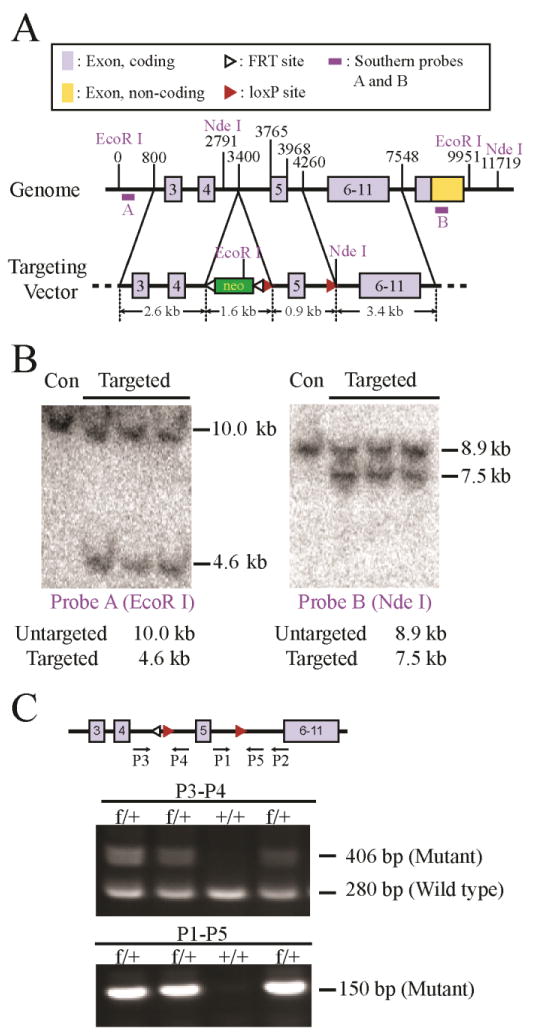
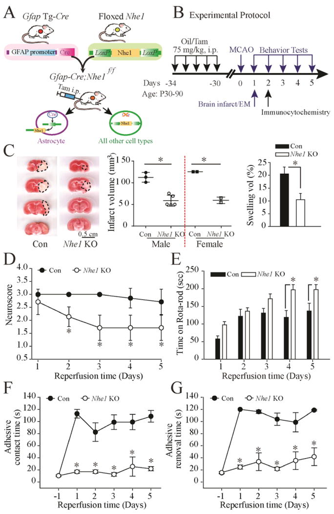
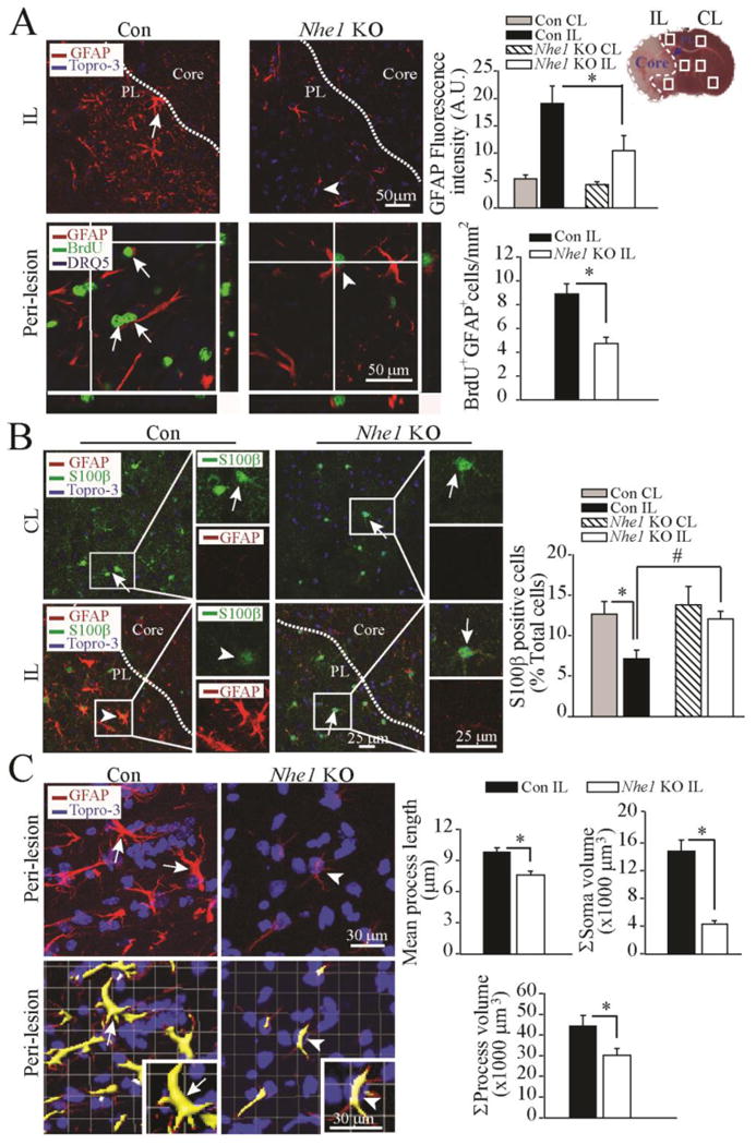
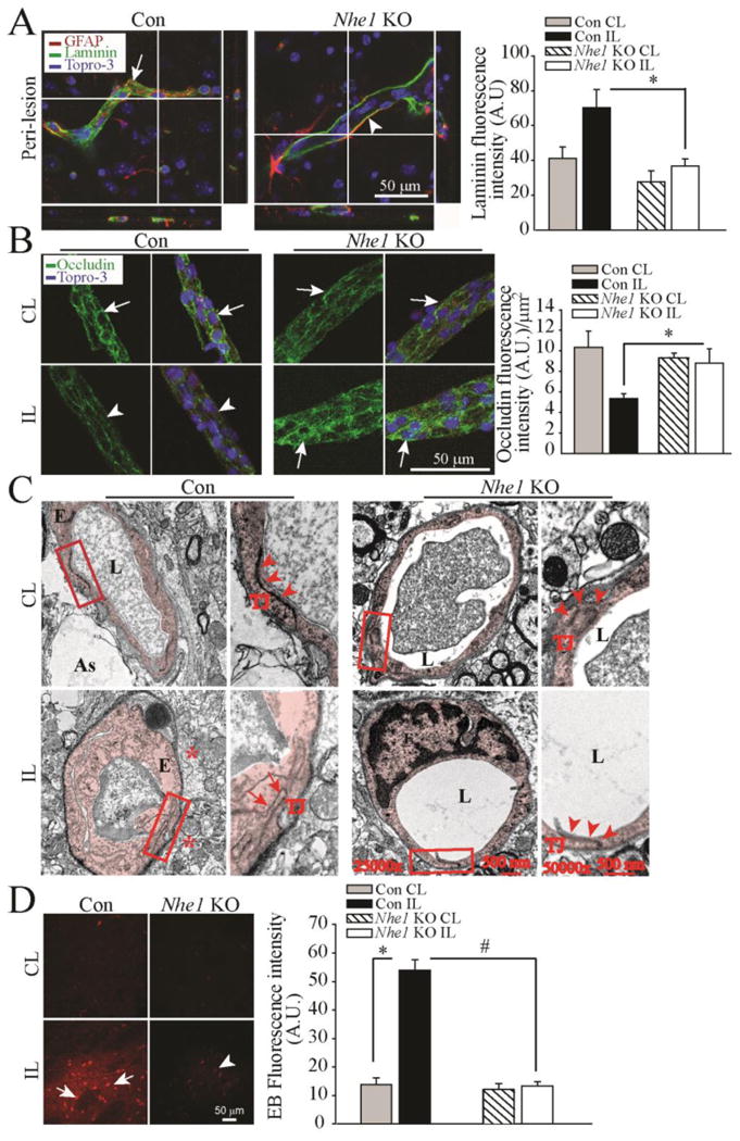
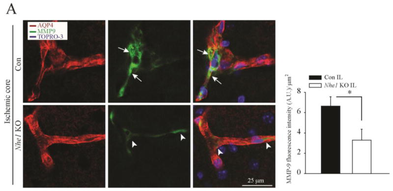
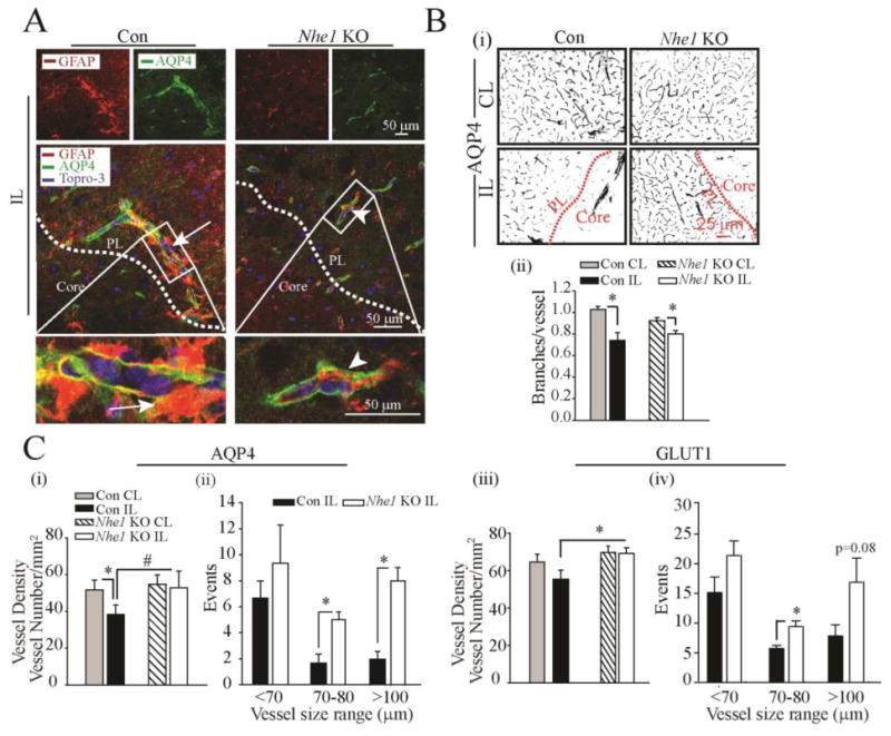
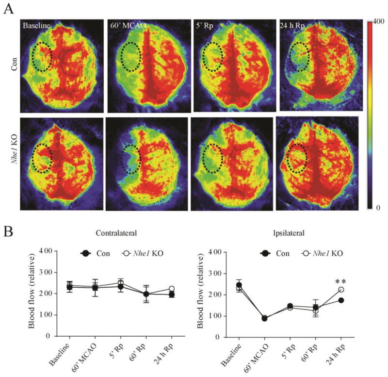
Stimulation of Na+ /H+ exchanger isoform 1 (NHE1) in astrocytes causes ionic dysregulation under ischemic conditions. In this study, we created a Nhe1flox/flox (Nhe1f/f ) mouse line with exon 5 of Nhe1 flanked with two loxP sites and selective ablation of Nhe1 in astrocytes was achieved by crossing Nhe1f/f mice with Gfap-CreERT2 Cre-recombinase mice. Gfap-CreERT2+/- ;Nhe1f/f mice at postnatal day 60-90 were treated with either corn oil or tamoxifen (Tam, 75 mg/kg/day, i.p.) for 5 days. After 30 days post-injection, mice underwent transient middle cerebral artery occlusion (tMCAO) to induce ischemic stroke. Compared with the oil-vehicle group (control), Tam-treated Gfap-CreERT2+/- ;Nhe1f/f (Nhe1 KO) mice developed significantly smaller ischemic infarction, less edema, and less neurological function deficits at 1-5 days after tMCAO. Immunocytochemical analysis revealed less astrocytic proliferation, less cellular hypertrophy, and less peri-lesion gliosis in Nhe1 KO mouse brains. Selective deletion of Nhe1 in astrocytes also reduced cerebral microvessel damage and blood-brain barrier (BBB) injury in ischemic brains. The BBB microvessels of the control brains show swollen endothelial cells, opened tight junctions, increased expression of proinflammatory protease MMP-9, and significant loss of tight junction protein occludin. In contrast, the Nhe1 KO mice exhibited reduced BBB breakdown and normal tight junction structure, with increased expression of occludin and reduced MMP-9. Most importantly, deletion of astrocytic Nhe1 gene significantly increased regional cerebral blood flow in the ischemic hemisphere at 24 hr post-MCAO. Taken together, our study provides the first line of evidence for a causative role of astrocytic NHE1 protein in reactive astrogliosis and ischemic neurovascular damage.

Keywords: MMP; astrocyte end-feet; blood-brain barrier; cerebral edema; gliosis; neurovascular unit.


Figures

Figure 1
Figure 1. Creation of Nhe1f/f conditional knockout mice
A. Nhe1 conditional knockout strategy and position of southern blots probes. A targeting construct was made with two loxP sites flanking exon 5 with a PGKneo cassette flanked by two FRT sites located in intron 4. The construct was transfected into ES cells and the clones with appropriate homologous recombination were identified by Southern blot analysis using probes A and B, which were from regions 5′ and 3′, respectively, to the fragments used to prepare the construct. B. Genomic DNA from three targeted clones and untargeted ES cells (Con) was digested with EcoR1 or Nde I and the products were detected by Southern blot analysis using probes A and B, respectively. C. Diagram of the targeted allele after deletion of the PGKneo cassette and PCR genotyping of Nhe1f/+ and Nhe1+/+ animals using P1 – P5 PCR Primers. Primers P3 and P4 amplify a 406-bp product in the floxed allele, and a 280-bp product in the wild type gene. Primers P1and P5 (which includes sequences from the LoxP site) amplifies a 150-bp product in the floxed allele only.
Figure 2
Figure 2. Deletion of Nhe1 in Gfap-CreER+/-;Nhe1 f/f mice decreases ischemic infarct, swelling, and improves neurological function
A. Breeding scheme for generation of astrocyte-specific Nhe1 knockout mice. Nhe1f/f mice were crossed with Gfap-CreER+/- transgenic mice to generate Gfap-CreER+/-;Nhe1f/f mice. B. Experimental protocol. Gfap-CreER+/-;Nhe1f/f mice at postnatal days P60-90 were treated with either corn oil or Tam in corn oil (75mg/kg/day, ip) for 5 days. After 30 days, ischemic stroke was induced by transient middle cerebral artery occlusion (MCAO) at P90-120. Biochemical or neurological assays were conducted at 1-5 days post-stroke. C. Representative TTC-stained coronal sections of control (oil-) and Nhe1 KO (Tam-treated) brains at 48 h reperfusion. Infarct volume and % of hemispheric swelling were shown. Data are mean ± SD. n = 5-7 (Oil: 3 male, 2 female; Tam: 5 male, 2 female). *p < 0.05 vs. oil. D. Neurological scores. Data are mean ± SD. n = 7 (Con: 1 male; 6 female; Nhe1 KO: 5 male, 2 female). * p < 0.05 vs. oil. E. Rotarod test with accelerating speed. Data are mean ± SEM. n = 9-11 (Con: 2 male, 7 female; Nhe1 KO: 6 male, 5 female). * p < 0.05 vs. oil. F. Contact time of adhesive test. Data are mean ± SEM. n = 7 (Con: 4 male, 3 female; Nhe1 KO: 4 male, 3 female). * p < 0.05 vs. oil. G. Removal time of adhesive test. Data are mean ± SEM. n = 7 (the same cohort). * p < 0.05 vs. Con.
Figure 3
Figure 3. Deletion of Nhe1 in Gfap-CreER+/-;Nhe1f/f mice inhibits reactive gliosis and astrocytic BrdU labeling
A. Representative images of GFAP+ astrocytes were shown in the peri-infarct zones of the Con or Nhe1 KO mice. A TTC-stained brain section at 48 h after MCAO was marked for the ischemic core, and sample collection areas (white box) in the peri-lesion tissue (PL, dotted lines) and contralateral control. Arrows: Increased GFAP expression or BrdU labeling. Arrowhead: Reduced GFAP expression or BrdU labeling. Data are mean ± SEM, n = 3-4. * p < 0.05 vs. Con. B. Representative images of GFAP, S100β, and To-pro-3 staining in the CL and IL cortex regions of the Con and Nhe1 KO mice. Arrows: Preserved S100 β expression. Arrowheads: loss of S100 β in GFAP+ astrocytes. Data are ± SEM, n = 3. *p < 0.05 vs. CL; # p < 0.05 vs. Con. C. Representative immunofluorescent Z-stack images of brain sections showing changes of GFAP+ reactive astrocyte morphology in the Con and Nhe1 KO mice. Lower panel shows cropped view of 3D rendering. Arrows: increased soma area of GFAP+ cells. Arrowheads: Reduced soma area of GFAP+ cells. Summary data of changes of GFAP+ cell processes, mean process length, and soma volume were shown. Data are mean ± SEM, n = 6. * p < 0.05 vs. Con.
Figure 4
Figure 4. Deletion of Nhe1 in Gfap-CreER+/-;Nhe1f/f mice promotes less cerebral vessel damage and improved BBB morphology after ischemic stroke
A. Representative confocal Z-stack images of laminin-stained microvessels in the Con and Nhe1 KO ischemic brains after 48 h reperfusion. Arrow: accumulation of degraded laminin. Arrowheads: less laminin accumulation. Summary data of laminin fluorescence intensity are mean ± SEM, n=4. *p < 0.05 vs Con. B. Representative confocal images of tight junction protein occludin stained vessels isolated from Con and Nhe1 KO ischemic brains at 48 h reperfusion. Arrows: Preserved occludin expression. Arrow heads: Reduced occludin expression. Data are mean ± SEM, n=4 *p < 0.05 vs Con C. Transmission electron microscopy images of cerebral vessels in the peri-lesion areas from the Con and Nhe1 KO mouse brains at 24 h reperfusion. Extensive perivascular damage, swollen endothelial cells and opened tight junctions were shown. Arrow: opened tight junctions. Arrowhead: intact tight junctions. As: Astrocyte endfeet; L: capillary lumen; E: endothelial cell; TJ: tight junctions. Box: to be enlarged. D. Changes of the BBB permeability in the ischemic brain parenchyma at 48 h reperfusion. EB extravasation was detected in the IL hemispheres of the Con (arrow) but not in the Nhe1 KO mice (arrowhead). Data are mean ± SEM, n=3. *p < 0.05 vs CL. #p < 0.05 vs Con.
Figure 5
Figure 5. Selective deletion of Nhe1 in Gfap-CreER+/-;Nhe1f/f mice inhibits MMP9 expression in cerebral vessels
A. Representative confocal images of MMP-9 and AQP4 expression in the Con and Nhe1 KO ischemic brains at 48 h reperfusion. Increased perivascular MMP9 expression (green) is associated with AQP4 signals (red) only in the Con but not in Nhe1 KO mice. Arrows: Increased perivascular MMP-9 expression. Arrowheads: Absence of perivascular MMP9 expression. Summary data of MMP-9 fluorescence intensity are mean ± SEM, n=5. *p < 0.05 vs Con.
Figure 6
Figure 6. Deletion of Nhe1 in Gfap-CreER+/-;Nhe1f/f mice reduced perivascular damage and preserved microvessel density after ischemic stroke
A. Representative confocal Z-stack images of AQP4-stained microvessels in the Con and Nhe1 KO ischemic brains at 48 h reperfusion. Increased numbers of perivascular reactive GFAP+ astrocytes (red) were associated with AQP4 stained microvessels (green), more clearly shown in the Con than in the KO mice. Arrows: increased perivascular reactive astrogliosis. Arrowheads: reduced perivascular reactive astrogliosis. White box: enlarged in the lower panel. B. Representative binary images of AQP4-labeled microvessels show different cerebral microvessel density in the peri-lesion regions of the Con and Nhe1 KO mice at 48 h reperfusion (i). Summary data of branches/vessel (ii). C. Summary data of vessel density and size distribution of AQP4-stained vessels are shown in (i) and (ii). Summary data of GLUT1 vessel density (iii), and size distribution of GLUT1-stained vessels (iv) were shown. Data are mean ± SEM, n=3-5. *p < 0.05 vs CL; #p < 0.05 vs Con.
Figure 7
Figure 7. Selective deletion of GFAP+ astrocytic Nhe1 improves rCBF in ischemic brains
A. Representative images of Laser Speckle analysis of blood perfusion in the Con and Nhe1 KO mice. Baseline and occlusion images show areas of red as high blood perfusion and areas of blue as low blood perfusion. Graph indicates quantification of blood perfusion in MCA regions of interest (black dotted circle) in the CL and IL hemispheres. B. A significantly higher improvement in the rCBF was observed in the IL hemisphere of Nhe1 KO brains at 24 h of reperfusion. n = 3-7. ** p < 0.01 compared to baseline measurements.

References

    1. Alvarez JI, Katayama T, Prat A. Glial influence on the blood brain barrier. Glia. 2013;61:1939–1958. - PMC - PubMed
    1. Amantea D, Certo M, Russo R, Bagetta G, Corasaniti MT, Tassorelli C. Early reperfusion injury is associated to MMP2 and IL-1beta elevation in cortical neurons of rats subjected to middle cerebral artery occlusion. Neuroscience. 2014;277:755–763. - PubMed
    1. Amiry-Moghaddam M, Otsuka T, Hurn PD, Traystman RJ, Haug FM, Froehner SC, Adams ME, Neely JD, Agre P, Ottersen OP, Bhardwaj A. An alpha-syntrophin-dependent pool of AQP4 in astroglial end-feet confers bidirectional water flow between blood and brain. Proc Natl Acad Sci U S A. 2003;100:2106–2111. - PMC - PubMed
    1. Anderson MA, Burda JE, Ren Y, Ao Y, O'Shea TM, Kawaguchi R, Coppola G, Khakh BS, Deming TJ, Sofroniew MV. Astrocyte scar formation aids central nervous system axon regeneration. Nature. 2016;532:195–200. - PMC - PubMed
    1. Argaw AT, Gurfein BT, Zhang Y, Zameer A, John GR. VEGF-mediated disruption of endothelial CLN-5 promotes blood-brain barrier breakdown. Proc Natl Acad Sci U S A. 2009;106:1977–1982. - PMC - PubMed

Publication types

MeSH terms

Substances