

Quantitative Analysis of Mouse Dural Afferent Neurons Expressing TRPM8, VGLUT3, and NF200
- PMID: 28925503
- PMCID: PMC5750063
- DOI: 10.1111/head.13188
Quantitative Analysis of Mouse Dural Afferent Neurons Expressing TRPM8, VGLUT3, and NF200
Abstract
Objective: To quantify the abundance of dural afferent neurons expressing transient receptor potential channel melastatin 8 (TRPM8), vesicular glutamate transporter 3 (VGLUT3), and neurofilament 200 (NF200) in adult mice.
Background: With the increasing use of mice as a model system to study headache mechanisms, it is important to understand the composition of dural afferent neurons in mice. In a previous study, we have measured the abundance of mouse dural afferent neurons that express neuropeptide calcitonin gene-related peptide as well as two TRP channels TRPV1 and TRPA1, respectively. Here, we conducted quantitative analysis of three other dural afferent subpopulations in adult mice.
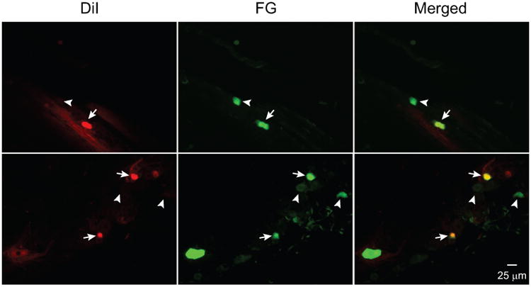
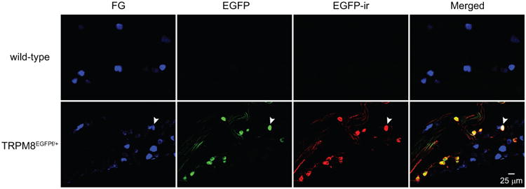
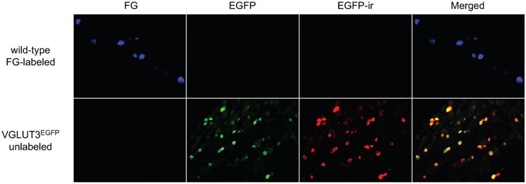
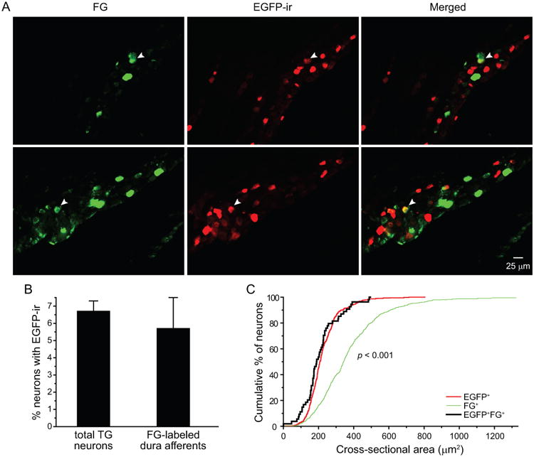
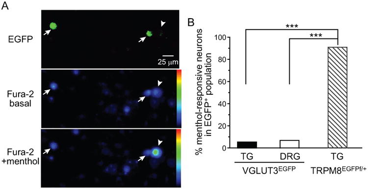
Methods: We used the fluorescent tracer Fluoro-Gold to retrogradely label dural afferent neurons in adult mice expressing enhanced green fluorescent protein in discrete subpopulations of trigeminal ganglion (TG) neurons. Mechanoreceptors with myelinated fibers were identified by NF200 immunoreactivity. We also conducted Ca2+ -imaging experiments to test the overlap between TRPM8 and VGLUT3 expression in mouse primary afferent neurons (PANs).
Results: The abundance of TRPM8-expressing neurons in dural afferent neurons was significantly lower than that in total TG neurons. The percentages of dural afferent neurons expressing VGLUT3 and NF200 were comparable to those of total TG neurons, respectively. TRPM8 agonist menthol evoked Ca2+ influx in less than 7% VGLUT3-expressing PANs in adult mice.
Conclusions: TG neurons expressing TRPM8, VGLUT3, and NF200 all innervate adult mouse dura. TRPM8 and VGLUT3 are expressed in distinct subpopulations of PANs in adult mice. These results provide an anatomical basis to investigate headache mechanisms in mouse models.
Keywords: NF200; TRPM8; VGlut3; dural afferent neurons; headache; migraine.
© 2017 American Headache Society.
Conflict of interest statement
Conflict of Interest: None
Figures


Similar articles
-
Tetrodotoxin-Sensitive Sodium Channels Mediate Action Potential Firing and Excitability in Menthol-Sensitive Vglut3-Lineage Sensory Neurons.J Neurosci. 2019 Sep 4;39(36):7086-7101. doi: 10.1523/JNEUROSCI.2817-18.2019. Epub 2019 Jul 12. J Neurosci. 2019. PMID: 31300524 Free PMC article.
-
Expression of the transient receptor potential channels TRPV1, TRPA1 and TRPM8 in mouse trigeminal primary afferent neurons innervating the dura.Mol Pain. 2012 Sep 12;8:66. doi: 10.1186/1744-8069-8-66. Mol Pain. 2012. PMID: 22971321 Free PMC article.
-
Function and postnatal changes of dural afferent fibers expressing TRPM8 channels.Mol Pain. 2015 Jun 26;11:37. doi: 10.1186/s12990-015-0043-0. Mol Pain. 2015. PMID: 26111800 Free PMC article.
-
The TRPM8 channel as a potential therapeutic target for bladder hypersensitive disorders.J Smooth Muscle Res. 2022;58(0):11-21. doi: 10.1540/jsmr.58.11. J Smooth Muscle Res. 2022. PMID: 35354708 Free PMC article. Review.
-
TRPM8 and Migraine.Headache. 2016 Oct;56(9):1406-1417. doi: 10.1111/head.12948. Epub 2016 Sep 16. Headache. 2016. PMID: 27634619 Free PMC article. Review.
Cited by
-
Tetrodotoxin-Sensitive Sodium Channels Mediate Action Potential Firing and Excitability in Menthol-Sensitive Vglut3-Lineage Sensory Neurons.J Neurosci. 2019 Sep 4;39(36):7086-7101. doi: 10.1523/JNEUROSCI.2817-18.2019. Epub 2019 Jul 12. J Neurosci. 2019. PMID: 31300524 Free PMC article.
-
Secretome as a Tool to Treat Neurological Conditions: Are We Ready?Int J Mol Sci. 2023 Nov 20;24(22):16544. doi: 10.3390/ijms242216544. Int J Mol Sci. 2023. PMID: 38003733 Free PMC article. Review.
-
TRP Channels as Potential Targets for Sex-Related Differences in Migraine Pain.Front Mol Biosci. 2018 Aug 14;5:73. doi: 10.3389/fmolb.2018.00073. eCollection 2018. Front Mol Biosci. 2018. PMID: 30155469 Free PMC article. Review.
-
Dysregulation of the peripheral glutamatergic system: A key player in migraine pathogenesis?Cephalalgia. 2021 Oct;41(11-12):1249-1261. doi: 10.1177/03331024211017882. Epub 2021 Jun 20. Cephalalgia. 2021. PMID: 34148407 Free PMC article. Review.
-
The neurotrophic factor artemin and its receptor GFRα3 mediate migraine-like pain via the ion channel TRPM8.bioRxiv [Preprint]. 2024 Sep 23:2024.09.09.611532. doi: 10.1101/2024.09.09.611532. bioRxiv. 2024. Update in: Cephalalgia. 2024 Nov;44(11):3331024241297679. doi: 10.1177/03331024241297679. PMID: 39314341 Free PMC article. Updated. Preprint.
References
-
- Goadsby PJ, Charbit AR, Andreou AP, Akerman S, Holland PR. Neurobiology of migraine. Neuroscience. 2009;161:327–341. - PubMed
-
- Pietrobon D, Moskowitz MA. Pathophysiology of migraine. Annual review of physiology. 2013;75:365–391. - PubMed
-
- Strassman AM, Raymond SA, Burstein R. Sensitization of meningeal sensory neurons and the origin of headaches. Nature. 1996;384:560–564. - PubMed
-
- Newsom J, Holt JL, Neubert JK, Caudle R, Ahn AH. A high density of TRPM8 expressing sensory neurons in specialized structures of the head; Society for Neuroscience Meeting Abstract; 2012.
MeSH terms
Substances
Grants and funding
LinkOut - more resources
Full Text Sources
Other Literature Sources
Miscellaneous
