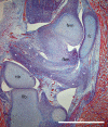
The fibular meniscus of the kangaroo as an adaptation against external tibial rotation during saltatorial locomotion
- PMID: 28925568
- PMCID: PMC5696135
- DOI: 10.1111/joa.12683
The fibular meniscus of the kangaroo as an adaptation against external tibial rotation during saltatorial locomotion
Abstract
The kangaroo knee is, as in other species, a complex diarthrodial joint dependent on interacting osseous, cartilaginous and ligamentous components for its stability. While principal load bearing occurs through the femorotibial articulation, additional lateral articulations involving the fibula and lateral fabella also contribute to the functional arrangement. Several fibrocartilage and ligamentous structures in this joint remain unexplained or have been misunderstood in previous studies. In this study, we review the existing literature on the structure of the kangaroo 'knee' before providing a new description of the gross anatomical and histological structures. In particular, we present strong evidence that the previously described 'femorofibular disc' is best described as a fibular meniscus on the basis of its gross and histological anatomy. Further, we found it to be joined by a distinct tendinous tract connecting one belly of the m. gastrocnemius with the lateral meniscus, via a hyaline cartilage cornu of the enlarged lateral fabella. The complex of ligaments connecting the fibular meniscus to the surrounding connective tissues and muscles appears to provide a strong resistance to external rotation of the tibia, via the restriction of independent movement of the proximal fibula. We suggest this may be an adaptation to resist the rotational torque applied across the joint during bipedal saltatory locomotion in kangaroos.
Keywords: cyamella; fabella; femorofibular joint; menisci; rotational torque.
© 2017 Anatomical Society.
Figures


Similar articles
-
Anatomy of the proximal tibiofibular joint.Knee Surg Sports Traumatol Arthrosc. 2006 Mar;14(3):241-9. doi: 10.1007/s00167-005-0684-z. Epub 2005 Dec 22. Knee Surg Sports Traumatol Arthrosc. 2006. PMID: 16374587
-
Anatomy of the anterolateral ligament of the knee.J Anat. 2013 Oct;223(4):321-8. doi: 10.1111/joa.12087. Epub 2013 Aug 1. J Anat. 2013. PMID: 23906341 Free PMC article.
-
Microstructural and compositional features of the fibrous and hyaline cartilage on the medial tibial plateau imply a unique role for the hopping locomotion of kangaroo.PLoS One. 2013 Sep 18;8(9):e74303. doi: 10.1371/journal.pone.0074303. eCollection 2013. PLoS One. 2013. PMID: 24058543 Free PMC article.
-
Meniscofibular ligament: how much do we know about this structure of the posterolateral corner of the knee: anatomical study and review of literature.Surg Radiol Anat. 2020 Oct;42(10):1203-1208. doi: 10.1007/s00276-020-02459-x. Epub 2020 Mar 29. Surg Radiol Anat. 2020. PMID: 32227270
-
The fibular collateral ligament of the knee: a detailed review.Clin Anat. 2014 Jul;27(5):789-97. doi: 10.1002/ca.22301. Epub 2013 Nov 19. Clin Anat. 2014. PMID: 24948572 Review.
Cited by
-
Intra-skeletal vascular density in a bipedal hopping macropod with implications for analyses of rib histology.Anat Sci Int. 2021 Jun;96(3):386-399. doi: 10.1007/s12565-020-00601-8. Epub 2021 Jan 22. Anat Sci Int. 2021. PMID: 33481185
-
Towards a Dynamic Model of the Kangaroo Knee for Clinical Insights into Human Knee Pathology and Treatment: Establishing a Static Biomechanical Profile.Biomimetics (Basel). 2019 Jul 25;4(3):52. doi: 10.3390/biomimetics4030052. Biomimetics (Basel). 2019. PMID: 31349696 Free PMC article.
References
-
- Alexander RM, Bennett MB (1987) Some principles of ligament function, with examples from the tarsal joints of the sheep (Ovis aries). J Zool 211, 487–504.
-
- Allen MJ, Houlton JEF, Adams SB, et al. (1998) The surgical anatomy of the stifle joint in sheep. Vet Surg 27, 596–605. - PubMed
-
- Andersson KI (2004) Elbow‐joint morphology as a guide to forearm function and foraging behaviour in mammalian carnivores. Zool J Linn Soc 142, 91–104.
MeSH terms
LinkOut - more resources
Full Text Sources
Other Literature Sources

