

A multiscale imaging and modelling dataset of the human inner ear
- PMID: 28925991
- PMCID: PMC5604133
- DOI: 10.1038/sdata.2017.132
A multiscale imaging and modelling dataset of the human inner ear
Abstract
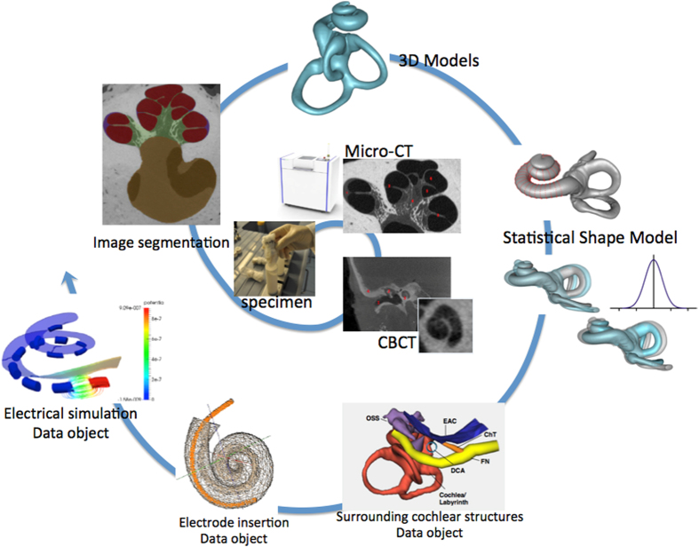
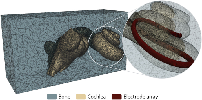
Understanding the human inner ear anatomy and its internal structures is paramount to advance hearing implant technology. While the emergence of imaging devices allowed researchers to improve understanding of intracochlear structures, the difficulties to collect appropriate data has resulted in studies conducted with few samples. To assist the cochlear research community, a large collection of human temporal bone images is being made available. This data descriptor, therefore, describes a rich set of image volumes acquired using cone beam computed tomography and micro-CT modalities, accompanied by manual delineations of the cochlea and sub-compartments, a statistical shape model encoding its anatomical variability, and data for electrode insertion and electrical simulations. This data makes an important asset for future studies in need of high-resolution data and related statistical data objects of the cochlea used to leverage scientific hypotheses. It is of relevance to anatomists, audiologists, computer scientists in the different domains of image analysis, computer simulations, imaging formation, and for biomedical engineers designing new strategies for cochlear implantations, electrode design, and others.
Conflict of interest statement
Scanco Medical AG has used its own scanners μCT 50 and μCT 100 for the acquisition of the μCT data.
Figures


References
Data Citations
-
- 2017. SICAS Medical Image Repository. https://doi.org/10.22016/smir.o.121756 - DOI
-
- 2017. SICAS Medical Image Repository. https://doi.org/10.22016/smir.o.121755 - DOI
-
- 2017. SICAS Medical Image Repository. https://doi.org/10.22016/smir.o.121754 - DOI
-
- 2017. SICAS Medical Image Repository. https://doi.org/10.22016/smir.o.121753 - DOI
-
- 2017. SICAS Medical Image Repository. https://doi.org/10.22016/smir.o.121752 - DOI
References
-
- Braun K., Böhnke F. & Stark T. Three-dimensional representation of the human cochlea using micro-computed tomography data: Presenting an anatomical model for further numerical calculations. Acta Otolaryngol. 132, 11 (2012). - PubMed
-
- Frijns J. H., Briaire J. J. & Grote J. J. The importance of human cochlear anatomy for the results of modiolus-hugging multichannel cochlear implants. Otol. Neurotol. 22, 340–349 (2001). - PubMed
-
- Bekesy G. Direct observation of the vibrations of the cochlear partition under a microscope. Acta Otolaryngol. 42, 197–201 (1952). - PubMed
Publication types
MeSH terms
LinkOut - more resources
Full Text Sources
Other Literature Sources
