. 2017 Sep 19;7(9):e1235.
doi: 10.1038/tp.2017.208.

A critical period for antidepressant-induced acceleration of neuronal maturation in adult dentate gyrus

Affiliations

A critical period for antidepressant-induced acceleration of neuronal maturation in adult dentate gyrus

I Åmellem et al. Transl Psychiatry. .

Abstract

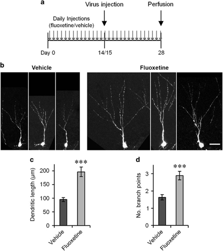
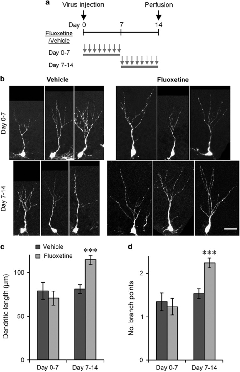
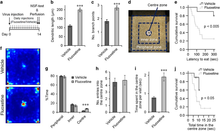
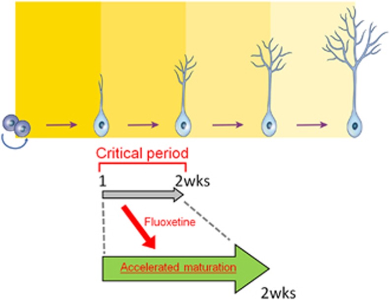
Selective serotonin reuptake inhibitors (SSRIs) are the most commonly used medications for mood and anxiety disorders, and adult neurogenesis in the dentate gyrus has been shown to be involved in the behavioral effects of SSRIs in mice. Studies have shown the varied effects of chronic treatment with SSRIs on adult neurogenesis. One such effect is the acceleration of neuronal maturation, which affects the functional integration of new neurons into existing neuronal circuitry. In this study, we labeled new neurons by using GFP-expressing retroviral vectors in mice and investigated the effect of an SSRI, fluoxetine, on these neurons at different time points after neuronal birth. Chronic treatment with fluoxetine accelerated the dendritic development of the newborn neurons and shifted the timing of the expression of the maturational marker proteins, doublecortin and calbindin. This accelerated maturation was observed even after sub-chronic treatment, only when fluoxetine was administered during the second week of neuronal birth. These results suggest the existence of a 'critical period' for the fluoxetine-induced maturation of new neurons. We propose that the modified functional integration of new neurons in the critical period may underlie the behavioral effects of fluoxetine by regulating anxiety-related decision-making processes.


Conflict of interest statement

The authors declare no conflict of interest.

Figures

Figure 1
Figure 1
Fluoxetine-induced transient increase in the dendritic arborization of new neurons. (a) Experimental time line. (b) Examples of GFP+ granule cells from the fluoxetine- or vehicle-treated mice 14, 21 and 28 days after virus injection. Scale bars, 25 μm (day 14), 50 μm (day 21 and 28). (c and d) Total dendritic length (c) and number of branch points (d) of GFP+ neurons on days 14, 21 and 28. ***P<0.005, two-tailed t-test with Bonaferroni correction.
Figure 2
Figure 2
The morphological effect on immature neurons is maintained after 4 weeks of fluoxetine treatment. (a) Experimental time line. (b) Example images of GFP+/Prox1+ neurons 2 weeks after virus injection. Scale bar, 25 μm. (c and d) Total dendritic length (c) and number of branch points (d) of GFP+/Prox1+ neurons on day 14. ***P<0.005, two-tailed t-test.
Figure 3
Figure 3
Fluoxetine treatment shifts maturational changes in the expression of DCX and calbindin in new neurons. The experimental time line is described in Figure 1a. (a) Representative images of two GFP+/Prox1+ cells each from the vehicle- and fluoxetine-treated mice on day 28. One of them expresses DCX. Red: DCX, Cyan: Prox1, Green: GFP. (b) Representative images of two GFP+/Prox1+ cells each from the vehicle- and fluoxetine-treated mice on day 28. Three of them express calbindin. Red: calbindin, Cyan: Prox1, Green: GFP. (c, d) Percentages of DCX+ cells in GFP+/Prox1+ cells (c) and calbindin+ cells in GFP+/Prox1+ cells (d) on days 14, 21, and 28. *P<0.05, Mann–Whitney U test with Bonaferroni correction. (e) Representative images of the granule cell layer from vehicle- and fluoxetine-treated mice. Red: DCX, Cyan: Prox1. (f) Representative images of the granule cell layer from the vehicle- and fluoxetine-treated mice. Red: calbindin, Cyan: Prox1. (g) Percentage of DCX+ cells and calbindin+ cells in Prox1+ cells on days 14, 21 and 28. Scale bars, 10 μm (a and b), 30 μm (e and f).
Figure 4
Figure 4
Fluoxetine-induced dendritic arborization requires treatment during the second week after neuronal birth. (a) Experimental time lines. (b) Example images of GFP+/Prox1+ neurons on day 14 with treatment on days 0–7 or days 7–14. Scale bar, 25 μm. (c) Total dendritic length (c) and number of branch points (d) of GFP+/Prox1+ neurons with treatment on days 0–7 or days 7–14. ***P<0.005, two-tailed t-test with Bonaferroni correction.
Figure 5
Figure 5
The morphological effect of fluoxetine treatment is associated with the behavioral effect in the novelty-suppressed feeding test. (a) Experimental time line. NSF-test: novelty-suppressed feeding test. (b and c) Total dendritic length (b) and number of branch points (c) of GFP+/Prox1+ neurons from the vehicle- and fluoxetine-treated mice. (d) Open field used for the novelty-suppressed feeding test and subdivision into the central, inner and peripheral zones. (e) Latency to eat the food pellet. The crosses indicate the time points at which data were censored because of the maximum time limit. (f) Percentage time spent in the central, inner and peripheral zones. (g) Color-coded average occupancy plots of the time that animals spent in individual positions. Warmer colors indicate a higher percentage of time spent in that position. There was no significant difference between the vehicle- and fluoxetine-treated mice in terms of the percentage time spent in the corners (P>0.05, two tailed t-test). (h) Number of entries into the central zone per 100 s. (i) Time spent in the central zone per visit. (j) Total time spent in the central zone. The crosses indicate the time points at which data were censored because the tests reached the maximum time limit. ***P<0.005, two-tailed t-test.
Figure 6
Figure 6
Schematic showing that fluoxetine treatment during the critical period of the second week after neuronal birth results in the accelerated maturation of new neurons.

References

    1. Kessler RC, Aguilar-Gaxiola S, Alonso J, Chatterji S, Lee S, Ormel J et al. The global burden of mental disorders: an update from the WHO World Mental Health (WMH) surveys. Epidemiol Psichiatria Soc 2009; 18: 23–33. - PMC - PubMed
    1. Smith K. Mental health: a world of depression. Nature 2014; 515: 181. - PubMed
    1. Blier P. The pharmacology of putative early-onset antidepressant strategies. Eur Neuropsychopharmacol: J Eur Coll Neuropsychopharmacol 2003; 13: 57–66. - PubMed
    1. Santarelli L, Saxe M, Gross C, Surget A, Battaglia F, Dulawa S et al. Requirement of hippocampal neurogenesis for the behavioral effects of antidepressants. Science 2003; 301: 805–809. - PubMed
    1. Surget A, Saxe M, Leman S, Ibarguen-Vargas Y, Chalon S, Griebel G et al. Drug-dependent requirement of hippocampal neurogenesis in a model of depression and of antidepressant reversal. Biol Psychiatry 2008; 64: 293–301. - PubMed

Publication types

MeSH terms

LinkOut - more resources