. 2018 Mar;53(3):135-142.
doi: 10.1097/RLI.0000000000000418.

Photon-Counting Computed Tomography for Vascular Imaging of the Head and Neck: First In Vivo Human Results

Photon-Counting Computed Tomography for Vascular Imaging of the Head and Neck: First In Vivo Human Results

Rolf Symons et al. Invest Radiol. 2018 Mar.

Abstract

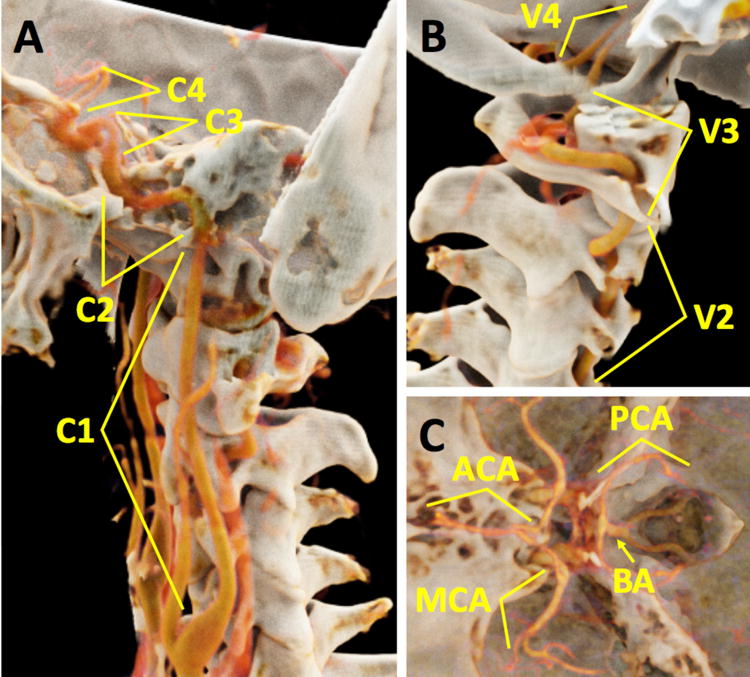
Purpose: The purpose of this study was to evaluate image quality of a spectral photon-counting detector (PCD) computed tomography (CT) system for evaluation of major arteries of the head and neck compared with conventional single-energy CT scans using energy-integrating detectors (EIDs).

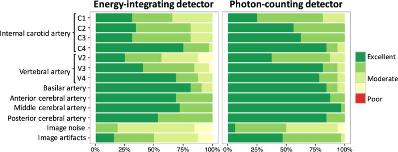
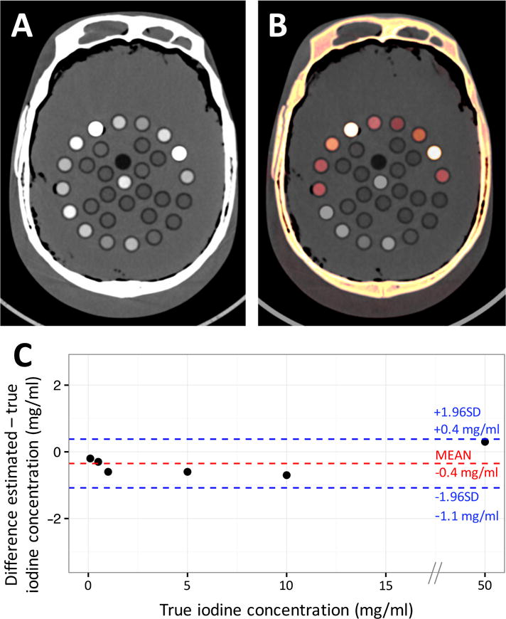
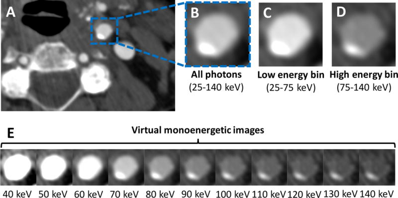
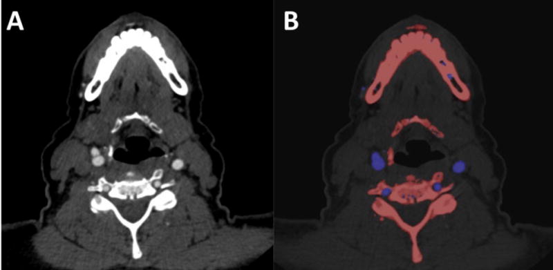
Methods: In this institutional review board-approved study, 16 asymptomatic subjects (7 men) provided informed consent and received both PCD and EID contrast-enhanced CT scans of the head and neck (mean age, 58 years; range, 46-75 years). Tube settings were (EID: 120 kVp/160 mA vs PCD: 140 kVp/108 mA) for all volunteers. Quantitative analysis included measurements of mean attenuation, image noise, and contrast-to-noise ratio (CNR). Spectral PCD data were used to reconstruct virtual monoenergetic images and iodine maps. A head phantom was used to validate iodine concentration measurements in PCD images only. Two radiologists blinded to detector type independently scored the image quality of different segments of the arteries, as well as diagnostic acceptability, image noise, and severity of artifacts of the PCD and EID images. Reproducibility was assessed with intraclass correlation coefficient. Linear mixed models that account for within-subject correlation of analyzed arterial segments were used. Linear regression and Bland-Altman analysis with 95% limits of agreement were used to calculate the accuracy of material decomposition.

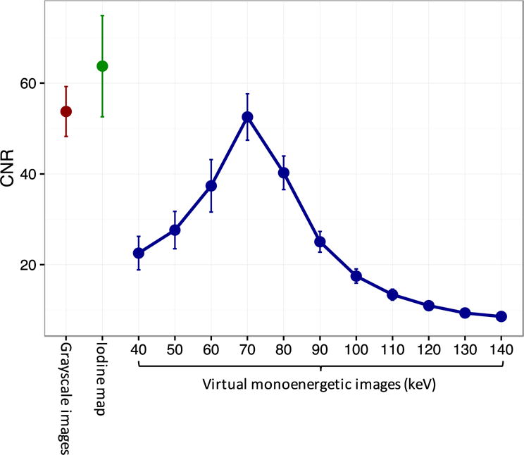
Results: Photon-counting detector image quality scores were significantly higher compared with EID image quality scores with lower image noise (P < 0.01) and less image artifacts (P < 0.001). Photon-counting detector image noise was 9.1% lower than EID image noise (8.0 ± 1.3 HU vs 8.8 ± 1.5 HU, respectively, P < 0.001). Arterial segments showed artifacts on EID images due to beam hardening that were not present on PCD images. On PCD images of the head phantom, there was excellent correlation (R = 0.998) between actual and calculated iodine concentrations without significant bias (bias: -0.4 mg/mL [95% limits of agreements: -1.1 to 0.4 mg/mL]). Iodine maps had 20.7% higher CNR compared with nonspectral PCD (65.2 ± 9.0 vs 54.0 ± 4.5, P = 0.01), and virtual monoenergetic image at 70 keV showed similar CNR to nonspectral images (52.6 ± 4.2 vs 54.0 ± 4.5, P = 0.39).

Conclusions: Photon-counting CT has the potential to improve the image quality of carotid and intracranial CT angiography compared with single-energy EID CT.


Figures

Figure 1
Figure 1
3D reconstructions in a 47-year-old male subject from a photon counting CT scan demonstrate the vessel segments that were analyzed in this study: (A) internal carotid artery (ICA) cervical segment (C1), ICA petrous segment (C2), ICA cavernous segment (C3), and ICA supraclinoid segment (C4); (B) vertebral artery (VA) foraminal segment (V2), VA atlantic segment (V3), and VA intradural segment (V4); (C) basilar artery (BA), anterior, middle and posterior cerebral arteries (ACA, MCA, and PCA). There is an intimate relationship between multiple vessel segments and surrounding bone (e.g., ICA C2, ICA C3, and VA V2). 3D volume rendering technique was performed on Cinematic Rendering v1.0 (Siemens Healthcare GmbH, Forchheim, Germany).
Figure 2
Figure 2
Qualitative image scores for the energy-integrating detector (EID) and photon-counting detector (PCD) images of the internal carotid artery (ICA) cervical segment (C1), ICA petrous segment (C2), ICA cavernous segment (C3), ICA supraclinoid segment (C4), vertebral artery (VA) foraminal segment (V2), VA atlantic segment (V3), VA intradural segment (V4), basilar artery (BA), anterior cerebral artery (ACA), middle cerebral artery (MCA), and posterior cerebral artery (PCA). PCD scores were significantly higher for the ICA C2, ICA C3, V2, V3, ACA, MCA, and PCA segments (all P<0.05) with significantly lower subjective image noise (P<0.01) and less image artifacts (P<0.001).
Figure 3
Figure 3
Example energy-integrating detector (EID) (A) and photon-counting detector (PCD) (B) curved multiplanar reconstructions (MPR) of the internal carotid artery (ICA) in a 55-year-old female. Artifactual areas of low density within the ICA petrous segment (C2) which may be mistaken for pathology are seen on the EID images but no on the PCD images (arrows). (C) Graph demonstrates changes in Hounsfield Unit (HU) values (means ± standard errors) in the ICA and MCA for all subjects. HU values are normalized to the cervical ICA (C1) segment. Mean EID HU values in the ICA C2, ICA C3, and ICA C4 segments were 26.3 HU, 30.4 HU, and 14.6 HU lower than HU values in the ICA C1 segment (all P<0.001), whereas mean PCD HU values did not change significantly (all P>0.05).
Figure 4
Figure 4
Example photon-counting detector (PCD) grayscale image (A) and iodine map overlay image (B) of the head phantom with multiple calibrated test tubes filled with fat (vegetable oil), water, hydroxyapatite, and different aqueous solutions of iodine (50, 10, 5, 1, 0.5, and 0.1 mg/ml) and ferrous sulfate (15, 10, and 5 mg/ml). (C) Bland-Altman plot shows agreement between the true iodine concentration and those estimated through PCD material decomposition. No significant bias was found with narrow 95% limits of agreement (LOAs) (bias: −0.4 mg/ml [95% LOAs: −1.1;0.4 mg/ml]).
Figure 5
Figure 5
(A) Example grayscale photon-counting detector (PCD) image reconstructed from all detected photons (25–140 keV) at the level of the proximal cervical internal carotid artery (ICA C1) in a 73-year-old female demonstrates mild eccentric calcified plaque. (B) Zoomed-in image of the ICA C1 with corresponding low (C) and high (D) energy bin images. Based on the specific behavior of materials at different photon energy levels, images can be decomposed into their constituent materials (e.g., iodine versus calcium) and virtual monoenergetic images (E) can be reconstructed to enhance facilitate plaque detection [window center: 145; window width: 800].
Figure 6
Figure 6
(A) Example photon-counting detector (PCD) grayscale images at the level of the cervical internal carotid artery (ICA C1) in a 75-year-old female. (B) Material classification overlay image enhances the differentiation between the arteries (iodine, blue) and the adjacent bone (calcium, red).
Figure 7
Figure 7
Contrast-to-noise-ratio (CNR) for differentiation between the cervical segment of the internal carotid artery (ICA C1) and the surrounding fat. Iodine map CNR was 20.7% higher than grayscale image CNR and could be used to enhance vessel differentiation and to quantify arterial enhancement (mean ± standard error: 65.2±9.0 vs 54.0±4.5, respectively, P=0.01).

References

    1. Borisch I, Boehme T, Butz B, et al. Screening for carotid injury in trauma patients: image quality of 16-detector-row computed tomography angiography. Acta radiol. 2007;48(7):798–805. - PubMed
    1. Malhotra AK, Camacho M, Ivatury RR, et al. Computed tomographic angiography for the diagnosis of blunt carotid/vertebral artery injury: a note of caution. Ann Surg. 2007;246(4):632–643. - PubMed
    1. Saba L, Sanfilippo R, Pirisi R, et al. Multidetector-row CT angiography in the study of atherosclerotic carotid arteries. Neuroradiology. 2007;49(8):623–637. - PubMed
    1. Brooks RA, Di Chiro G. Beam hardening in x-ray reconstructive tomography. Phys Med Biol. 1976;21(3):390. - PubMed
    1. Zatz LM, Alvarez RE. An Inaccuracy in Computed Tomography: The Energy Dependence of CT Values. Radiology. 1977;124(1):91–97. - PubMed

Publication types

MeSH terms