

Functional characterization of zebrafish orthologs of the human Beta 3-Glucosyltransferase B3GLCT gene mutated in Peters Plus Syndrome
- PMID: 28926587
- PMCID: PMC5604996
- DOI: 10.1371/journal.pone.0184903
Functional characterization of zebrafish orthologs of the human Beta 3-Glucosyltransferase B3GLCT gene mutated in Peters Plus Syndrome
Abstract
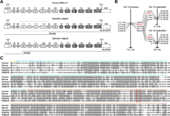
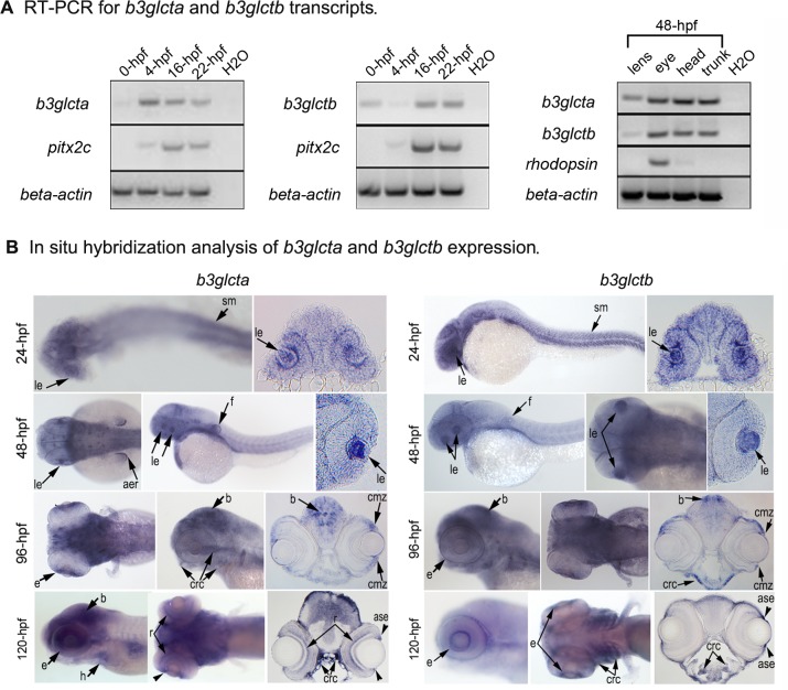
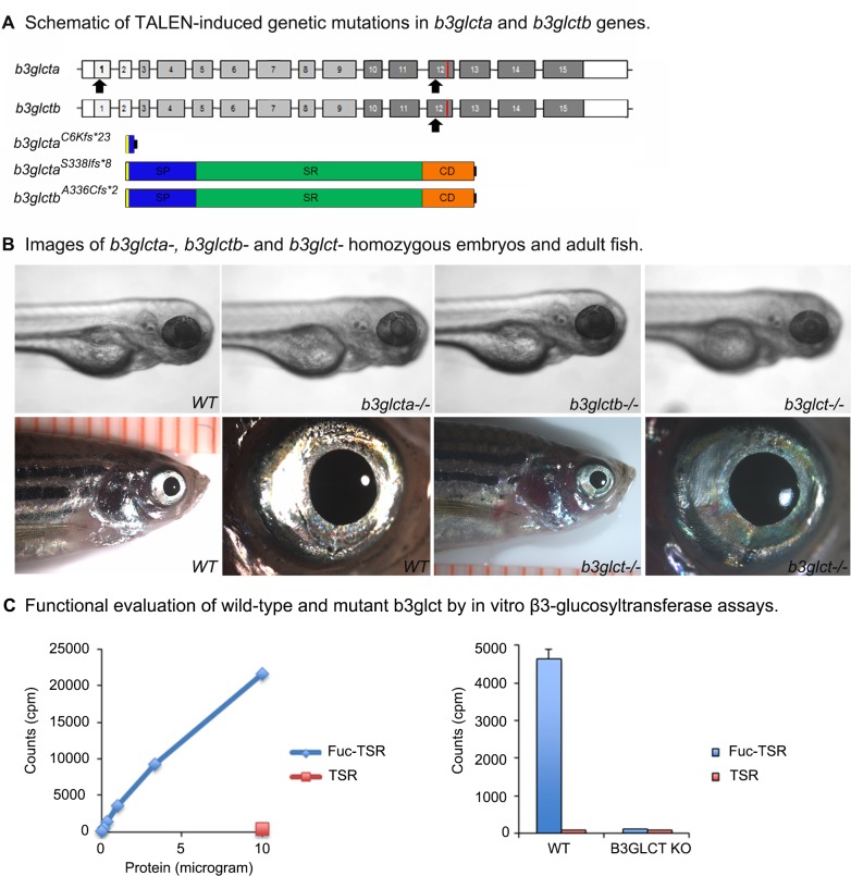
Peters Plus Syndrome (PPS) is a rare autosomal recessive disease characterized by ocular defects, short stature, brachydactyly, characteristic facial features, developmental delay and other highly variable systemic defects. Classic PPS is caused by loss-of-function mutations in the B3GLCT gene encoding for a β3-glucosyltransferase that catalyzes the attachment of glucose via a β1-3 glycosidic linkage to O-linked fucose on thrombospondin type 1 repeats (TSRs). B3GLCT was shown to participate in a non-canonical ER quality control mechanism; however, the exact molecular processes affected in PPS are not well understood. Here we report the identification and characterization of two zebrafish orthologs of the human B3GLCT gene, b3glcta and b3glctb. The b3glcta and b3glctb genes encode for 496-aa and 493-aa proteins with 65% and 57% identity to human B3GLCT, respectively. Expression studies demonstrate that both orthologs are widely expressed with strong presence in embryonic tissues affected in PPS. In vitro glucosylation assays demonstrated that extracts from wildtype embryos contain active b3glct enzyme capable of transferring glucose from UDP-glucose to an O-fucosylated TSR, indicating functional conservation with human B3GLCT. To determine the developmental role of the zebrafish genes, single and double b3glct knockouts were generated using TALEN-induced genome editing. Extracts from double homozygous b3glct-/- embryos demonstrated complete loss of in vitro b3glct activity. Surprisingly, b3glct-/- homozygous fish developed normally. Transcriptome analyses of head and trunk tissues of b3glct-/- 24-hpf embryos identified 483 shared differentially regulated transcripts that may be involved in compensation for b3glct function in these embryos. The presented data show that both sequence and function of B3GLCT/b3glct genes is conserved in vertebrates. At the same time, complete b3glct deficiency in zebrafish appears to be inconsequential and possibly compensated for by a yet unknown mechanism.
Conflict of interest statement
Figures
Similar articles
-
ADAMTS9 and ADAMTS20 are differentially affected by loss of B3GLCT in mouse model of Peters plus syndrome.Hum Mol Genet. 2019 Dec 15;28(24):4053-4066. doi: 10.1093/hmg/ddz225. Hum Mol Genet. 2019. PMID: 31600785 Free PMC article.
-
Peters plus syndrome mutations affect the function and stability of human β1,3-glucosyltransferase.J Biol Chem. 2021 Jul;297(1):100843. doi: 10.1016/j.jbc.2021.100843. Epub 2021 May 28. J Biol Chem. 2021. PMID: 34058199 Free PMC article.
-
Peters plus syndrome mutations disrupt a noncanonical ER quality-control mechanism.Curr Biol. 2015 Feb 2;25(3):286-295. doi: 10.1016/j.cub.2014.11.049. Epub 2014 Dec 24. Curr Biol. 2015. PMID: 25544610 Free PMC article.
-
Ocular Phenotype of Peters-Plus Syndrome.Cornea. 2022 Feb 1;41(2):219-223. doi: 10.1097/ICO.0000000000002889. Cornea. 2022. PMID: 34629439 Review.
-
Peters'-plus syndrome is a congenital disorder of glycosylation caused by a defect in the beta1,3-glucosyltransferase that modifies thrombospondin type 1 repeats.Ann Med. 2009;41(1):2-10. doi: 10.1080/07853890802301975. Ann Med. 2009. PMID: 18720094 Review.
Cited by
-
ADAMTS9 and ADAMTS20 are differentially affected by loss of B3GLCT in mouse model of Peters plus syndrome.Hum Mol Genet. 2019 Dec 15;28(24):4053-4066. doi: 10.1093/hmg/ddz225. Hum Mol Genet. 2019. PMID: 31600785 Free PMC article.
-
O-fucosylation stabilizes the TSR3 motif in thrombospondin-1 by interacting with nearby amino acids and protecting a disulfide bond.J Biol Chem. 2022 Jun;298(6):102047. doi: 10.1016/j.jbc.2022.102047. Epub 2022 May 18. J Biol Chem. 2022. PMID: 35597280 Free PMC article.
-
Integrated genome-based probiotic relevance and safety evaluation of Lactobacillus reuteri PNW1.PLoS One. 2020 Jul 20;15(7):e0235873. doi: 10.1371/journal.pone.0235873. eCollection 2020. PLoS One. 2020. PMID: 32687505 Free PMC article.
-
The first review on prenatal drug exposure and ocular malformation occurrence.Front Pediatr. 2024 Sep 4;12:1379875. doi: 10.3389/fped.2024.1379875. eCollection 2024. Front Pediatr. 2024. PMID: 39296666 Free PMC article. Review.
-
Hydrocephalus in mouse B3glct mutants is likely caused by defects in multiple B3GLCT substrates in ependymal cells and subcommissural organ.Glycobiology. 2021 Sep 9;31(8):988-1004. doi: 10.1093/glycob/cwab033. Glycobiology. 2021. PMID: 33909046 Free PMC article.
References
-
- Maillette de Buy Wenniger-Prick LJ, Hennekam RC: The Peters' plus syndrome: a review. Ann Genet 2002, 45(2):97–103. - PubMed
-
- Reis LM, Tyler RC, Abdul-Rahman O, Trapane P, Wallerstein R, Broome D, et al.: Mutation analysis of B3GALTL in Peters Plus syndrome. Am J Med Genet A 2008, 146A(20):2603–2610. doi: 10.1002/ajmg.a.32498 - DOI - PMC - PubMed
-
- Weh E, Reis LM, Tyler RC, Bick D, Rhead WJ, Wallace S, et al.: Novel B3GALTL mutations in classic Peters plus syndrome and lack of mutations in a large cohort of patients with similar phenotypes. Clin Genet 2014, 86(2):142–148. doi: 10.1111/cge.12241 - DOI - PMC - PubMed
-
- Harissi-Dagher M, Colby K: Anterior segment dysgenesis: Peters anomaly and sclerocornea. Int Ophthalmol Clin 2008, 48(2):35–42. doi: 10.1097/IIO.0b013e318169526c - DOI - PubMed
-
- Lesnik Oberstein SA, Kriek M, White SJ, Kalf ME, Szuhai K, den Dunnen JT, et al.: Peters Plus syndrome is caused by mutations in B3GALTL, a putative glycosyltransferase. Am J Hum Genet 2006, 79(3):562–566. doi: 10.1086/507567 - DOI - PMC - PubMed
MeSH terms
Substances
Supplementary concepts
Grants and funding
LinkOut - more resources
Full Text Sources
Other Literature Sources
Molecular Biology Databases
