Review
. 2017 Sep 18;6(3):43.
doi: 10.3390/pathogens6030043.

Emerging Roles of Heparanase in Viral Pathogenesis

Affiliations
Review

Emerging Roles of Heparanase in Viral Pathogenesis

Neel Thakkar et al. Pathogens. .

Abstract

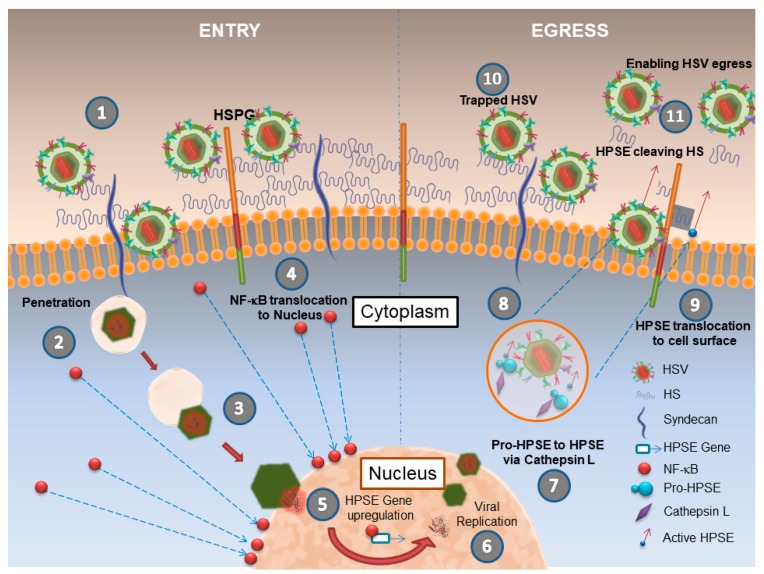
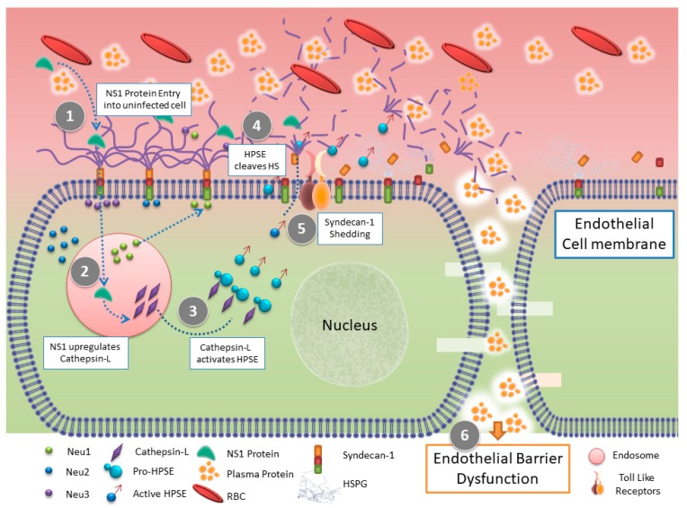
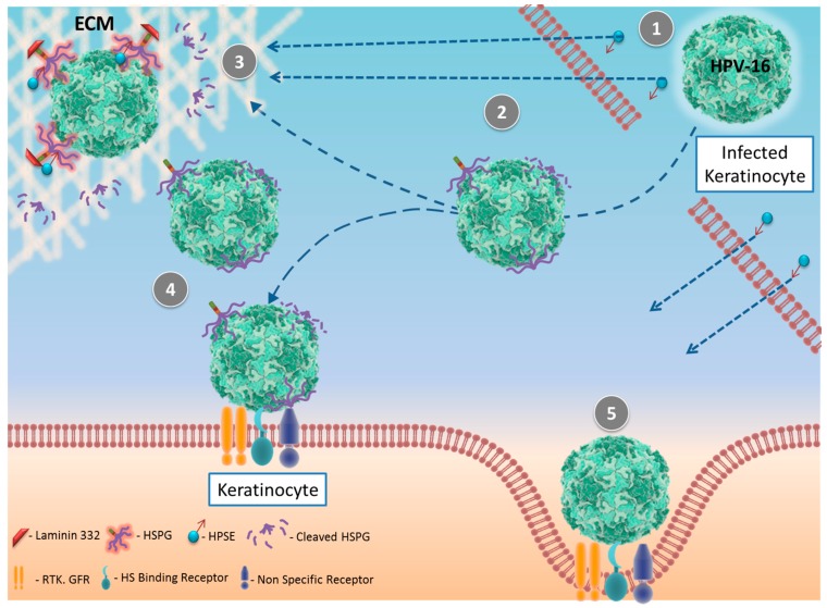
Heparan sulfate (HS) is ubiquitously expressed on mammalian cells. It is a polysaccharide that binds growth factors, cytokines, and chemokines, and thereby controls several important physiological functions. Ironically, many human pathogens including viruses interact with it for adherence to host cells. HS functions can be regulated by selective modifications and/or selective cleavage of the sugar chains from the cell surface. In mammals, heparanase (HPSE) is the only known enzyme capable of regulating HS functions via a selective endoglycosidase activity that cleaves polymeric HS chains at internal sites. During homeostasis, HPSE expression and its endoglycosidase activity are tightly regulated; however, under stress conditions, including infection, its expression may be upregulated, which could contribute directly to the onset of several disease pathologies. Here we focus on viral infections exemplified by herpes simplex virus, dengue virus, human papillomavirus, respiratory syncytial virus, adenovirus, hepatitis C virus, and porcine respiratory and reproductive syncytial virus to summarize recent advances in understanding the highly significant, but emerging roles, of the enzyme HPSE in viral infection, spread and pathogenesis.

Keywords: heparan sulfate; heparanase; herpes simplex virus.


Conflict of interest statement

The authors declare no conflicts of interest.

Figures

Figure 1
Figure 1
Schematics of HPSE’s role in HSV infection. (1) Virions attach to HSPGs including syndecans on a host cell. (2) Capsid penetration occurs at the plasma membrane (via membrane fusion or endocytosis). (3) The capsid is released into the cytoplasm, which reaches the nuclear membrane for docking and release of viral DNA in the nucleus for replication. (4) Sensing viral invasion, NF-kB translocates to the nucleus and (5) signals HPSE overexpression. (6) New capsids containing viral genomes mature and bud out of the nucleus. (7–8) Enveloped capsids with viral glycoproteins are exocytosed. The same vesicles are also suggested to carry Pro-HPSE, which is eventually activated by cathepsin-L. (9) The enzymatically active HPSE then translocates to the cell surface where it cleaves off heparan sulfate chains (10) to help release any surface bound virions. (11) removal of heparan sulfate enables unrestricted release of HSV-1 virions.
Figure 2
Figure 2
Schematic of HPSE functions in epithelial barrier dysfunction during DENV infection. (1) DENV NS1 protein from an infected cell binds to the surface of an uninfected human pulmonary microvascular endothelial cells (HPMEC) and upregulates the expression and translocation of endothelial sialidases to the cell membrane.(2) DENV NS1 enhances the activity of cathepsin-L and the expression of pro-HPSE. (3) Cathepsin L. activity on pro-HPSE converts it into an active form (4) which leads to the cleavage of heparan sulfate chains on the endothelial glycocalyx layer (EGL) and results in (5) The shedding of Syndecan-1. (6) Puerta-Guardo et al suggest that, together all these processes lead to EGL disruption on endothelial cell surface in-turn resulting in endothelial barrier dysfunction and hyperpermeability that occurs in severe dengue disease.
Figure 3
Figure 3
Schematics of HPSE’s role in HPV 16 infection. (1) Mammalian heparanase from surrounding cells or extrinsically added bacterial heparinase III (2) cleaves heparan sulfate proteoglycan (HSPG) chains attached to the surface of HPV16 or (3) reaches the extracellular membrane (ECM) where HPV16 particles attached to the ECM via HSPG are cleaved and released. (4) Released HPV16 viruses are infectious and (5) infect surrounding keratinocytes during a wound healing in vivo model, as shown by Surviladze et al [39].

References

    1. Sarrazin S., Lamanna W.C., Esko J.D. Heparan sulfate proteoglycans. Cold Spring Harb. Perspect. Biol. 2011;3:1–33. doi: 10.1101/cshperspect.a004952. - DOI - PMC - PubMed
    1. Rivara S., Milazzo F.M., Giannini G. Heparanase: A rainbow pharmacological target associated to multiple pathologies including rare diseases. Future Med. Chem. 2016;8:647–680. doi: 10.4155/fmc-2016-0012. - DOI - PubMed
    1. Agelidis A.M., Shukla D. Cell entry mechanisms of HSV: What we have learned in recent years. Future Virol. 2015;10:1145–1154. doi: 10.2217/fvl.15.85. - DOI - PMC - PubMed
    1. Kreuger J., Kjellén L. Heparan Sulfate Biosynthesis: Regulation and Variability. J. Histochem. Cytochem. 2012;60:898–907. doi: 10.1369/0022155412464972. - DOI - PMC - PubMed
    1. Shukla D., Spear P.G. Herpesviruses and heparan sulfate: An intimate relationship in aid of viral entry. J. Clin. Investig. 2001;108:503–510. doi: 10.1172/JCI200113799. - DOI - PMC - PubMed

LinkOut - more resources