. 2017 Sep;14(3):3755-3759.
doi: 10.3892/ol.2017.6576. Epub 2017 Jul 15.

An early superficial non-ampullary duodenal tumor cured with endoscopic submucosal dissection: A case report

Affiliations

An early superficial non-ampullary duodenal tumor cured with endoscopic submucosal dissection: A case report

Yi Wei Fu et al. Oncol Lett. 2017 Sep.

Abstract

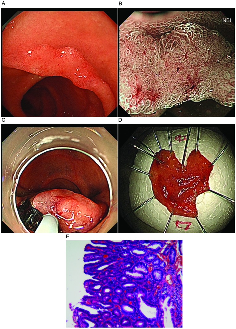
Early superficial non-ampullary duodenal tumors are particularly rare, the clinical manifestations, including typical endoscopic or imaging features, and treatment methods are not well-characterized. The present case report describes a case of an asymptomatic 74-year-old male who presented to the Taizhou People's Hospital (Taizhou, China) for a regular health screening, where a primary superficial non-ampullary duodenal tumor was identified. Upper endoscopy revealed ~1.2 cm lesion in the second portion of the duodenum. Chromoscopy and magnification endoscopy indicated an early cancer characteristic. Subsequent endoscopic submucosal dissection was performed to remove the lesion. Histopathology validated that the lesion was a high-grade intro-epithelial neoplasm without lymph node or blood vessel invasion.

Keywords: endoscopic submucosal dissection; high grade intro-epithelial neoplasm; magnifying endoscopy; narrow band imaging; superficial non-ampullary duodenal tumor.


Figures

Figure 1.
Figure 1.
An early superficial non-ampullary duodenal tumor. (A) A flat elevated lesion (1.2 cm) was visible in the second portion of the duodenum, using conventional endoscopy with white light imaging. (B) Surface pattern was preserved in one region and is absent in another region, and an unclassified vascular pattern was exhibited following magnifying endoscopy with narrow band imaging. (C) The endoscopic submucosal dissection method was performed to cure the lesion. (D) The whole lesion was cut from duodenum. (E) The lesion was diagnosed as a category 4/5 tumor by hematoxylin and eosin staining (magnification, ×100).

References

    1. Alwmark A, Andersson A, Lasson A. Primary carcinoma of the duodenum. Ann Surg. 1980;191:13–18. doi: 10.1097/00000658-198001000-00003. - DOI - PMC - PubMed
    1. Yokoyama T, Saito D, Kondo H, Kido M, Hosokawa K, Shirao K, Yokota T, Yamaguchi H, Oguro Y, Ishikawa T, et al. Endoscopic diagnosis of malignant lesions of the duodenum. Stomach Int. 1993;28:641–649.
    1. Vasen HF, Möslein G, Alonso A, Aretz S, Bernstein I, Bertario L, Blanco I, Bülow S, Burn J, Capella G, et al. Guidelines for the clinical management of familial adenomatous polyposis (FAP) Gut. 2008;57:704–713. doi: 10.1136/gut.2007.136127. - DOI - PubMed
    1. Perzin KH, Bridge MF. Adenomas of the small intestine: A clinicopathologic review of 51 cases and a study of their relationship to carcinoma. Cancer. 1981;48:799–819. doi: 10.1002/1097-0142(19810801)48:3<799::AID-CNCR2820480324>3.0.CO;2-Q. - DOI - PubMed
    1. Bakaeen FG, Murr MM, Sarr MG, Thompson GB, Farnell MB, Nagorney DM, Farley DR, van Heerden JA, Wiersema LM, Schleck CD, Donohue JH. What prognostic factors are important in duodenal adenocarcinoma? Arch Surg. 2000;135:635–641. doi: 10.1001/archsurg.135.6.635. - DOI - PubMed