. 2017 Sep 30;40(9):643-654.
doi: 10.14348/molcells.2017.0030. Epub 2017 Sep 20.

The Roles of the SNARE Protein Sed5 in Autophagy in Saccharomyces cerevisiae

Affiliations

The Roles of the SNARE Protein Sed5 in Autophagy in Saccharomyces cerevisiae

Shenshen Zou et al. Mol Cells. .

Abstract

Autophagy is a degradation pathway in eukaryotic cells in which aging proteins and organelles are sequestered into double-membrane vesicles, termed autophagosomes, which fuse with vacuoles to hydrolyze cargo. The key step in autophagy is the formation of autophagosomes, which requires different kinds of vesicles, including COPII vesicles and Atg9-containing vesicles, to transport lipid double-membranes to the phagophore assembly site (PAS). In yeast, the cis-Golgi localized t-SNARE protein Sed5 plays a role in endoplasmic reticulum (ER)-Golgi and intra-Golgi vesicular transport. We report that during autophagy, sed5-1 mutant cells could not properly transport Atg8 to the PAS, resulting in multiple Atg8 dots being dispersed into the cytoplasm. Some dots were trapped in the Golgi apparatus. Sed5 regulates the antero-grade trafficking of Atg9-containing vesicles to the PAS by participating in the localization of Atg23 and Atg27 to the Golgi apparatus. Furthermore, we found that overexpression of SFT1 or SFT2 (suppressor of sed5 ts) rescued the autophagy defects in sed5-1 mutant cells. Our data suggest that Sed5 plays a novel role in autophagy, by regulating the formation of Atg9-containing vesicles in the Golgi apparatus, and the genetic interaction between Sft1/2 and Sed5 is essential for autophagy.

Keywords: Atg9-containing vesicles; Sed5; Sft1/2; autophagy; golgi apparatus.


Figures

Fig. 1
Fig. 1
The Cvt pathway and autophagy were defective in sed5-1 mutant cells. (A) GFP-Atg8 processing was blocked in sed5-1 mutant cells. GFP-Atg8 was integrated into the chromosomes of WT, atg1Δ and sed5-1 mutant cells. For each strain, the cells were grown at 26°C to an early log phase in YPD medium and then either washed and transferred to SD-N medium at 26°C or pretreated for 30 min at 34°C before being washed and transferred to SD-N medium at 34°C. The cultures were collected at 0 h, 1 h, 2 h, and 4 h after SD-N treatment. GFP-Atg8 cleavage was determined in the cell lysates using immunoblot analysis with anti-GFP antibodies (Pgk1 served as a loading control). The band intensities of blots from three independent experiments were quantified with IMAGEJ software (National Institutes of Health), and the percentages of GFP-Atg8 and GFP were plotted. The graphs represent the average of three experiments. **P < 0.01; N.S., no significance. (B) Ape1 maturation was blocked in sed5-1 mutant cells under non-starvation (YPD medium) and starvation (SD-N medium) conditions. Cells were grown as described in the Materials and Methods, and Ape1 processing was determined by immunoblot analysis with anti-Ape1 antibodies. Quantification of the Ape1 processing assay was performed using IMAGEJ software. The graphs represent the average of three experiments. *p < 0.05; **P < 0.01. (C) Pho8Δ60 alkaline phosphatase (ALP) activity in the sed5-1 mutant. ALP activity was determined in the lysates of wild-type, atg1Δ (as a negative control), and sed5-1 mutant cells. This assay was performed as described in the Materials and Methods. Error bars represent the SD of three independent experiments. **P < 0.01.
Fig. 2
Fig. 2
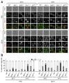
Atg8 was mislocalized in the Cvt pathway and autophagy in sed5-1 mutant cells. (A) Abnormal Atg8 localization in the Cvt pathway and autophagy in sed5-1 mutant cells. WT and mutant cells were tagged with GFP-Atg8 integration plasmids. The vacuolar membranes were stained with FM 4-64 as described in the Materials and Methods. The experiments were repeated twice and representative results from a single experiment are presented. Scale bars, 5 μm. (B) Quantification of the percentage of cells with GFP-Atg8 dots in three categories: 0, 1 and multiple dots (≥ 2 dots per cell). At least 300 cells were counted in at least three fields for each strain. Error bars represent standard deviation.
Fig. 3
Fig. 3
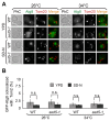
Atg8 was not trapped in mitochondria. (A) GFP-Atg8 and Tom20-RFP were integrated into the chromosomes of WT and sed5-1 cells. The cells were grown and examined as described in the Materials and Methods. Scale bars, 5 μm. (B) Quantitation of the percentage of GFP-Atg8 dots colocalized with the Tom20-RFP puncta in (A). At least 300 cells were counted in at least three fields for each strain. Error bars represent SD. N.S., no significance.
Fig. 4
Fig. 4
(A) Most multiple GFP-Atg8 dots were mislocalized with the PAS in the Cvt pathway and autophagy in sed5-1 mutants. GFP-Atg8 and RFP-Ape1 were integrated into the chromosomes of WT, atg1Δ and sed5-1 mutant cells. The cells were grown and examined as described in the Materials and Methods. Scale bars, 5 μm. (B) Quantitation of the percentage of GFP-Atg8 dots colocalized with the RFP-Ape1 puncta in (A). At least 300 cells were counted in at least three fields for each strain. Error bars represent SD. **P < 0.01.
Fig. 5
Fig. 5
Sed5 acts in autophagy under starvation condition. Multiple GFP-Atg8 dots disappeared in the sed5-1 mutants in which the ATG5 gene was deleted, but not in those in which ATG9, ATG14 and TRS85 were deleted, under the starvation condition at both 26°C and 34°C. The cells were cultured and detected as described in the Materials and Methods. Scale bars, 5 μm. The percentage of cells with GFP-Atg8 dots in three categories: 0, 1 and multiple dots ( 2 dots per cell), were quantified. At least 300 cells were counted in at least three fields for each strain. Error bars represent SD.
Fig. 6
Fig. 6
Multiple GFP-Atg8 dots were trapped in the Golgi apparatus of sed5-1 mutants in the Cvt pathway and autophagy. (A–C) WT and sed5-1 mutant cells co-expressing GFP-Atg8 with DsRed-HDEL, Sec13-tdTomato or Cop1-tdTomato were treated as described in the Materials and Methods. Experiments were repeated 3 times, and the results shown are from a single experiment. Scale bars, 5 μm. Arrows indicate colocalization between GFP-Atg8 and Cop1-tdTomato. The percentage of the percent of GFP-Atg8 dots colocalized with the red puncta in (A–C) was quantified. At least 300 cells were counted in at least three fields for each strain. Error bars represent standard deviation. **P < 0.01; N.S., no significance.
Fig. 7
Fig. 7
The Atg9 complex could not formed in the Golgi apparatus of sed5-1 mutants during autophagy. (A–C) WT and sed5-1 mutant cells co-expressing Cop1-tdTomato with Atg9-3XGFP, Atg23-2XGFP and Atg27-2XGFP were treated as described in the Materials and Methods. Experiments were repeated 3 times, and the results shown are from a single experiment. Scale bars, 5 μm. Arrows indicate the colocalization of green and red puncta. The percentage of Atg9-3XGFP that colocalized with Cop1-tdTomato in (A) was quantified. The percentage of cells with Atg27-2XGFP dots in two categories, 0 and ≥ 1 dot in (B), was quantified. The percentage of cells with GFP-Atg8 dots in three categories, 0, 1 and multiple dots (≥ 2 dots per cell) in (C), was quantified. All the statistics included more than 300 cells in at least three fields for each strain. Error bars represent standard deviation. N.S., no significance.
Fig. 8
Fig. 8
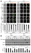
Sft1 and Sft2 suppressed the autophagy defects in sed5-1 mutant cells. (A) Sft1 and Sft2 suppressed the defective transport of GFP-Atg8 in the sed5-1 mutant strain. Empty vector (pRS424), Sft1 or Sft2 was transformed into the sed5-1 strain. The cells were grown as described in the Materials and Methods. Experiments were repeated 3 times, and the results shown are from a single experiment. Quantification of the percentage of cells with GFP-Atg8 either outside the vacuole or diffused in the vacuole is shown. At least 300 cells were counted in at least 3 fields for each strain. Error bars represent SD. (B) GFP-Atg8 degradation was recovered when Sft1 and Sft2 were overexpressed in the sed5-1 mutant strain. The GFP-Atg8 processing assay was performed. Quantification of the GFP-Atg8 processing assay was performed using IMAGEJ software. The graphs represent the average of three experiments. *p < 0.05; **P < 0.01; N.S., no significance.
Fig. 9
Fig. 9
Model for Sed5 during autophagy. Sed5 was required for Atg23 and Atg27 localization to the Golgi apparatus, wherein the Atg9 complex (including Atg9, Atg27 and Atg23) forms and regulates Atg9-containing vesicles formation and transport to the PAS.

Similar articles

Cited by

References

    1. Baba M., Osumi M., Scott S.V., Klionsky D.J., Ohsumi Y. Two distinct pathways for targeting proteins from the cytoplasm to the vacuole/lysosome. J Cell Biol. 1997;139:1687–1695. - PMC - PubMed
    1. Backues S.K., Orban D.P., Bernard A., Singh K., Cao Y., Klionsky D.J. Atg23 and Atg27 act at the early stages of Atg9 trafficking in S. cerevisiae. Traffic. 2015;16:172–190. - PMC - PubMed
    1. Banfield D.K., Lewis M.J., Pelham H.R. A SNARE-like protein required for traffic through the Golgi complex. Nature. 1995;375:806–809. - PubMed
    1. Barlowe C., Orci L., Yeung T., Hosobuchi M., Hamamoto S., Salama N., Rexach M.F., Ravazzola M., Amherdt M., Schekman R. COPII, a membrane coat formed by Sec proteins that drive vesicle budding from the endoplasmic reticulum. Cell. 1994;77:895–907. - PubMed
    1. Cai H., Yu S., Menon S., Cai Y., Lazarova D., Fu C., Reinisch K., Hay J.C., Ferro-Novick S. TRAPPI tethers COPII vesicles by binding the coat subunit Sec23. Nature. 2007;445:941–944. - PubMed

MeSH terms

LinkOut - more resources