

Beyond classic dermoscopic patterns of dermatofibromas: a prospective research study
- PMID: 28927449
- PMCID: PMC5605998
- DOI: 10.1186/s13256-017-1429-6
Beyond classic dermoscopic patterns of dermatofibromas: a prospective research study
Abstract
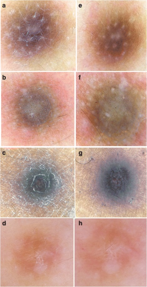
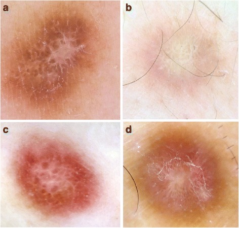
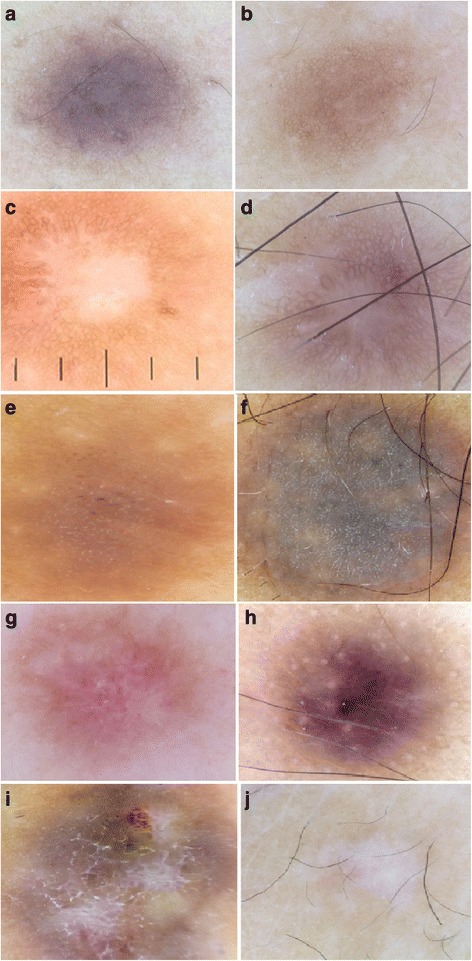
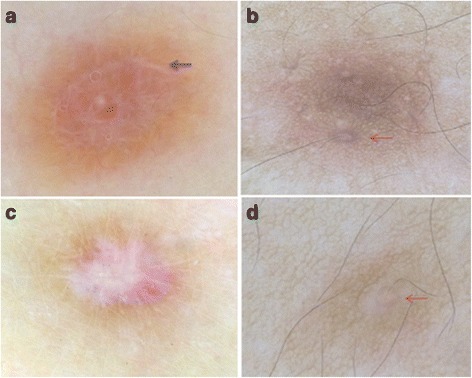
Background: The usual stereotypical dermoscopic pattern associated with dermatofibromas is a pigment network and central white patch. However, this pattern may be difficult to diagnose in some variant cases. We aimed to describe dermoscopic patterns of dermatofibroma according to its histopathological subtypes, with special emphasis on new and rare dermoscopic features.
Methods: This prospective study, which was conducted between September 2015 and May 2016 in the Department of Dermatology, University Hospital Hassan II, Fez, Morocco, included 100 cases of dermatofibroma confirmed on clinical and histological grounds. Each lesion was scored for classic, previously reported, or new dermoscopic features.
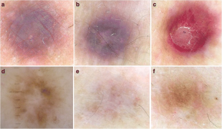
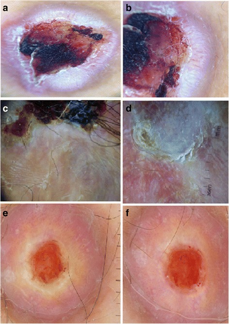
Results: All our Moroccan patients had a dark skin phototype (Fitzpatrick scale types IV and V). A total of 14 morphological dermoscopic structures were distinguished, and 17 dermoscopic patterns were observed, with the most common pattern being the central white patch and peripheral pigment network (21%). New patterns observed in our study were a white ring around an ulceration (6%), a pigment network with a pigmented ring around follicular openings (2%), and a discreet peripheral network and starlike white patch (3%). A patchy network with white patches was significantly noted in atrophic dermatofibroma (p = 0.01); vascularization was described in both aneurysmal and hemosiderotic dermatofibromas (p = 0.002); and a white ring around an ulceration was noted in aneurysmal dermatofibroma (p < 0.001).
Conclusions: We provide a description of dermoscopic patterns of dermatofibroma according to its histological subtypes in a dark skin phototype, along with a new report of a white ring around an ulceration as a significant pattern in aneurysmal dermatofibroma.
Keywords: Dermatofibroma; Dermoscopy; New dermoscopic patterns; Rare dermoscopic patterns; Variants.
Conflict of interest statement
Ethics approval and consent to participate
Ethical approval was obtained from the ethics committee of the University Hospital Center Hassan II in Fez, Morocco, and all the subjects were informed of the conditions related to the study.
Consent for publication
Written informed consent was obtained from the patients for publication of this report and any accompanying images. A copy of the written consent is available for review by the Editor-in-Chief of this journal.
Competing interests
The authors declare that they have no competing interests.
Publisher’s Note
Springer Nature remains neutral with regard to jurisdictional claims in published maps and institutional affiliations.
Figures
Comment in
-
Dermoscopic patterns and features of dermatofibroma in darker skin phototypes.Int J Dermatol. 2022 Aug;61(8):e282-e286. doi: 10.1111/ijd.15979. Epub 2021 Nov 24. Int J Dermatol. 2022. PMID: 34817858 No abstract available.
Similar articles
-
Dermoscopy of lipidised dermatofibroma: A morphological study of 13 cases.Australas J Dermatol. 2019 May;60(2):e127-e131. doi: 10.1111/ajd.12956. Epub 2018 Nov 26. Australas J Dermatol. 2019. PMID: 30478949
-
Different dermoscopic faces of dermatofibromas.J Am Acad Dermatol. 2007 Sep;57(3):401-6. doi: 10.1016/j.jaad.2006.10.984. Epub 2007 Jun 8. J Am Acad Dermatol. 2007. PMID: 17560684
-
Dermoscopic findings of haemosiderotic and aneurysmal dermatofibroma: report of six patients.Br J Dermatol. 2006 Feb;154(2):244-50. doi: 10.1111/j.1365-2133.2005.06844.x. Br J Dermatol. 2006. PMID: 16433792
-
Unique histopathologic features of the eyelid dermatofibroma.Orbit. 2019 Aug;38(4):274-278. doi: 10.1080/01676830.2018.1513045. Epub 2018 Sep 5. Orbit. 2019. PMID: 30183445 Review.
-
Atrophic Dermatofibroma: A Comprehensive Literature Review.Dermatol Ther (Heidelb). 2019 Sep;9(3):449-468. doi: 10.1007/s13555-019-0309-y. Epub 2019 Jul 23. Dermatol Ther (Heidelb). 2019. PMID: 31338755 Free PMC article. Review.
Cited by
-
Clinical and Dermoscopic Patterns of Basal Cell Carcinoma and Its Mimickers in Skin of Color: A Practical Summary.Medicina (Kaunas). 2024 Aug 24;60(9):1386. doi: 10.3390/medicina60091386. Medicina (Kaunas). 2024. PMID: 39336428 Free PMC article. Review.
-
Dermoscopic patterns of dermatofibroma in 72 Chinese patients.Chin Med J (Engl). 2019 Sep 5;132(17):2121-2122. doi: 10.1097/CM9.0000000000000406. Chin Med J (Engl). 2019. PMID: 31425360 Free PMC article. No abstract available.
-
A case of atrophic dermatofibroma: a possible role of matrix metalloproteinase-2.An Bras Dermatol. 2024 Mar-Apr;99(2):284-286. doi: 10.1016/j.abd.2022.06.011. Epub 2023 Nov 27. An Bras Dermatol. 2024. PMID: 38016889 Free PMC article. No abstract available.
-
International Dermoscopy Society (IDS) Criteria for Skin Tumors: Validation for Skin of Color Through a Delphi Expert Consensus by the "Imaging in Skin of Color" IDS Task Force.Dermatol Pract Concept. 2023 Jan 1;13(1):e2023067. doi: 10.5826/dpc.1301a67. Dermatol Pract Concept. 2023. PMID: 36892378 Free PMC article.
-
A Secure Framework toward IoMT-Assisted Data Collection, Modeling, and Classification for Intelligent Dermatology Healthcare Services.Contrast Media Mol Imaging. 2022 Jun 29;2022:6805460. doi: 10.1155/2022/6805460. eCollection 2022. Contrast Media Mol Imaging. 2022. PMID: 35845738 Free PMC article.
References
-
- Crisan D, Gheuca Solovastru L, Crisan M, Badea R. Cutaneous histiocytoma – histological and imaging correlations. A case report. Med Ultrason. 2014;16(3):268–70. - PubMed
-
- Parish LC, Yazdanian S, Lambert WC, Lambert PC. Dermatofibroma: a curious tumor. Skinmed. 2012; 10(5):268-70. - PubMed
-
- Parish LC, Yazdanian S, Lambert WC, Lambert PC. Dermatofibroma: a curious tumor. Skinmed. 2012;10(5):268–70. - PubMed
MeSH terms
LinkOut - more resources
Full Text Sources
Other Literature Sources
Medical
