. 2017 Sep 19;10(1):430.
doi: 10.1186/s13071-017-2367-2.

Microarchitecture of the tsetse fly proboscis

Affiliations

Microarchitecture of the tsetse fly proboscis

Wendy Gibson et al. Parasit Vectors. .

Abstract

Background: Tsetse flies (genus Glossina) are large blood-sucking dipteran flies that are important as vectors of human and animal trypanosomiasis in sub-Saharan Africa. Tsetse anatomy has been well described, including detailed accounts of the functional anatomy of the proboscis for piercing host skin and sucking up blood. The proboscis also serves as the developmental site for the infective metacyclic stages of several species of pathogenic livestock trypanosomes that are inoculated into the host with fly saliva. To understand the physical environment in which these trypanosomes develop, we have re-examined the microarchitecture of the tsetse proboscis.

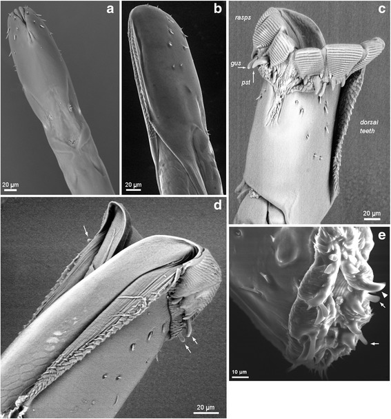
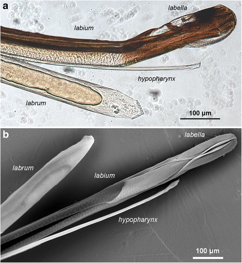
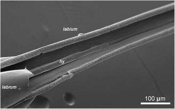
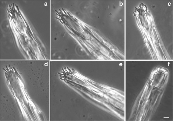
Results: We examined proboscises from male and female flies of Glossina pallidipes using light microscopy and scanning electron microscopy (SEM). Each proboscis was removed from the fly head and either examined intact or dissected into the three constituent components: Labrum, labium and hypopharynx. Our light and SEM images reaffirm earlier observations that the tsetse proboscis is a formidably armed weapon, well-adapted for piercing skin, and provide comparative data for G. pallidipes. In addition, the images reveal that the hypopharynx, the narrow tube that delivers saliva to the wound site, ends in a remarkably ornate and complex structure with around ten finger-like projections, each adorned with sucker-like protrusions, contradicting previous descriptions that show a simple, bevelled end like a hypodermic needle. The function of the finger-like projections is speculative; they appear to be flexible and may serve to protect the hypopharynx from influx of blood or microorganisms, or control the flow of saliva. Proboscises were examined after colonisation by Trypanosoma congolense savannah. Consistent with the idea that colonisation commences in the region nearest the foregut, the highest densities of trypanosomes were found in the region of the labrum proximal to the bulb, although high densities were also found in other regions of the labrum. Trypanosomes were visible through the thin wall of the hypopharynx by both light microscopy and SEM.

Conclusions: We highlight the remarkable architecture of the tsetse proboscis, in particular the intricate structure of the distal end of the hypopharynx. Further work is needed to elucidate the function of this intriguing structure.

Keywords: Blood-sucking; Glossina; Haematophagous; Hypopharynx; Labellum; Labium; Labrum; Proboscis; Trypanosoma congolense; Tsetse.


Conflict of interest statement

Ethics approval and consent to participate

Not applicable

Consent for publication

Not applicable

Competing interests

The authors declare that they have no competing interests.

Publisher’s Note

Springer Nature remains neutral with regard to jurisdictional claims in published maps and institutional affiliations.

Figures

Fig. 1
Fig. 1
Skin piercing machinery - light microscopy. The tip of the Glossina pallidipes proboscis showing the arrays of rasps and teeth used to penetrate through the skin to find blood. Light microscopy images of the tip of the proboscis, inverted (a) and everted (b). The dark pigmented region is on the ventral side of the labella and there is a long array of teeth on the dorsal side. These dorsal teeth are on the left in the lateral view shown in a; the rasps near the tip of the proboscis are clearly visible through the labellum wall, with gustatory sensilla and denticles protruding slightly from the tip. In b the everted labella are seen in ventral view, with dorsal teeth visible on both sides. Both sets of rasps, each subdivided into three, are visible, and now the gustatory sensilla (gus) and prostomal teeth are exposed in a ring at the base of the rasps
Fig. 2
Fig. 2
Skin piercing machinery - scanning electron microscopy. The tip of the Glossina pallidipes proboscis showing the arrays of rasps and teeth used to penetrate through the skin. Scanning electron microscopy images of the tip of the proboscis, inverted (a, b), everted (c, d) and partially everted (e). a Ventral view showing the small array of ventral teeth where the two labella meet centrally. b Dorso-lateral view showing the dorsal teeth in the groove formed by the closely opposed labella. c Everted labella in ventral view. The sets of rasps, each subdivided into three, are prominent; the small array of ventral teeth can be seen where the two labella meet centrally, as well as one of the larger arrays of dorsal teeth on the right-hand labellum. The gustatory sensilla (gus) and prestomal teeth (pst) are exposed at the base of the rasps. d Everted labella in dorsal view revealing the split between the labella on the dorsal side. Rasps and dorsal teeth are visible, and three of the eight gustatory sensilla are indicated (arrows). e Partially everted labella; ventral surface is at the top. Rasps visible within; two gustatory sensilla (arrows) and one prostomal tooth (arrowhead) are indicated
Fig. 3
Fig. 3
Component parts of the proboscis. The proboscis of Glossina pallidipes separated into its three component parts: labrum, labium and hypopharynx. a Light microscopy. b SEM, dorsal view of labium. The labrum interlocks with the labium approximately 400 μm from its distal tip, so that a continuous food canal is formed. The tip of the hypopharynx lies beyond the tip of the labrum, but does not reach the tip of the proboscis formed by the labella
Fig. 4
Fig. 4
Labial groove. SEM image of proboscis with part of labrum removed showing the position of the hypopharynx (hy) within the groove in the labium. The lateral flanges of the hypopharynx can be clearly seen
Fig. 5
Fig. 5
Distal tip of the hypopharynx. Structure of the hypopharynx tip. a-f Distal tip of hypopharynx by light microscopy. The hypopharynx ends in approximately ten finger-like projections of variable length. In f, the fingers are not obvious and appear to be retracted or curled up; this was the appearance in about one third of specimens examined. The opening of the salivary duct can be seen clearly in a. Scale-bars: 2 μm
Fig. 6
Fig. 6
Microarchitecture of the hypopharynx tip. SEM images of the distal tip of the hypopharynx showing the complex structure of the finger-like projections. Five different specimens are shown (a-e). The opening of the salivary duct can be seen clearly in d. In e the tip of the hypopharynx is resting on the labium; each lateral flange ends in a broad tip. Scale-bars: a-d, 2 μm; e, 5 μm
Fig. 7
Fig. 7
Trypanosome-infected proboscises. SEM images of tsetse mouthparts infected with Trypanosoma congolense savannah. a Distal end of hypopharynx. Trypanosomes can be seen densely packed inside the lumen. b Part of hypopharynx overlaying the labrum. Trypanosomes are visible on the internal wall of the labrum, and also as silhouettes within the hypopharynx. c Cluster of trypanosomes attached to the wall of the labrum close to a hair-like mechanoreceptor. Scale-bars: a, 10 μm; b, 20 μm; c, 5 μm

References

    1. Buxton PA. The natural history of tsetse flies. Memoir 10 London School of Hygiene and Tropical Medicine. London: HK Lewis; 1955.
    1. Rogers DJ, Robinson TP. Tsetse distribution. In: Maudlin I, Holmes PH, Miles MA, editors. The Trypanosomiases. Wallingford: CAB International; 2004. pp. 139–179.
    1. Krenn HW, Aspock H. Form, function and evolution of the mouthparts of blood-feeding Arthropoda. Arthropod Struct Dev. 2012;41:101–118. doi: 10.1016/j.asd.2011.12.001. - DOI - PubMed
    1. Weitz B. The feeding habits of Glossina. Bull WHO. 1963;28:711–729. - PMC - PubMed
    1. Bitome-Essono P-Y, Ollomo B, Arnathau C, Durand P, Mokoudoum ND, Yacka-Mouele Y, et al. Tracking zoonotic pathogens using blood-sucking flies as ‘flying syringes’. elife. 2017;6:e22069. doi: 10.7554/eLife.22069. - DOI - PMC - PubMed

MeSH terms

LinkOut - more resources