Review
. 2017 Oct;42(10):824-843.
doi: 10.1016/j.tibs.2017.06.008. Epub 2017 Sep 17.

Biochemistry of Mitochondrial Coenzyme Q Biosynthesis

Affiliations
Review

Biochemistry of Mitochondrial Coenzyme Q Biosynthesis

Jonathan A Stefely et al. Trends Biochem Sci. 2017 Oct.

Abstract

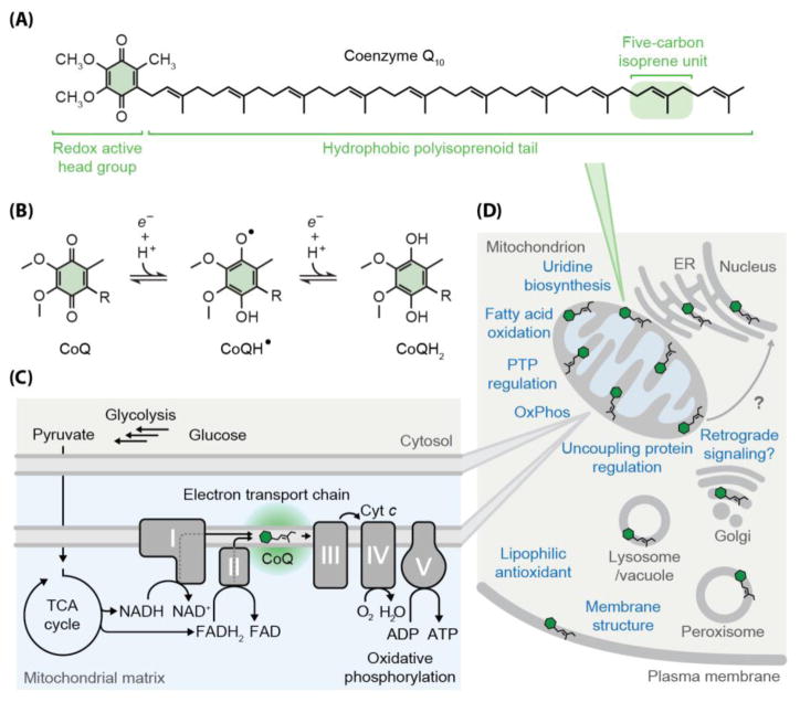
Coenzyme Q (CoQ, ubiquinone) is a redox-active lipid produced across all domains of life that functions in electron transport and oxidative phosphorylation and whose deficiency causes human diseases. Yet, CoQ biosynthesis has not been fully defined in any organism. Several proteins with unclear molecular functions facilitate CoQ biosynthesis through unknown means, and multiple steps in the pathway are catalyzed by currently unidentified enzymes. Here we highlight recent progress toward filling these knowledge gaps through both traditional biochemistry and cutting-edge 'omics' approaches. To help fill the remaining gaps, we present questions framed by the recently discovered CoQ biosynthetic complex and by putative biophysical barriers. Mapping CoQ biosynthesis, metabolism, and transport pathways has great potential to enhance treatment of numerous human diseases.

Keywords: CoQ-synthome; biosynthesis; coenzyme Q; complex Q; lipids; metabolon; mitochondria; mitochondrial disease; oxidative phosphorylation; protein complex; ubiquinone.


Figures

Figure I
Figure I. CoQ Deficiency in Human Health and Disease
(A) Network of phenotypes and genes associated with primary CoQ deficiency. Lines indicate phenotypes reported to be caused by mutations in the indicate gene(s). (B) Diseases, conditions, drugs, and genes linked to secondary CoQ deficiency. (C) Candidate disease genes for potential primary CoQ deficiencies that have not yet been reported to exist in patients. “(?)” indicates an unproven ortholog relationship to a yeast gene that has been linked to CoQ biosynthesis. Additional uncharacterized (orphan) genes may also be linked to CoQ biosynthesis (e.g., the gene that encodes the unidentified C1 hydroxylase).
Figure I
Figure I. Strategies for Treatment of CoQ Deficiency
(A) Efficient uptake of exogenous CoQ into rat tissues after intraperitoneal injection is limited to white blood cells, liver, and spleen. Lesser uptake occurs in other tissues. Data in figure from Bentinger et al [178]. (B) Exogenous CoQ must be transported across multiple lipid membranes and multiple aqueous compartments to reach the mitochondrial inner membrane, but the mechanisms facilitating this CoQ transport are unclear. (C) An example of the “CoQ intermediate bypass strategy” for treating select CoQ deficiencies. 4-hydroxybenzoate (4-HB) is the natural CoQ head group precursor. 2,4-dihydroxybenzoate (2,4-DHB) be used as an alternative head group precursor to bypass a defect in COQ7 [181].
Figure I
Figure I. A Multi-Omic Approach to Mitochondrial Biochemistry
Overview of methods used for multi-omic investigation of mitochondrial pathways, using CoQ biosynthesis as a key example. The cartoon indicates gaps in knowledge (?) that were recently filled using a multi-omic approach (Hfd1p) [34, 42] or that are targets for linking a currently undefined mitochondrial uncharacterized protein (MXP) to CoQ biosynthesis.
Figure 1
Figure 1. Chemical and Biological Functions of Coenzyme Q
(A) Chemical structure of coenzyme Q10 (CoQ10). (B) Reduction-oxidation reactions of the CoQ head group. “R” indicates the polyisoprenoid tail. (C) Cartoon indicating the central role of CoQ in the electron transport chain and mitochondrial oxidative phosphorylation (OxPhos). I–V, OxPhos complexes I–V; Cyt c, cytochrome c; TCA cycle, tricarboxylic acid cycle. (D) Overview of the widespread cellular functions of CoQ. Within mitochondria, CoQ supports and/or regulates the mitochondrial permeability transition pore (PTP) [10], mitochondrial uncoupling proteins [9], uridine biosynthesis [7], fatty acid oxidation [8], and OxPhos. More broadly, CoQ functions as a lipophilic antioxidant [–14] and a membrane stabilizer [15]. In plants, plastoquinone functions as a retrograde signal from chloroplasts to the nucleus [16], suggesting that mitochondrial CoQ could play an analogous role.
Figure 2
Figure 2. Current Model for the Eukaryotic CoQ Biosynthesis Pathway
Scheme of eukaryotic CoQ biosynthesis with currently unidentified enzymes indicated by question marks. The primary CoQ pathway, which is conserved from yeast to humans, is depicted. A secondary CoQ pathway in yeast uses para-aminobenzoate (pABA) as the head group precursor instead of 4-hydroxybenzoate (4-HB) [39, 40], and Coq6p and Coq9p subsequently support C4-deamination via C4-hydroxylation [64, 140]. ‘R’ indicates the polyisoprenoid tail (Figure 1A), which would likely be anchored in the mitochondrial inner membrane. “+” symbol by the arrow from COQ8 and COQ9 indicates a hypothesized supportive role for the indicated reaction. AADAT, mitochondrial alpha-aminoadipate aminotransferase; ALDH3A1, aldehyde dehydrogenase 3A1; FDXR, adrenodoxin reductase; FDX2 (FDX1L), mitochondrial ferredoxin 2 (ferredoxin 1-like); PDSS1, prenyl (decaprenyl) diphosphate synthase subunit 1; TAT, tyrosine aminotransferase; DMAPP, dimethylallyl pyrophosphate; GPP, geranyl pyrophosphate; FPP, farnesyl pyrophosphate; IPP, isopentenyl pyrophosphate; 4-HPP, 4-hydroxyphenylpyruvate; 4-HBz, 4-hydroxybenzaldehyde; PPHB, polyprenyl-hydroxybenzoate; PPDHB, polyprenyl-dihydroxybenzoate; PPVA, polyprenyl-vanillic acid; DDMQ, demethoxy-demethyl-coenzyme Q; DMQ, demethoxy-coenzyme Q; DMeQ, demethyl-coenzyme Q.
Figure 3
Figure 3. Models Framing Outstanding Questions and Knowledge Gaps in CoQ Biochemistry
(A) Model framing questions about complex Q assembly, regulation of CoQ biosynthesis, and mechanisms for CoQ transport. (B) Model for a biophysical barrier to accessing lipophilic CoQ intermediates, highlighting the currently unclear mechanism for solving this problem. CoQ and hydrophobic CoQ intermediates are predicted to reside near the lipid bilayer midplane (i) with some movement (gray arrows) toward the bilayer surface (ii), but not beyond the glycerol backbones [129, 130]. Biochemical mechanisms for moving CoQ intermediates past the layer of glycerol backbones and polar lipid head groups are unclear. (C) Models framing unanswered questions about complex Q biochemistry. (D) Summary of the currently incomplete state of complex Q structural biology. X-ray structures have been reported for Coq5p (PDB IDs 4OBW and 4OBX) [105], COQ8A (ADCK3) (PDB IDs 4PED and 5I35) [86] [87], and COQ9 (PBD ID 4RHP) [116].

References

    1. Crane FL, et al. Isolation of a quinone from beef heart mitochondria. Biochim Biophys Acta. 1957;25:220–221. - PubMed
    1. Morton RA. Ubiquinone. Nature. 1958;182:1764–1767. - PubMed
    1. Lester RL, et al. Coenzyme Q: a new group of quinones. Journal of the American Chemical Society. 1958;80:4751–4752.
    1. Wolf DEHCH, Trenner NR, Arison BH, Shunk CH, Linn BO, McPherson JF, Folkers K. Coenzyme Q. I. Structure Studies on the Coenzyme Q Group. J Am Chem Soc. 1958;80:4752.
    1. Mitchell P. Protonmotive redox mechanism of the cytochrome b-c1 complex in the respiratory chain: protonmotive ubiquinone cycle. FEBS Lett. 1975;56:1–6. - PubMed

Publication types