. 2017 Dec 8;121(12):1370-1378.
doi: 10.1161/CIRCRESAHA.117.311456. Epub 2017 Sep 19.

PDK4 Inhibits Cardiac Pyruvate Oxidation in Late Pregnancy

Affiliations

PDK4 Inhibits Cardiac Pyruvate Oxidation in Late Pregnancy

Laura X Liu et al. Circ Res. .

Abstract

Rationale: Pregnancy profoundly alters maternal physiology. The heart hypertrophies during pregnancy, but its metabolic adaptations, are not well understood.

Objective: To determine the mechanisms underlying cardiac substrate use during pregnancy.

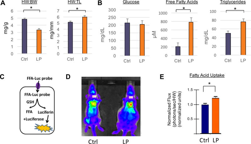
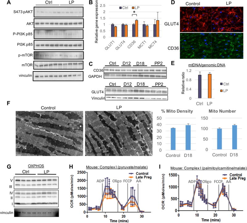
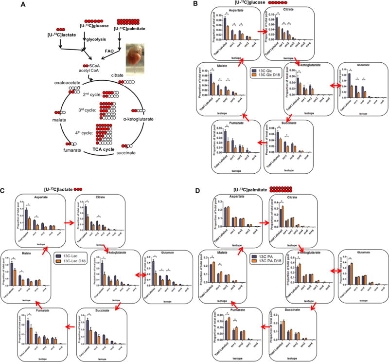
Methods and results: We use here 13C glucose, 13C lactate, and 13C fatty acid tracing analyses to show that hearts in late pregnant mice increase fatty acid uptake and oxidation into the tricarboxylic acid cycle, while reducing glucose and lactate oxidation. Mitochondrial quantity, morphology, and function do not seem altered. Insulin signaling seems intact, and the abundance and localization of the major fatty acid and glucose transporters, CD36 (cluster of differentiation 36) and GLUT4 (glucose transporter type 4), are also unchanged. Rather, we find that the pregnancy hormone progesterone induces PDK4 (pyruvate dehydrogenase kinase 4) in cardiomyocytes and that elevated PDK4 levels in late pregnancy lead to inhibition of PDH (pyruvate dehydrogenase) and pyruvate flux into the tricarboxylic acid cycle. Blocking PDK4 reverses the metabolic changes seen in hearts in late pregnancy.

Conclusions: Taken together, these data indicate that the hormonal environment of late pregnancy promotes metabolic remodeling in the heart at the level of PDH, rather than at the level of insulin signaling.

Keywords: glucose; heart; metabolism; mitochondria; pregnancy; progesterone.


Figures

Figure 1
Figure 1. Cardiac fatty acid uptake and accumulation increases in late gestational mice
(A) Morphometric parameters including heart weight to body weight (HW:BW) and heart weight to tibia length (HW:TL) ratios in non-pregnant controls (n=5, blue) and late pregnant mice (n=5, orange). (B) Plasma glucose, free fatty acid, and triglyceride concentrations in late pregnant and non- pregnant mice (n≥5 for all groups). (C) Diagram of bioluminescence detection of free fatty acid probe conjugated to luciferin (FFA- Luc) upon uptake into mouse hearts. (D–E) In vivo bioluminescence image of non-pregnant (D, left) and late pregnant mouse hearts (D, right), and quantification (E) of photon flux at time of saturation (n≥6 for all groups). All data are represented as mean ±SEM; *, p < 0.05 compared with control.
Figure 2
Figure 2. Isolated hearts from late gestational mice exhibit endogenous substrate switch of relative decreased oxidation of glucose and lacate, and increased oxidation of fatty acid
(A) Schematic of the formation of different labeled isotopomer species of TCA metabolites after utilization of 5mM uniformly labeled glucose ([U-13C]glucose), 1.5mM ([U-13C]lactate) or 0.4mM bovine serum albumin (BSA)-conjugated palmitate ([U-13C]palmitate). Red circles indicate labeled carbons and white circles are unlabeled carbons. (B–D) [U-13C]glucose (B), ([U-13C]lactate) (C), or [U-13C]palmitate (D) was perfused, along with unlabeled remaining fuels, via Langendorff on late pregnant and non-pregnant mouse hearts for 30 minutes before detection of TCA metabolites by mass spectrometry (n≥3 for all groups). Data are represented as mean ±SEM; *, p < 0.05 compared with control.
Figure 3
Figure 3. Insulin signaling, GLUT4 and CD36 expression and translocation, and mitochondrial function are unaltered in late gestational mouse hearts
(A) Immunoblots of late pregnant and non-pregnant mouse hearts. Quantitation is presented in Online Figure IIA. (B) Quantitative, real-time PCR (qRT-PCR) analyses in hearts from mice during late gestation or non-pregnant controls (n=7 for all groups). (C) Immunoblots of CD36 and GLUT4 expression in late pregnant mouse hearts compared to control. Quantification is presented in Online Figure IIE. (D) Immunohistochemistry of GLUT4 and CD36 in mouse hearts from late gestational period vs. control. (E) Mitochondrial copy number determined by assessing the ratio of mitochondrial DNA to genomic DNA in control or late pregnant mouse hearts (n=3 for all groups). (F) Transmission electron micrographs demonstrating mitochondria among sarcomeres (left), and quantification of mitochondrial density and number (right). (G) Immunoblot of ETC component subunits. ATP5A (C V), UQCRC2 (C III), MT-CO1 (C IV), SDHB (C II) and NDUFB8 (C I). (H and I) Respiration of mitochondria isolated from indicated mice, assessed with pyruvate/malate (G) or palmitoylcarnitine/malate (H) as substrate (n=3 for all groups). Oligo: Oligomycin. FCCP: Carbonyl cyanide-4-(trifluoromethoxy)phenylhydrazone. AA: Antimycin A. All data represented as mean ±SEM.
Figure 4
Figure 4. The gestational hormone progesterone induces PDK4, and PDK4 inhibition reverses the endogenous substrate switch in hearts from late gestational mice
(A) qRT-PCR analysis of the indicated genes in hearts from pregnant mice (n=7 for all groups). (B) qRT-PCR analysis of hearts from different time points during pregnancy (n=3 for all groups). (C) Representative immunoblots and respective quantification of phospho-PDH at serine 232, 293 and 300 in late pregnant mouse hearts versus control (n=8 for all groups). (D and E) qRT-PCR from NRVMs treated with different pregnancy hormones (D) or pre-treated with mifepristone for 1 hour before incubation with progesterone (E) for 8 hours (n=3 for all groups). Data are represented as mean ±SD. (F) [U-13C]glucose was perfused via Langendorff on late pregnant and non-pregnant mouse hearts, in the presence or absence of DCA, for 30 minutes before detection of TCA metabolites by mass spectrometry (n=4 for all groups). (G) Echocardiographic parameters in nulliparous (NP) versus D18 pregnant mice, in the presence or absence of DCA (n=4 for all groups). LVIDd: left ventricle internal diameter in diastole. FS: fractional shortening. LVMI: left ventricular mass index. RWT: rear wall thickness. All data are represented as mean ±SEM, unless otherwise noted; *, p < 0.05 compared with control; **, p < 0.05 compared with progesterone.

Comment in

References

    1. Lain KY, Catalano PM. Metabolic changes in pregnancy. Clin Obstet Gynecol. 2007;50:938–48. - PubMed
    1. Barbour LA, McCurdy CE, Hernandez TL, Kirwan JP, Catalano PM, Friedman JE. Cellular mechanisms for insulin resistance in normal pregnancy and gestational diabetes. Diabetes care. 2007;30(Suppl 2):S112–9. - PubMed
    1. Kirwan JP, Hauguel-De Mouzon S, Lepercq J, Challier JC, Huston-Presley L, Friedman JE, Kalhan SC, Catalano PM. TNF-alpha is a predictor of insulin resistance in human pregnancy. Diabetes. 2002;51:2207–13. - PubMed
    1. Melchiorre K, Sharma R, Thilaganathan B. Cardiac structure and function in normal pregnancy. Curr Opin Obstet Gynecol. 2012;24:413–21. - PubMed
    1. Chung E, Leinwand LA. Pregnancy as a cardiac stress model. Cardiovascular research. 2014;101:561–70. - PMC - PubMed