. 2017 Oct 3;114(40):10642-10647.
doi: 10.1073/pnas.1708194114. Epub 2017 Sep 19.

Structure insight of GSDMD reveals the basis of GSDMD autoinhibition in cell pyroptosis

Affiliations

Structure insight of GSDMD reveals the basis of GSDMD autoinhibition in cell pyroptosis

Siyun Kuang et al. Proc Natl Acad Sci U S A. .

Abstract

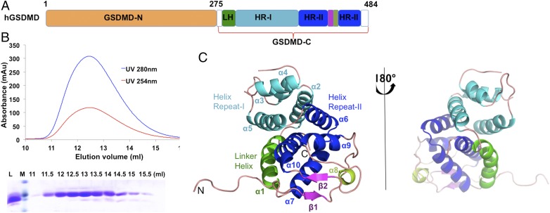
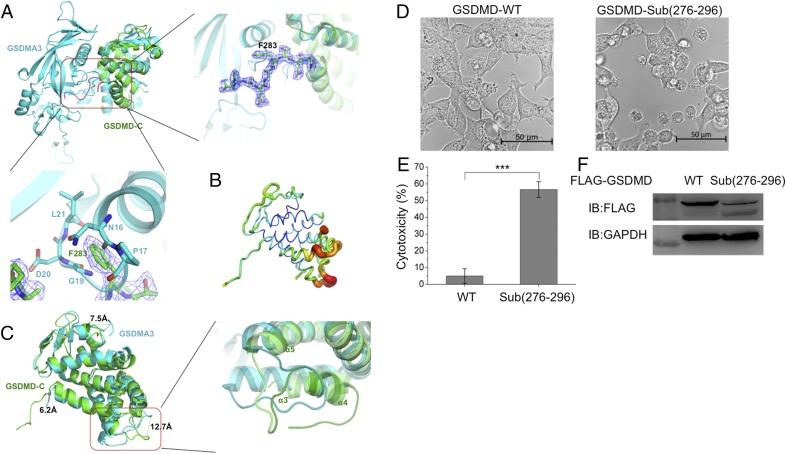
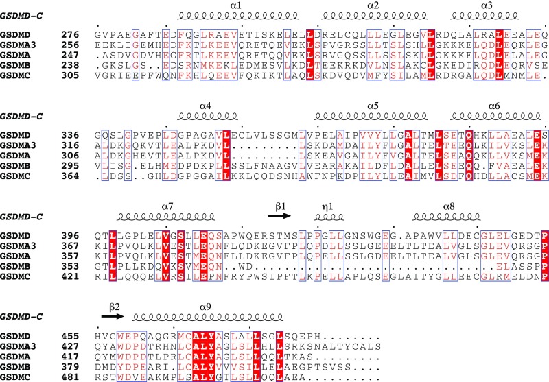
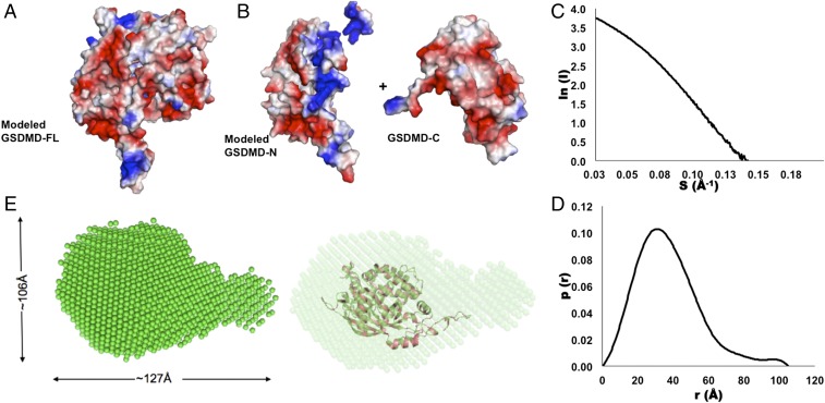
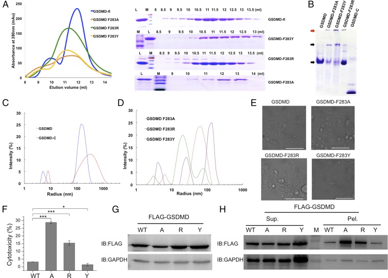
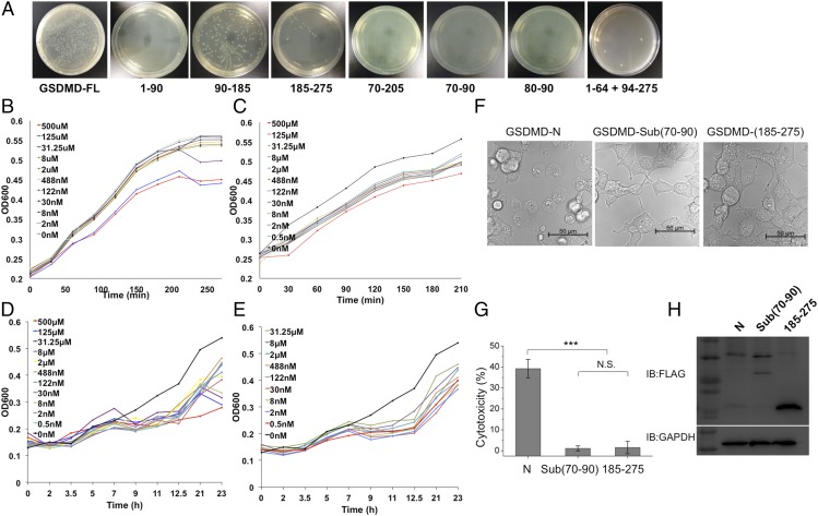
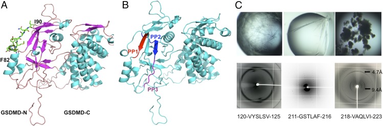
Recent findings have revealed that the protein gasdermin D (GSDMD) plays key roles in cell pyroptosis. GSDMD binds lipids and forms pore structures to induce pyroptosis upon microbial infection and associated danger signals. However, detailed structural information for GSDMD remains unknown. Here, we report the crystal structure of the C-terminal domain of human GSDMD (GSDMD-C) at 2.64-Å resolution. The first loop on GSDMD-C inserts into the N-terminal domain (GSDMD-N), which helps stabilize the conformation of the full-length GSDMD. Substitution of this region by a short linker sequence increased levels of cell death. Mutants F283A and F283R can increase protein heterogeneity in vitro and are capable of undergoing cell pyroptosis in 293T cells. The small-angle X-ray-scattering envelope of human GSDMD is consistent with the modeled GSDMD structure and mouse GSDMA3 structure, which suggests that GSDMD adopts an autoinhibited conformation in solution. The positive potential surface of GSDMD-N covered by GSDMD-C is exposed after being released from the autoinhibition state and can form high-order oligomers via a charge-charge interaction. Furthermore, by mapping different regions of GSDMD, we determined that one short segment is sufficient to kill bacteria in vitro and can efficiently inhibit cell growth in Escherichia coli and Mycobacterium Smegmatis These findings reveal that GSDMD-C acts as an auto-inhibition executor and GSDMD-N could form pore structures via a charge-charge interaction upon cleavage by caspases during cell pyroptosis.

Keywords: antibacterial activity; autoinhibition; crystal structure; gasdermin D.


Conflict of interest statement

The authors declare no conflict of interest.

Figures

Fig. 1.
Fig. 1.
Crystal structure of human GSDMD-C. (A) Schematics of human GSDMD domains. GSDMD is composed of two domains, the N-terminal domain, GSDMD-N, and the C-terminal domain, GSDMD-C. GSDMD-C includes four subdomains: the linker helix (LH, green), the helix repeat-I bundle (HR-I, cyan), the helix repeat repeat-II bundle (HR-II, blue), and the intermediated β-strand insertion (purple). (B) Gel filtration profile (Top) and SDS/PAGE analysis (Bottom) of GSDMD-C. GSDMD-C was eluted at a peak of 12.6 mL on a Superdex G-75 10/300 column. L, loading sample; M, protein marker. (C) A ribbon representation of the GSDMD-C structure, colored by domain structure. Secondary structures are labeled in accordance with A.
Fig. 2.
Fig. 2.
Structural comparison between human GSDMD-C and mouse GSDMA3 reveals that the linker region (276–296) is important for cell pyroptosis. (A, Upper Left) GSDMD-C (green) was superimposed on the C-terminal domain of GSDMA3 (cyan). Both structures showed similar overall folds, with a major difference existing in the linker region between the N-terminal and C-terminal domains. (Upper, Right) Enlarged view of the boxed area at the left showing residue F283 inserted into the pocket formed by α1, β3, and α5 of GSDMA3. (Lower) This insertion might result in severe clashes with adjacent residues located on the loop region (residues 16–21) of GSDMA3. The 2Fo − Fc electron density map was contoured to 1.5σ. (B) The B-factor distribution of human GSDMD-C. The wider and darker red tubing indicates higher B-factor. (C, Left) The structure superimposition revealed three major differences between human GSDMD-C and mouse GSDMA3. (Right) One inserted helix α4 is absent on the mouse GSDMA3 structure, in which it forms a flexible loop. (D) Live images of GSDMD and its loop mutant. 293T cells were transfected with FLAG-tagged WT GSDMD or GSDMD-Sub(276–296) (in which the 276–296 region was replaced by a short linker sequence GSGGGS). (E) The lactate dehydrogenase (LDH) assay shows that GSDMD-Sub(276–296) has pyroptosis-induced activity. Asterisks indicate significance: ***P < 0.001. (F) Expression levels of GSDMD and GSDMD-Sub(276–296) in 293T cells.
Fig. S1.
Fig. S1.
Structure-based multiple-sequence alignment of human GSDMD-C with other gasdermin family proteins. GSDMA3 is from mouse; other proteins are from human. Red backgrounds indicate identical residues, and red type indicates highly conserved residues.
Fig. 3.
Fig. 3.
The potential surface distribution and SAXS analysis of human full-length GSDMD. (A and B) The electronic potential surfaces of human GSDMD-C (A) and the modeled GSDMD-FL and GSDMD-N structures (B). Red indicates negative potential, and blue indicates positive potential. (C and D) The experimental scattering curve (C) and the distance distribution function curve (D). (E, Left) The SAXS envelope using the ab initio molecular modeling has a gourd shape. (Right) The modeled structure of GSDMD-FL was fitted into the SAXS envelope.
Fig. 4.
Fig. 4.
The properties of WT GSDMD and its mutants. (A) The gel-filtration profiles of WT and mutants (Left) and the corresponding SDS/PAGE result (Right). L, loading sample; M, protein marker. (B) The native PAGE of human GSDMD and its mutants. The low molecular weight proteins and the aggregated proteins are identified by black and red arrows, respectively. (C and D) DLS analysis of human GSDMD-C and full-length GSDMD (C) and the mutants of GSDMD (D). (E) Live images of GSDMD and its F283A, F283R, and F283Y mutants. 293T cells were transfected with FLAG-tagged WT GSDMD or its mutants. (Scale bars, 50 μm.) (F) The LDH assay shows that the F283A or F283R mutation induced cell pyroptosis. A, F283A mutation; R, F283R mutation; Y, F283Y mutation. Asterisks indicate significance: *P < 0.05; ***P < 0.001. (G) The whole-cell protein expression levels of GSDMD and its mutants in 293T cells. (H) Expression levels of GSDMD and its mutants with supernatant/pellet fractionation, corresponding with G. Mutant A has a higher protein expression level in precipitation even though the whole-cell expression is lower than in WT or other GSDMD mutants.
Fig. 5.
Fig. 5.
Identification of the antibacterial peptides of GSDMD. (A) The E. coli cells transformed with WT and truncated GSDMD were incubated at 37 °C for 20 h. The data showed that the short regions (residues 80–90 and residues 70–90) are sufficient to inhibit bacterial growth. (B and C) The effects of two synthesized peptides, FHFYDAMDGQI (B) and AEPDVQRGRSFHFYDAMDGQI (C), on the growth of E. coli host strains. The peptide sequences are the same as the two regions in GSDMD. (D and E) The effects of two synthesized peptides, FHFYDAMDGQI (D) and AEPDVQRGRSFHFYDAMDGQI (E) on the growth of M. smegmatis host strains. Serial dilutions of the peptides with final concentrations of 500 μM, 125 μM, 31.25 μM, 8 μM, 2 μM, 488 nM, 122 nM, 30 nM, 8 nM, 2 nM, and 0 nM were added to the LB media (B and C) or to Middlebrook 7H9 broth with OADC enrichment (D and E) cultures. The growth curves were determined by measuring the OD at 600 nm. (F) Live images of GSDMD-N and its mutants. 293T cells were transfected with FLAG-tagged GSDMD-N, Sub(70–90) (in which residues 70–90 were replaced with a linker sequence GSGGGS), or the truncated version 185–275. (G) The LDH assay shows that substitution of residues 70–90 abolished pyroptosis-induced activity. ***P < 0.001; N.S., not significant. (H) Expression levels of GSDMD-N and its mutants in 293T cells.
Fig. 6.
Fig. 6.
The amyloid-forming segments of human GSDMD. (A) The loop region (residues 82–90) is shown by sticks on the modeled GSDMD structure. (B) The three amyloid-forming segments are labeled as PP1 (120-VYSLSV-125, red), PP2 (211-GSTLAF-216, blue), and PP3 (218-VAQLVI-223, purple), respectively. (C) The three short segments are prone to form amyloid fibrils. (Upper) Needle-like crystals. (Lower) X-ray diffraction patterns. The perpendicular rings at 4.7 Å and 9.4 Å represent a typical amyloid diffraction pattern. (Magnification: Upper, 80×; Lower, 2×.)

References

    1. Shi J, et al. Cleavage of GSDMD by inflammatory caspases determines pyroptotic cell death. Nature. 2015;526:660–665. - PubMed
    1. Kayagaki N, et al. Caspase-11 cleaves gasdermin D for non-canonical inflammasome signalling. Nature. 2015;526:666–671. - PubMed
    1. Shi J, Gao W, Shao F. Pyroptosis: Gasdermin-mediated programmed necrotic cell death. Trends Biochem Sci. 2017;42:245–254. - PubMed
    1. He WT, et al. Gasdermin D is an executor of pyroptosis and required for interleukin-1beta secretion. Cell Res. 2015;25:1285–1298. - PMC - PubMed
    1. Ding J, et al. Pore-forming activity and structural autoinhibition of the gasdermin family. Nature. 2016;535:111–116. - PubMed

Publication types

MeSH terms

Associated data

LinkOut - more resources