Review
. 2017 Sep 19;8(5):e01009-17.
doi: 10.1128/mBio.01009-17.

Picornaviruses and Apoptosis: Subversion of Cell Death

Affiliations
Review

Picornaviruses and Apoptosis: Subversion of Cell Death

Sarah N Croft et al. mBio. .

Abstract

Infected cells can undergo apoptosis as a protective response to viral infection, thereby limiting viral infection. As viruses require a viable cell for replication, the death of the cell limits cellular functions that are required for virus replication and propagation. Picornaviruses are single-stranded RNA viruses that modify the host cell apoptotic response, probably in order to promote viral replication, largely as a function of the viral proteases 2A, 3C, and 3CD. These proteases are essential for viral polyprotein processing and also cleave cellular proteins. Picornavirus proteases cleave proapoptotic adaptor proteins, resulting in downregulation of apoptosis. Picornavirus proteases also cleave nucleoporins, disrupting the orchestrated manner in which signaling pathways use active nucleocytoplasmic trafficking, including those involved in apoptosis. In addition to viral proteases, the transmembrane 2B protein alters intracellular ion signaling, which may also modulate apoptosis. Overall, picornaviruses, via the action of virally encoded proteins, exercise intricate control over and subvert cell death pathways, specifically apoptosis, thereby allowing viral replication to continue.

Keywords: apoptosis; innate immunity; picornavirus; proteases; virus-host interactions.


Figures

FIG 1
FIG 1
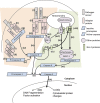
Antiviral apoptotic pathways. Induction of the extrinsic apoptotic pathway (pink shaded area) is initiated by the binding of a picornavirus-associated molecular pattern to a pathogen receptor (i), e.g., dsRNA to TLR3, MDA-5, or RIG-I. Subsequently, adaptor proteins TRIF, FADD, RIPK1, and TRADD are recruited directly to the receptor or via IPS-1 in the case of RIG-I and MDA-5 (ii), forming the death receptor complexes. Procaspase-8 is recruited to the death receptor complexes (iii) and undergoes autocatalysis (iv) to active caspase 8. Caspase 8 then cleaves and activates caspase 3 (v), which translocates to the nucleus (vi) and activates proteins to execute apoptotic morphological changes (vii). The cleavage of Bid by caspase 8 (xii) produces truncated Bid (tBid), which induces Bax and Bak (xiii) to insert into the outer mitochondrial membrane, allowing cytochrome c to translocate into the cytoplasm (ix). APAF-1 binds cytochrome c (x), and procaspase-9 is recruited to the complex, undergoing autocatalysis to caspase 9 (xi). Caspase 9 cleaves procaspase-3 to caspase 3 (v), which translocates to the nucleus (vi) and activates proteins to execute apoptotic morphological changes (vii). TBK1 and IKKε can also be recruited to IPS-1 (xv), and once recruited, phosphorylate IRF-3. Phospho-IRF-3 translocates to the outer mitochondrial membrane (xvi), complexing with Bax to form a pore (viii), releasing cytochrome c into the cytoplasm (ix) and inducing a procaspase-9-dependent apoptotic signaling cascade. Abbreviations: TLR3, Toll-like receptor 3; TRIF, TIR domain-containing adaptor-inducing beta interferon; RIPK1, receptor-interacting protein kinase 1; FADD, Fas-associated protein with death domain; TRADD, tumor necrosis factor receptor type 1-associated death domain protein; RIG-I, retinoic acid-inducible gene 1; MDA-5, melanoma differentiation-associated protein 5; IPS-1, interferon-β promoter stimulator 1; TBK1, TANK-binding kinase 1; IKKε, inhibitor of nuclear factor κB kinase epsilon; IRF-3, interferon regulatory factor 3; Bak, Bcl-2 homologous antagonist/killer; Bax, Bcl-2-like protein 4; Bid, BH3-interacting-domain death agonist; tBid, truncated Bid; APAF-1, apoptotic protease activating factor 1.
FIG 2
FIG 2
Effects of picornavirus infections on intrinsic apoptosis pathways. (A) Caspase 9-dependent intrinsic apoptosis is initiated by the release of mitochondrial cytochrome c into the cytoplasm as described in the legend to Fig. 1. Procaspase-9 is cleaved in PV infection (indicated in red), resulting in a lack of function and inability to activate caspase 3, thus inhibiting intrinsic apoptosis. (B) The abrupt release of Ca2+ from the endoplasmic reticulum (ER) is a protective response following ER stress. The released Ca2+ is taken up into the mitochondrial matrix via RyR and IP3R channels. Increase of Ca2+ above a threshold level in the mitochondrial matrix induces an increase in mitochondrial membrane permeability, allowing the cytoplasmic release of cytochrome c, inducing intrinsic apoptosis. Picornaviral 2B protein inserts into the ER, forming an ion channel (indicated in red), allowing Ca2+ to leak into the cytoplasm, and decreasing the amount of Ca2+ available to translocate to the mitochondria, thus inhibiting intrinsic apoptosis. (C) RIG-I and MDA-5, when activated, associate with IPS-1 and recruit TBK1 and IKKε into a death receptor complex at the mitochondria. TBK1 and IKKε act by phosphorylating IRF-3 dimers which then induce a pore in the mitochondrial membrane, allowing release of cytochrome c into the cytoplasm, inducing intrinsic apoptosis. RIG-I, MDA-5, and IPS-1 are cleaved during infection (indicated in red) by several picornaviruses, inhibiting intrinsic apoptosis. P, phosphate; EchoV, echovirus.
FIG 3
FIG 3
Effects of picornavirus infections on extrinsic apoptosis pathways. (A) Sensing of dsRNA by RIG-I/MDA-5 leads to the formation of a death receptor complex at the mitochondria through the adaptor protein IPS-1. Subsequent ubiquination of RIPK1 is recognized by the IKKγ (Nemo) subunit of the IKKα-IKKβ-IKKγ complex, leading to phosphorylation and degradation of IκB and release of NF-κB that translocates into the nucleus to upregulate expression of proapoptotic genes. Cleavage of RIG-I and IPS-1 by various picornaviruses (indicated in red) disrupts extrinsic apoptosis signaling. Foot-and-mouth disease virus (FMDV) infection results in cleavage of IKKγ, abrogating NF-kB activation and attenuating transcriptional activation of proapoptotic factors. (B) Sensing of dsRNA by TLR3 and subsequent recruitment of TRIF initiates the formation of a death receptor complex, leading to induction of extrinsic apoptosis as described in the legend to Fig. 1. TRIF is cleaved in infection by multiple picornaviruses (indicated in red) resulting in disruption of the death receptor complex and inhibition of extrinsic apoptosis. P, phosphate; DD, death domain; RHIM, RIP homotypic interaction motif.
FIG 4
FIG 4
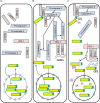
Effects of picornavirus infections on nuclear pore and Sam68. (A) Sam68 (yellow and green) shuttles between the cytoplasm and nucleus, being located predominately in the nucleus in healthy cells, with cytoplasmic cell death proteins in precursor, inactive form. (B) With the detection of dsRNA by RIG-I/MDA-5 or TLR-3, procaspase-8 is recruited to the complex formed, inducing activation of caspase 8 and recruitment of death receptor proteins, e.g., FADD and RIPK1. Death receptor complex signaling is dependent on recruitment of cytoplasmic Sam68. (C) Disruption of the NPC in picornavirus infection (i) (indicated by broken outline of NPCs) allows passive movement of Sam68 between the nucleus and the cytoplasm (as indicated by the red double-headed arrow). The NLS motif of Sam68 (green) is cleaved in picornavirus infection (ii), resulting in reduced recruitment of RIPK1 to the FADD complex, rendering it inactive. Loss of the NLS motif also inhibits the active import of Sam68 into the nucleus (ii), retaining it in the cytoplasm. Last, the viral genomic element, IRES, binds Sam68 within the cytoplasm (iii), allowing viral replication.

References

    1. Deszcz L, Seipelt J, Vassilieva E, Roetzer A, Kuechler E. 2004. Antiviral activity of caspase inhibitors: effect on picornaviral 2A proteinase. FEBS Lett 560:51–55. doi:10.1016/S0014-5793(04)00069-9. - DOI - PubMed
    1. Belov GA, Romanova LI, Tolskaya EA, Kolesnikova MS, Lazebnik YA, Agol VI. 2003. The major apoptotic pathway activated and suppressed by poliovirus. J Virol 77:45–56. doi:10.1128/JVI.77.1.45-56.2003. - DOI - PMC - PubMed
    1. Walker EJ, Younessi P, Fulcher AJ, McCuaig R, Thomas BJ, Bardin PG, Jans DA, Ghildyal R. 2013. Rhinovirus 3C protease facilitates specific nucleoporin cleavage and mislocalisation of nuclear proteins in infected host cells. PLoS One 8:e71316. doi:10.1371/journal.pone.0071316. - DOI - PMC - PubMed
    1. Gustin KE, Sarnow P. 2002. Inhibition of nuclear import and alteration of nuclear pore complex composition by rhinovirus. J Virol 76:8787–8796. doi:10.1128/JVI.76.17.8787-8796.2002. - DOI - PMC - PubMed
    1. Walker EJ, Jensen LM, Croft S, Ghildyal R. 2015. Variation in the nuclear effects of infection by different human rhinovirus serotypes. Front Microbiol 6:875. doi:10.3389/fmicb.2015.00875. - DOI - PMC - PubMed

Publication types

LinkOut - more resources