. 2017 Nov 6;7(11):3731-3743.
doi: 10.1534/g3.117.300227.

The Ubiquitin Ligase (E3) Psh1p Is Required for Proper Segregation of both Centromeric and Two-Micron Plasmids in Saccharomyces cerevisiae

Affiliations

The Ubiquitin Ligase (E3) Psh1p Is Required for Proper Segregation of both Centromeric and Two-Micron Plasmids in Saccharomyces cerevisiae

Meredith B Metzger et al. G3 (Bethesda). .

Abstract

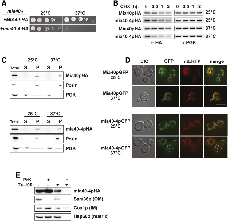
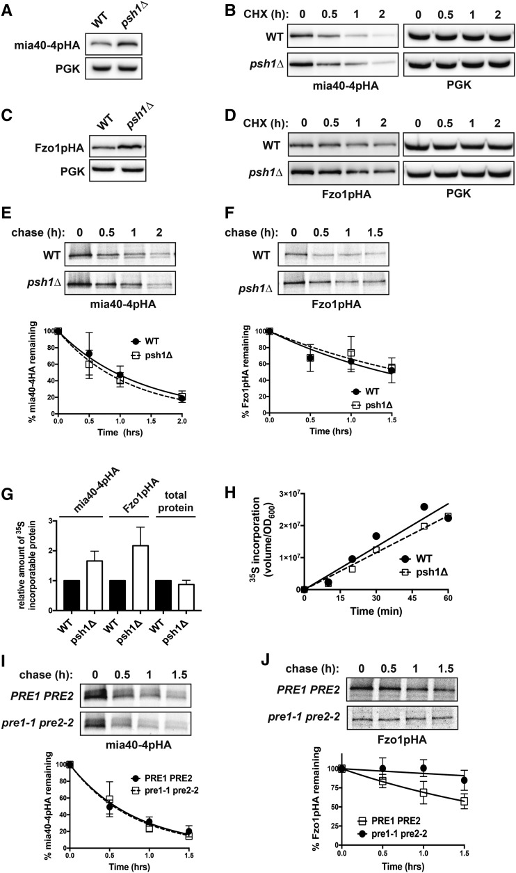
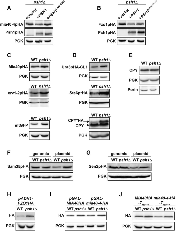
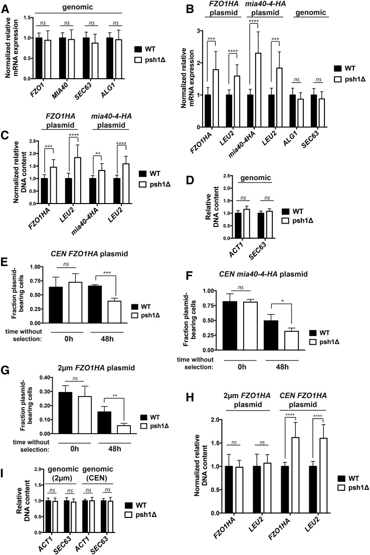
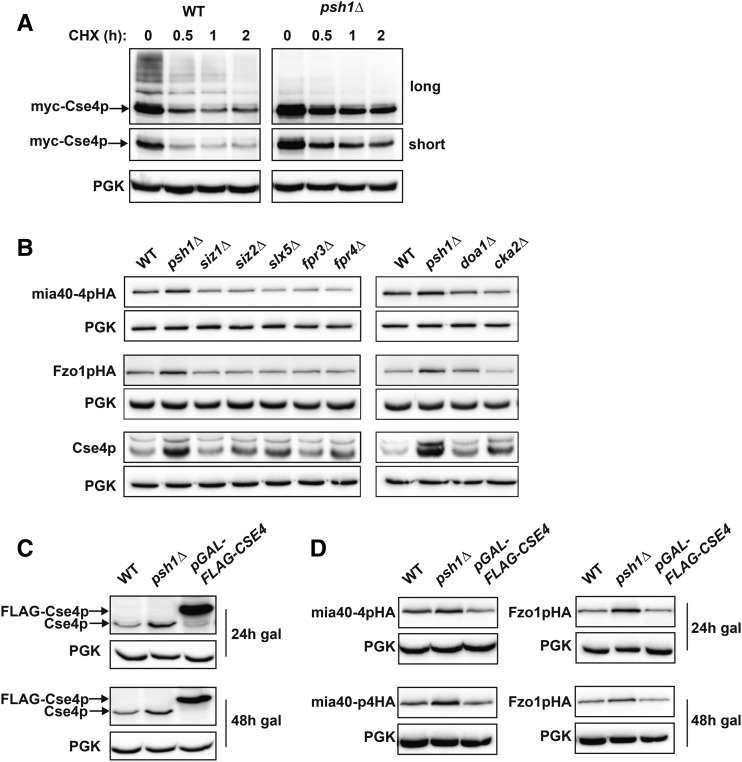
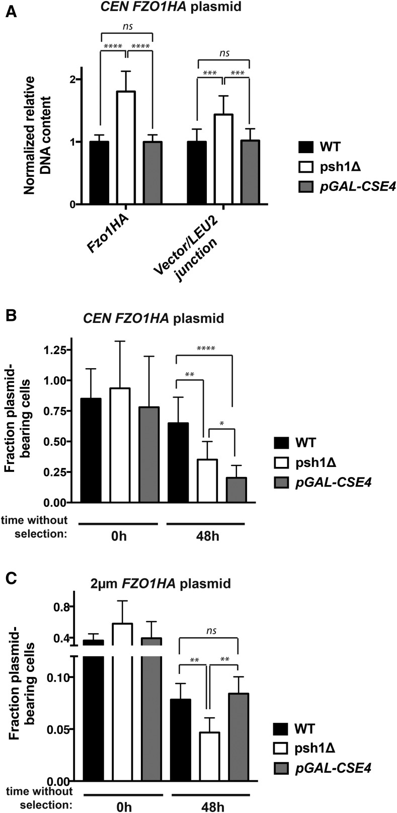
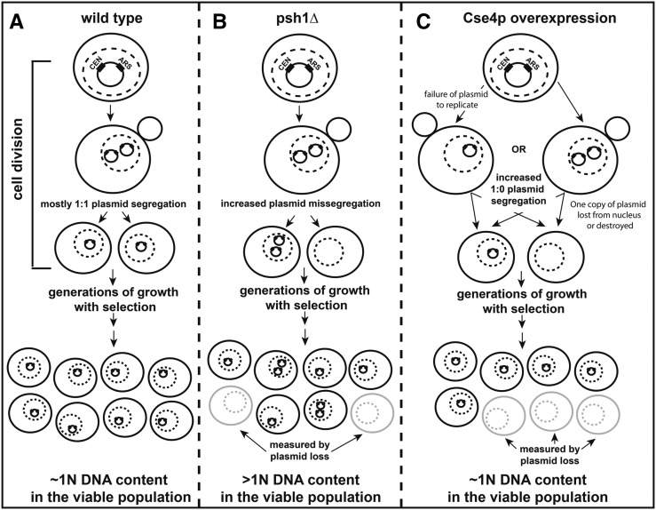
Protein degradation by the ubiquitin-proteasome system is essential to many processes. We sought to assess its involvement in the turnover of mitochondrial proteins in Saccharomyces cerevisiae We find that deletion of a specific ubiquitin ligase (E3), Psh1p, increases the abundance of a temperature-sensitive mitochondrial protein, mia40-4pHA, when it is expressed from a centromeric plasmid. Deletion of Psh1p unexpectedly elevates the levels of other proteins expressed from centromeric plasmids. Loss of Psh1p does not increase the rate of turnover of mia40-4pHA, affect total protein synthesis, or increase the protein levels of chromosomal genes. Instead, psh1Δ appears to increase the incidence of missegregation of centromeric plasmids relative to their normal 1:1 segregation. After generations of growth with selection for the plasmid, ongoing missegregation would lead to elevated plasmid DNA, mRNA, and protein, all of which we observe in psh1Δ cells. The only known substrate of Psh1p is the centromeric histone H3 variant Cse4p, which is targeted for proteasomal degradation after ubiquitination by Psh1p However, Cse4p overexpression alone does not phenocopy psh1Δ in increasing plasmid DNA and protein levels. Instead, elevation of Cse4p leads to an apparent increase in 1:0 plasmid segregation events. Further, 2 μm high-copy yeast plasmids also missegregate in psh1Δ, but not when Cse4p alone is overexpressed. These findings demonstrate that Psh1p is required for the faithful inheritance of both centromeric and 2 μm plasmids. Moreover, the effects that loss of Psh1p has on plasmid segregation cannot be accounted for by increased levels of Cse4p.

Keywords: 2 μm plasmid; CEN plasmid; Cse4p; plasmid missegregation; ubiquitination.


Figures

Figure 1
Figure 1
mia40-4pHA is an unstable mitochondrial protein. (A) Ten-fold serial dilutions of mia40Δ cells expressing mia40-4-HA or MIA40-HA from CEN plasmids at permissive (25°) or nonpermissive (37°) temperatures were spotted to minimal media lacking leucine. (B) WT yeast were treated with CHX at 25 or 37° for the indicated times to assess the degradation of CEN plasmid-expressed mia40-4pHA or Mia40pHA. Proteins were detected by immunoblotting with HA antibody. Phosphoglycerate kinase (PGK) served as a protein loading control. (C) Lysates from cells expressing Mia40pHA (top) or mia40-4pHA (bottom) from CEN plasmids at 25 or 37° were fractionated into mitochondrial pellets (P) and postmitochondrial supernatants (S). Fractions were subject to immunoblotting with antibodies to HA, Porin, or PGK. (D) Differential interference contrast (DIC) and fluorescence microscopy at 25 and 37° of cells coexpressing mia40-4pGFP or Mia40pGFP and a mitochondrial matrix-targeted RFP variant, mtERFP. Bar, 10 μm and “merge” is an overlay of GFP and RFP channels. (E) Mitochondria isolated as in (C) were treated with proteinase K (PrK) alone, or in combination with Triton X-100 (Tx-100), and subject to immunoblotting with antibodies specific to the OM protein Sam35p, the IM protein Cox1p, or the matrix protein Hsp60p.
Figure 2
Figure 2
Loss of Psh1p affects the steady-state levels of mia40-4pHA and Fzo1pHA, without affecting their rates of turnover or total cellular protein levels. (A) The steady-state protein level of CEN plasmid-expressed mia40-4pHA was assessed at 37° in WT and psh1Δ cells by immunoblotting with HA antibody. PGK serves as a control for equal loading. (B) CHX chase for the indicated times assessing turnover of mia40-4pHA expressed from a CEN plasmid in WT and psh1Δ yeast cells. Proteins were detecting by immunoblotting. (C) The steady-state protein levels of Fzo1pHA, analyzed as in (A) except at 30°. (D) CHX chase of Fzo1pHA, analyzed as in (B) except at 30°. (E) Representative 35S pulse-chase analysis to assess turnover of mia40-4pHA in WT and psh1Δ cells at 37° at the indicated time points. The mean of three independent experiments is graphed below, with error bars depicting the SD. (F) Representative 35S pulse-chase analysis of the turnover of Fzo1pHA in WT and psh1Δ at 30° at the indicated time points, analyzed and graphed as in (E). (G) Quantification of 35S incorporated into mia40-4pHA, Fzo1pHA, or total protein during a 30-min pulse with 35S-labeled methionine/cysteine in psh1Δ and WT strains. Values were normalized to the incorporation in the WT strain. The average and SD of three independent experiments is shown. (H) The rate of total protein synthesis in WT and psh1Δ strains was measured by analyzing 35S methionine/cysteine incorporation into total protein relative to cell density (OD600) over time. (I) Representative 35S pulse-chase analysis of the turnover of mia40-4pHA in a pre1-1 pre2-2 proteasome mutant strain and its PRE1 PRE2 isogenic WT strain, analyzed and graphed as in (E). (J) Representative 35S pulse-chase analysis of the turnover of Fzo1pHA in a pre1-1 pre2-2 proteasome mutant strain and its PRE1 PRE2 isogenic WT strain, analyzed and graphed as in (F).
Figure 3
Figure 3
Loss of Psh1p or its ubiquitin ligase activity affects the steady-state levels of many proteins when expressed from plasmids, but not when expressed from the chromosome. (A, B) The steady-state protein levels of CEN-plasmid expressed mia40-4pHA (A) or Fzo1pHA (B) were analyzed in psh1Δ cells coexpressing either vector, WT PSH1HA, or RING domain mutant PSH1HAC45S C50S growing at 37° (mia40-4pHA) or 30° (Fzo1pHA) by immunoblotting with HA antibody. Anti-PGK serves as a loading control. (C) Levels of the mitochondrial proteins Mia40pHA, erv1-2pHA, and mitochondrial-targeted GFP (mtGFP) expressed from CEN plasmids in WT and psh1Δ cells were assessed in WT and psh1Δ cells at 30° as in (A). (D) CEN plasmid-expressed Ura3pHA-CL1, Ste6p*HA, and CPY*HA were analyzed in WT and psh1Δ cells at 30° as in (A) except CPY*HA was visualized using anti-CPY. (E) Chromosomal proteins (CPY, PGK, and Porin) were analyzed in WT and psh1Δ cells by immunoblotting using antibodies specific to these targets. (F, G). Sam35pHA (F) or Sen2pHA (G) expressed from either the genome or a CEN plasmid were analyzed in WT and psh1Δ cells at 30° as in (A). (H–J) CEN plasmid-expressed Fzo1pHA under control of the ADH1 promoter (H) or mia40-4pHA and Mia40pHA under control of the GAL10 promoter (I) or with the native MIA40 3′ untranslated region (J) were assessed in psh1Δ and WT cells at 30° (Fzo1pHA) or 37° (mia40-4pHA), as in (A).
Figure 4
Figure 4
Loss of Psh1p increases mRNA levels of genes expressed from CEN plasmids due to higher plasmid DNA content achieved through plasmid missegregation. (A) Normalized mRNA expression of the endogenous, genome-expressed genes FZO1, MIA40, SEC63, or ALG1 in WT and psh1Δ cells grown in YPD medium, not expressing a plasmid, was determined using RT-qPCR analysis. Expression was normalized to ACT1 levels and is graphed relative to WT cells; error bars represent the SD of at least three biological replicates with three technical replicates each. (B) Normalized mRNA expression of FZO1HA, mia40-4HA, and LEU2 in WT and psh1Δ cells carrying the indicated CEN plasmid, and normalized mRNA expression of genomic SEC63 and ALG1 in WT and psh1Δ cells expressing the CEN FZO1HA plasmid were determined as in (A). (C) Normalized DNA levels of CEN plasmid FZO1HA, mia40-4HA, and LEU2 in DNA harvested from WT and psh1Δ cells carrying the indicated CEN plasmid as determined by qPCR on DNA samples. Plasmid DNA content was normalized to the genomic level of ACT1 and graphed as in (A). (D) DNA levels of genome-expressed ACT1 and SEC63 in the DNA used in (C) from cells expressing the FZO1HA CEN plasmid as determined by qPCR. Values are graphed relative to WT cells; error bars represent the SD of at least three biological replicates with three technical replicates each. (E–G) The fraction of FZO1HA CEN (E) mia40-4HA CEN (F) or FZO1HA 2 μm plasmid-bearing WT and psh1Δ cells after 0 and 48 hr without selection for the plasmid was assessed by comparing the number of cells able to grow on selective media to the cell number on YPD. Error bars represent the SD of at least three biological replicates. (H) Normalized DNA levels of 2 μm and CEN plasmid FZO1HA and LEU2 in WT and psh1Δ cells, determined as in (C). (I) DNA levels of genome-expressed ACT1 and SEC63 in the DNA used in (H) as determined by qPCR, graphed and analyzed as in (D). For (A–I) P-values are calculated from a two-tailed t-test and * P < 0.05, ** P < 0.01, *** P < 0.001, **** P < 0.0001, and ns = P > 0.05.
Figure 5
Figure 5
Overexpression of Cse4p does not phenocopy loss of Psh1p in increasing CEN plasmid protein levels. (A) CHX chase for the indicated times assessing turnover of myc-Cse4p in WT and psh1Δ yeast cells. Proteins were detecting by immunoblotting with myc antibody. Both long and short exposures are shown. Anti-PGK serves as a loading control. (B) The steady-state protein levels of CEN plasmid-expressed mia40-4pHA and Fzo1pHA, as well as endogenous genome-expressed Cse4p, were detected by immunoblotting with HA or Cse4p antibodies, in the indicated deletion strains. (C) Immunoblotting for Cse4p using Cse4p antibodies in WT cells, psh1Δ cells, or cells overexpressing Cse4p from the genome (pGAL-FLAG-CSE4) for the indicated time points after continuous galactose induction. (D) Immunoblotting using anti-HA for CEN plasmid-expressed mia40-4pHA or Fzo1pHA as in (C).
Figure 6
Figure 6
Overexpression of Cse4p increases CEN, but not 2 μm, plasmid loss and does not affect plasmid DNA content under selective growth conditions. (A) Normalized DNA levels of CEN FZO1HA plasmid were detected by qPCR using two plasmid-specific primer pairs (FZO1HA and vector/LEU2 junction) on DNA harvested from WT cells, psh1Δ cells, or cells overexpressing Cse4p from the chromosome (pGAL-FLAG-CSE4) for 24 hr in selective media. LEU2 primers could not be used, as the mutated leu2-3,112 allele is present in the genome of these strains. DNA content was normalized to ACT1 levels and graphed as in Figure 4C. See Figure S3A in File S1 legend for further detail. (B) The fraction of CEN FZO1HA plasmid-bearing was analyzed as in Figure 4D after 0 and 48 hr growth in media without selection for the CEN plasmid. Cells were grown in medium containing galactose for 24 hr prior to the shift to nonselective medium, and galactose induction was continued during growth without selection. Error bars represent the SD of at least three biological replicates. (C) The fraction of 2 μm FZO1HA plasmid-bearing WT cells, psh1Δ cells, or cells overexpressing Cse4p from the chromosome (pGAL-FLAG-CSE4) was analyzed as in (B) For (A–C) P-values are calculated from a two-tailed t-test; * P < 0.05, ** P < 0.01, *** P < 0.001, **** P < 0.0001, and ns = P > 0.05.
Figure 7
Figure 7
Models for the distinct mechanisms of plasmid missegregation in haploid psh1Δ and Cse4p-overexpressing cells. (A) In WT cells, CEN/ARS-containing yeast plasmids generally accurately replicate and segregate in a 1:1 manner during cell division, and thus have a low frequency of plasmid loss. After generations of growth in media selective for cells containing the plasmid, cells typically maintain one copy of the plasmid and are viable. Thus, the overall plasmid DNA content in the population remains close to 1N in haploid yeast cells. (B) In haploid psh1Δ cells, CEN/ARS-containing plasmids are replicated, but have an apparent increased propensity for missegregation. One cell may receive two copies of the plasmid, while the other receives none. Under selective growth conditions, cells without plasmid are inviable. Yeast with one or more copies of plasmid would be viable and continue to divide, leading to an overall increase in the DNA content (>1N) in the population after generations of growth with selection for the plasmid. Shown is a model for the simplest 2:0 missegregation of plasmid, but other mechanisms of missegregation (i.e., 4:0, 3:1, etc.) also become possible after initiating missegregation events have occurred. An increase in cells lacking plasmid is detected by the plasmid loss assay, which measures colony forming units on selective vs. nonselective media after generations of growth without selection for the plasmid. (C) In cells overexpressing Cse4p, there appears to be an increase in the incidence of 1:0 segregation of CEN/ARS-containing plasmids. This could occur because the plasmid fails to replicate properly, or if one copy of the plasmid is lost from the nucleus or degraded following replication. Regardless, the net result is that only one progeny cell receives a copy of plasmid. Again, the cell without plasmid would be inviable under selective growth conditions and this loss would be measured by the plasmid loss assay. The cell with one copy of the plasmid would remain viable and continue to divide, with the DNA content in the population remaining close to 1N after generations of growth in medium selective for the plasmid. For (A–C) the nucleus is indicated by a dotted line.

Similar articles

Cited by

References

    1. Akutsu M., Dikic I., Bremm A., 2016. Ubiquitin chain diversity at a glance. J. Cell Sci. 129: 875–880. - PubMed
    1. Amato A., Schillaci T., Lentini L., Di Leonardo A., 2009. CENPA overexpression promotes genome instability in pRb-depleted human cells. Mol. Cancer 8: 119. - PMC - PubMed
    1. Au W. C., Crisp M. J., DeLuca S. Z., Rando O. J., Basrai M. A., 2008. Altered dosage and mislocalization of histone H3 and Cse4p lead to chromosome loss in Saccharomyces cerevisiae. Genetics 179: 263–275. - PMC - PubMed
    1. Au W. C., Dawson A. R., Rawson D. W., Taylor S. B., Baker R. E., et al. , 2013. A novel role of the N terminus of budding yeast histone H3 variant Cse4 in ubiquitin-mediated proteolysis. Genetics 194: 513–518. - PMC - PubMed
    1. Bengtson M. H., Joazeiro C. A., 2010. Role of a ribosome-associated E3 ubiquitin ligase in protein quality control. Nature 467: 470–473. - PMC - PubMed

MeSH terms

Substances

LinkOut - more resources