. 2017 Sep 19;8(1):606.
doi: 10.1038/s41467-017-00728-9.

Agonist immunotherapy restores T cell function following MEK inhibition improving efficacy in breast cancer

Affiliations

Agonist immunotherapy restores T cell function following MEK inhibition improving efficacy in breast cancer

Sathana Dushyanthen et al. Nat Commun. .

Abstract

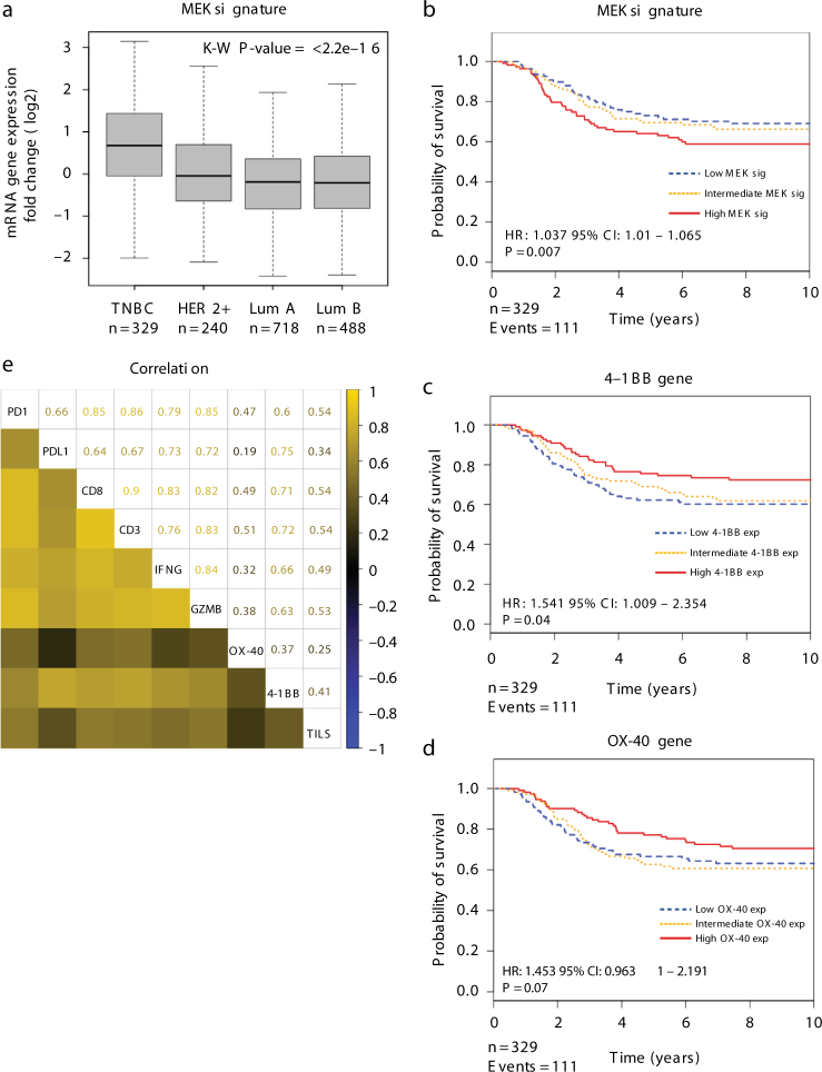
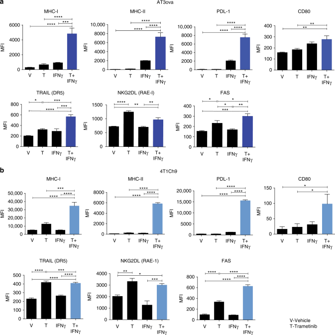
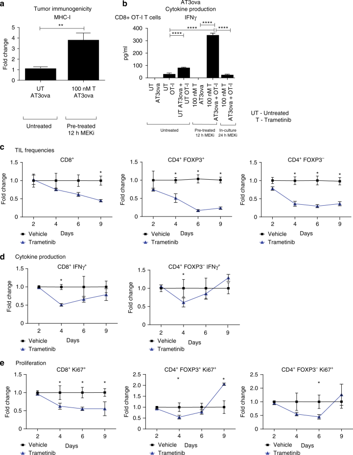
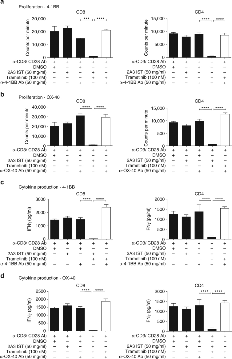
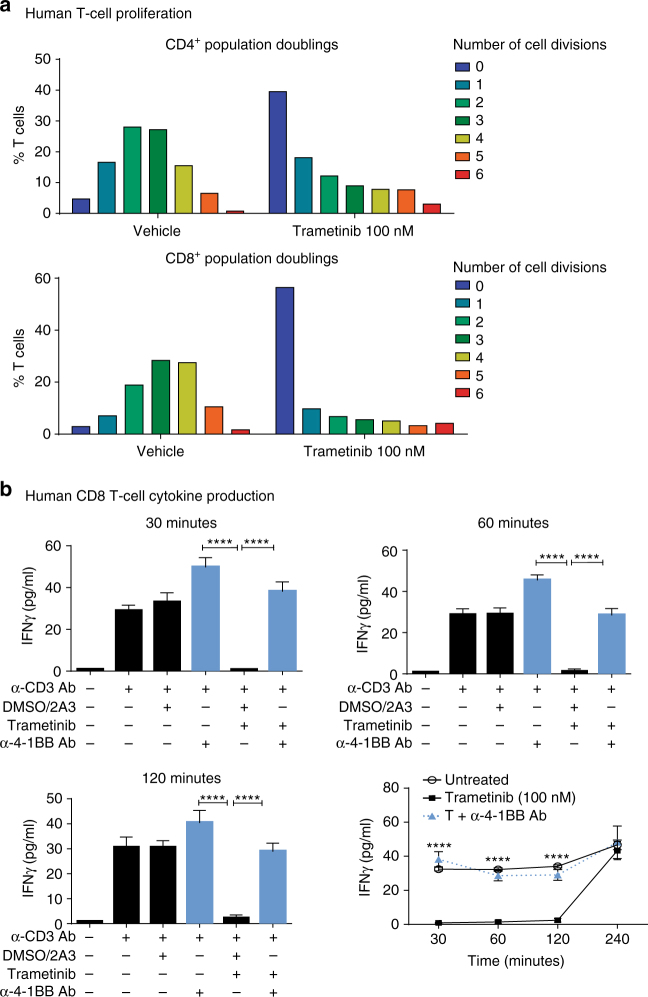
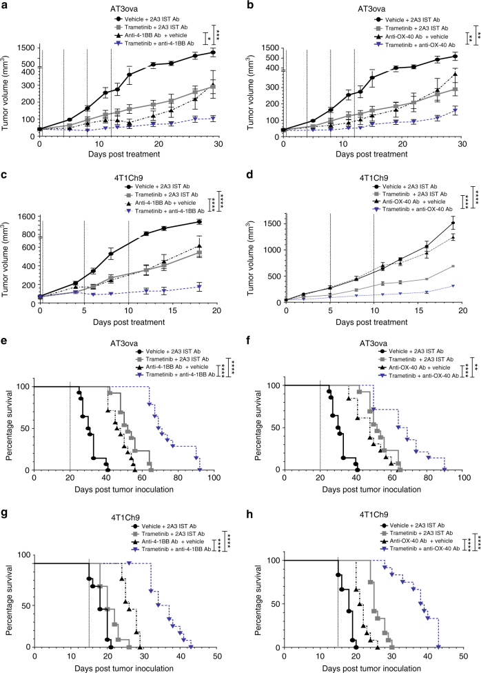
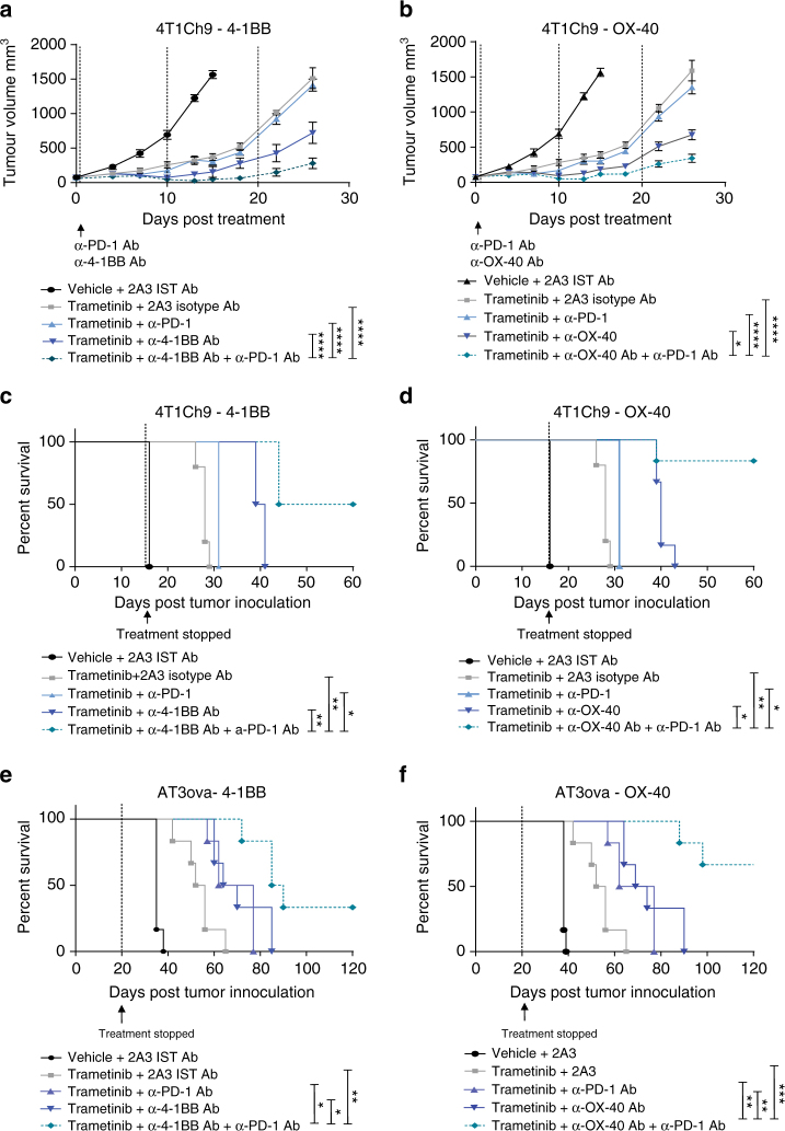
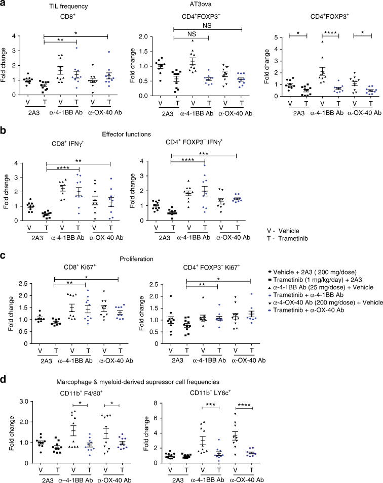
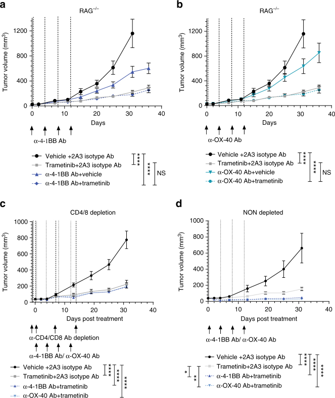
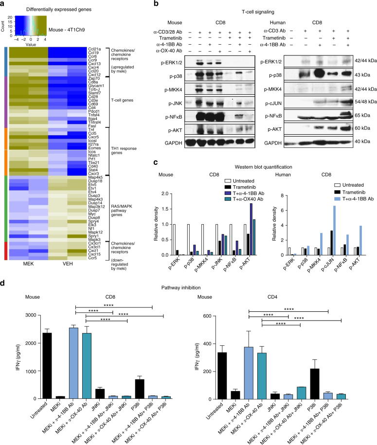
The presence of tumor-infiltrating lymphocytes in triple-negative breast cancers is correlated with improved outcomes. Ras/MAPK pathway activation is associated with significantly lower levels of tumor-infiltrating lymphocytes in triple-negative breast cancers and while MEK inhibition can promote recruitment of tumor-infiltrating lymphocytes to the tumor, here we show that MEK inhibition adversely affects early onset T-cell effector function. We show that α-4-1BB and α-OX-40 T-cell agonist antibodies can rescue the adverse effects of MEK inhibition on T cells in both mouse and human T cells, which results in augmented anti-tumor effects in vivo. This effect is dependent upon increased downstream p38/JNK pathway activation. Taken together, our data suggest that although Ras/MAPK pathway inhibition can increase tumor immunogenicity, the negative impact on T-cell activity is functionally important. This undesirable impact is effectively prevented by combination with T-cell immune agonist immunotherapies resulting in superior therapeutic efficacy.MEK inhibition in breast cancer is associated with increased tumour infiltrating lymphocytes (TILs), however, MAPK activity is required for T cells function. Here the authors show that TILs activity following MEK inhibition can be enhanced by agonist immunotherapy resulting in synergic therapeutic effects.


Conflict of interest statement

S.L.’s lab receives research funding from Novartis. The remaining authors declare no competing interests.

Figures

Fig. 1
Fig. 1
Clinical correlates of a MEK activation gene signature and 4-1BB and OX-40 gene expression in human TNBC. a Higher levels of the MEK gene signature in TNBC (n = 329, Kruskal–Wallis; P = 2.2e-16) compared with other breast cancer subtypes from the METABRIC data set. Box plot represents the lower, median and upper quartile, while whiskers represent the highest and lowest range for the upper and lower quartiles. P-value represents Kruskal–Wallis test bd Kaplan–Meier survival curves of TNBC patients according to tertiles of the b MEK signature (HR: 1.037, 95% CI: 1.01–1.065; P = 0.007), c 4-1BB gene (HR: 1.541 95% CI: 1.009–2.354; P = 0.04), and d OX-40 gene (HR: 1.453 95% CI: 0.9631–2.191; P = 0.07), stratified by low, intermediate, and high gene expression tertiles. P-values represent Cox regression analysis. e Gene correlations between the TILs signature, OX-40, 4-1BB, and key breast cancer-related immune prognostic genes from the TCGA database. Yellow high correlation, blue low correlation. P-values represent Pearson’s correlation coefficient
Fig. 2
Fig. 2
MEK inhibition increases tumor immunogenicity of AT3ova and 4T1Ch9 TNBC tumors in vitro. FACS analysis of MHC-I, MHC-II, PDL-1, CD80, TRAIL (DR5) NKG2DL (RAE-1), and Fas expression (MFI; mean fluorescence index) on the a AT3ova and b 4T1Ch9 murine TNBC cell lines following trametinib treatment alone (100 nM), IFNγ stimulation alone (5 ng/ml), or combination of trametinib treatment and IFNγ stimulation. Data are presented as mean ± SEM of triplicate samples. P-values represent one-way ANOVA and post hoc Fisher’s LSD tests. *P < 0.05, **P < 0.01, ***P < 0.001, ****P < 0.0001
Fig. 3
Fig. 3
MEK inhibition increases tumor immunogenicity but reduces T-cell effector functions. Co-culture studies undertaken with AT3ova tumor cells and CD8+ OT-I T cells; 12 h pre-treatment followed by co-culture, or 24 h co-culture with trametinib treatment. a FACS analysis of MHC-I expression of AT3ova tumor cells normalized to non-treated tumors. b IFNγ production from OT-I T cells. ce Mice (n = 3 per group) bearing AT3ova tumors were treated with vehicle (PEG 400/solutol) or trametinib (1 mg/kg/daily), and tumors were harvested on day 2, 4, 6, and 9 post treatment. Changes in c TIL frequency (CD8+, CD4+ FOXP3, CD4+ FOXP3+) as a proportion of CD45+ live cells, d cytokine production by T cells, and e proliferation of T cells measured by Ki67 expression were determined ex vivo by FACS analysis. Values were normalized to vehicle controls at each time point and data are expressed as fold change for the number of positive cells. Data are presented as mean ± SEM, and is a representative of three independent repeats. P-values represent unpaired t-tests at each time point and post hoc Fisher’s LSD tests. *P < 0.05, **P < 0.01, ***P < 0.001, ****P < 0.0001
Fig. 4
Fig. 4
Agonist immunotherapy treatment rescues inhibition of mouse T-cell function induced by MEK inhibition. Purified CD8+ or CD4+ murine T cells were stimulated with α-CD3 (1 µg/ml) and α-CD28 (0.5 µg/ml) antibodies and treated with vehicle (DMSO), 2A3 isotype, α-4-1BB or α-OX-40 antibody, trametinib alone, or combination of trametinib and agonist antibody. a, b Cell proliferation was measured by 3H-thymidine incorporation (added at 48 h) after 72 h of incubation with treatments. Proliferation in counts per minute (CPM) was measured for CD4+ and CD8+ T cells for a α-4-1BB antibody and b α-OX-40 antibody combinations. c, d IFNγ cytokine production (pg/ml) was measured from 72 h cultured supernatants via CBA analysis of c α-4-1BB antibody and d α-OX-40 antibody combinations in both CD8+ and CD4+ T cells, respectively. Experiments were performed in quadruplicate and is representative of 2–3 independent repeats. Controls groups are duplicated between panels as these experiments were performed concurrently. Data are presented as mean ± SEM. P-values represent one-way ANOVA and post hoc Fisher’s LSD tests. *P < 0.05, **P < 0.01, ***P < 0.001, ****P < 0.0001
Fig. 5
Fig. 5
Agonist immunotherapy rescues human T-cell effector function following MEK inhibition treatment. Purified naive CD4+ and CD8+ T cells were isolated from donor human PBMCs and used for proliferation and cytokine production assays. a Carboxyfluorescein succinimidyl ester (CFSE) dilution FACS analysis of human CD4+ and CD8+ T-cell population doublings after 96 h of stimulation with α-CD3 antibody in the presence of vehicle or trametinib (100 nM). Numbers represent the number of cell divisions. b IFNγ production of human CD8+CD45RA+ T cells following 16 h of α-CD3 (1 µg/ml) antibody pre-stimulation and treatment: unstimulated, untreated (α-CD3 only), trametinib (100 nM) only, α-4-1BB (50 µg/ml) antibody alone, or combination of trametinib and agonist antibody. Experiment was performed in quadruplicate and is representative of 2–3 independent repeats. Data are presented as mean ± SEM. P-values represent unpaired t-tests and one-way ANOVA, post hoc Fisher’s LSD tests. *P < 0.05, **P < 0.01, ***P < 0.001, ****P < 0.0001
Fig. 6
Fig. 6
Combined MEK inhibition and agonist immunotherapy leads to enhanced efficacy of treatment against established TNBCs. Mice (n = 6 per group) bearing established AT3ova a, b, e, f or 4T1Ch9 c, d, g, h tumors were treated with vehicle or trametinib via daily oral gavage for 20 days (AT3ova) or 15 days (4T1Ch9) and either 2A3 isotype control, α-4-1BB, or α-OX-40 antibody alone; three doses (4T1Ch9) or four doses (AT3ova) via IP injection on days 0, 4, 8, and 12 or combination of trametinib and agonists. ad Tumor growth volume (mm3) and eh survival (n = 12) were monitored. End point was determined as when tumors reached an ethical limit of 1400 mm3. Experiments are a representative of n = 2–3 replicates, with pooled mouse numbers for survival (mean tumor volume ± SEM). Controls groups are duplicated between panels as these experiments were performed concurrently. P-values represent two-way ANOVA, post hoc Tuckey’s tests for tumor growth, and log ranked (Mantel–Cox) test for survival proportions. *P < 0.05, **P < 0.01, ***P < 0.001, ****P < 0.0001
Fig. 7
Fig. 7
Triple combination of MEK inhibition and agonist immunotherapy plus anti PD-1 checkpoint blockade leads to enhanced efficacy in AT3ova and 4T1Ch9 tumor bearing mice. Mice (n = 6 per group) bearing established 4T1Ch9 (ad) or AT3ova (e, f) tumors were treated with a double combination of trametinib via daily oral gavage for 20 days (AT3ova) or 15 days (4T1Ch9) and either α-4-1BB, α-OX-40, or α-PD-1 antibody or triple combination of trametinib and either agonist (α-4-1BB, α-OX-40) in combination with α-PD-1 antibody; three doses (4T1Ch9) or four doses (AT3ova) via IP injection on days 0, 4, 8, and 12 or combination of trametinib and agonists. a, b Tumor growth volume (mm3) in the 4T1Ch9 model and survival (n = 6) in the 4T1Ch9 (c, d) and AT3ova (e, f) were monitored. End point was determined as when tumors reached an ethical limit of 1400 mm3 (mean tumor volume ± SEM). Controls groups are duplicated between panels as these experiments were performed concurrently. P-values represent two-way ANOVA, post hoc Tuckey’s tests for tumor growth, and log ranked (Mantel–Cox) test for survival proportions. *P < 0.05, **P < 0.01, ***P < 0.001, ****P < 0.0001
Fig. 8
Fig. 8
Agonist immunotherapy rescues T-cell effector functions in the presence of MEK inhibition. Mice bearing established AT3ova tumors were treated with vehicle, trametinib plus isotype control antibody and either α-4-1BB or α-OX-40 antibody alone; IP injection on day 0, or combination of trametinib and agonists. Changes in TIL populations (CD8+, CD4+ FOXP3, CD4+ FOXP3+ T cells, macrophage, and MDSCs) were determined ex vivo by FACS analysis 4 days post treatment. a TIL frequency as a proportion of CD45+ live cells, b IFNγ cytokine production by T cells, c proliferation of T cells measured by Ki67 expression, and d frequency of macrophage (CD11b+, F4/80+) and MDSC (CD11b+, Ly6c+) subsets. Values were normalized to vehicle controls in each experiment. Data are expressed as fold change ± SEM for the number of positive cells and represents n = 5–10 mice per group, pooled from two independent experiments. P-values represent one-way ANOVA, post hoc Fisher’s LSD tests.*P < 0.05, **P < 0.01, ***P < 0.001, ****P < 0.0001
Fig. 9
Fig. 9
MEK inhibition and agonist combination therapy in RAG and T-cell depletion models demonstrates the functional role of T cells in enhancing therapeutic responses. a, b RAG−/− mice (n = 8 per group) bearing established AT3ova tumors were treated with vehicle or trametinib via daily oral gavage for 20 days (AT3ova) and either 2A3 isotype control, α-4-1BB, or α-OX-40 antibody alone; four doses (AT3ova) via IP injection on days 0, 4, 8, and 12 or combination of trametinib and agonists. c WT C57BL/6 mice (n = 6 per group) bearing established AT3ova tumors were concurrently depleted of CD4 and CD8 T cells using depletion antibodies (200 µg/dose) on day −1, 0, 7, and 14 of treatment. d Matched non-depleted control groups of WT C57BL/6 mice (n = 6 per group) bearing established AT3ova tumors were also administered the same treatment schedule as described above for single and combination therapies. ad Tumor growth volume (mm3) was monitored. Controls groups are duplicated between panels as these experiments were performed concurrently. Vehicle and trametinib arms are duplicated in a and b. P-values represent two-way ANOVA, post hoc Tuckey’s tests for tumor growth. *P < 0.05, **P < 0.01, ***P < 0.001, ****P < 0.0001
Fig. 10
Fig. 10
Rescue of T-cell function by agonist antibodies occurs via activation of alternative MAPK signaling pathways independent of ERK. Ex vivo RNA extraction of trametinib-(1 mg/kg/day) treated 4T1Ch9 or vehicle-treated tumors was undertaken to perform Affymetrix microarray analysis. a Heat map representing upregulated and downregulated differentially expressed genes between vehicle- and MEKi-treated 4T1Ch9 tumors (duplicates/condition) based on adjusted P-value of <0.05. Hierarchical clustering was performed. b Mouse and human T-cell signaling was analyzed using purified CD8+ mouse T cells isolated from mouse spleen and activated for 16 h with α-CD3 (1 µg/ml) antibody. Treatment groups include: unstimulated or stimulation with α-CD3/CD28 antibody (0.5 µg/ml in mouse), trametinib (10 nM), α-4-1BB (50 µg/ml) or α-OX-40 (50 µg/ml) antibody alone, or combination of trametinib and agonist antibody for an incubation period of 72 h. Human T-cell signaling was analyzed using purified CD45RA+ CD8+ human T cells isolated from human PBMCs and activated for 16 h with α-CD3 (OKT3; 1 µg/ml) antibody. Treatment groups were as above, dosed for an incubation period of 5 min. b Western blot analysis was performed for phosphorylated proteins of T-cell signaling pathways; p-ERK (42/44 k Da), p-p38 (43 kDa), p-MKK3/6 (38/40 kDa), p-MKK4 (44 kDa), p-AKT (60 kDa), p-JNK (54 kDa), c-JUN (48 kDa), p-NFкB (65 kDa), and GAPDH (40 kDa). c Western blot quantitation based on relative density, normalized to untreated control. d CBA analysis of IFNγ production by α-CD3/CD28 antibody- (1 or 0.5 µg/ml) activated mouse CD4 and CD8 T cells following MEKi (100 nM) and agonist rescue (50 µg/ml) with inhibition of JNK and P38 pathways using P38 inhibitor (10 µM) and JNK inhibitor (10 µM) following 72 h of treatment. Experiment was performed in quadruplicate and is representative of 2–3 independent repeats. Data are presented as mean ± SEM. P-values represent one-way ANOVA, post hoc Fisher’s LSD tests. *P < 0.05, **P < 0.01, ***P < 0.001, ****P < 0.0001

References

    1. Vasaturo A, et al. T-cell landscape in a primary melanoma predicts the survival of patients with metastatic disease after their treatment with dendritic cell vaccines. Cancer Res. 2016;76:3496–3506. doi: 10.1158/0008-5472.CAN-15-3211. - DOI - PubMed
    1. Ladanyi A. Prognostic and predictive significance of immune cells infiltrating cutaneous melanoma. Pigment Cell Melanoma Res. 2015;28:490–500. doi: 10.1111/pcmr.12371. - DOI - PubMed
    1. McLaughlin J, et al. Quantitative assessment of the heterogeneity of PD-L1 expression in non-small-cell lung cancer. JAMA Oncol. 2016;2:46–54. doi: 10.1001/jamaoncol.2015.3638. - DOI - PMC - PubMed
    1. Zeng DQ, et al. Prognostic and predictive value of tumor-infiltrating lymphocytes for clinical therapeutic research in patients with non-small cell lung cancer. Oncotarget. 2016;7:13765–13781. doi: 10.18632/oncotarget.7282. - DOI - PMC - PubMed
    1. Galon J, et al. Type, density, and location of immune cells within human colorectal tumors predict clinical outcome. Science. 2006;313:1960–1964. doi: 10.1126/science.1129139. - DOI - PubMed

Publication types

MeSH terms